

Chronic exposures to low levels of estradiol and their effects on the ovaries and reproductive hormones: Comparison with aging
- PMID: 26779558
- PMCID: PMC4714780
- DOI: 10.4161/23273739.2014.967127
Chronic exposures to low levels of estradiol and their effects on the ovaries and reproductive hormones: Comparison with aging
Abstract
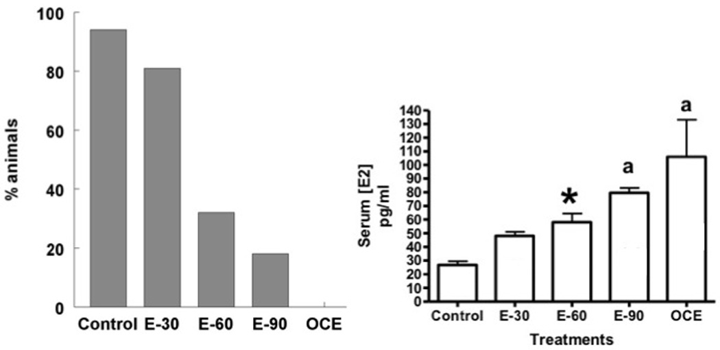
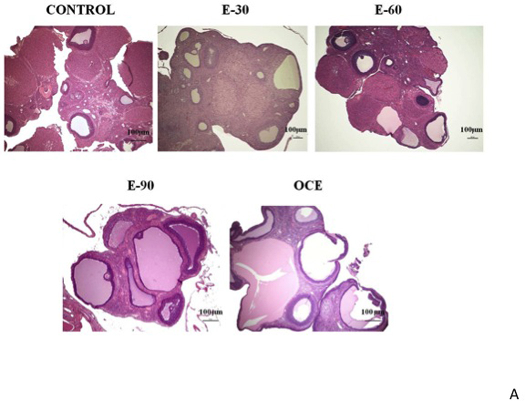
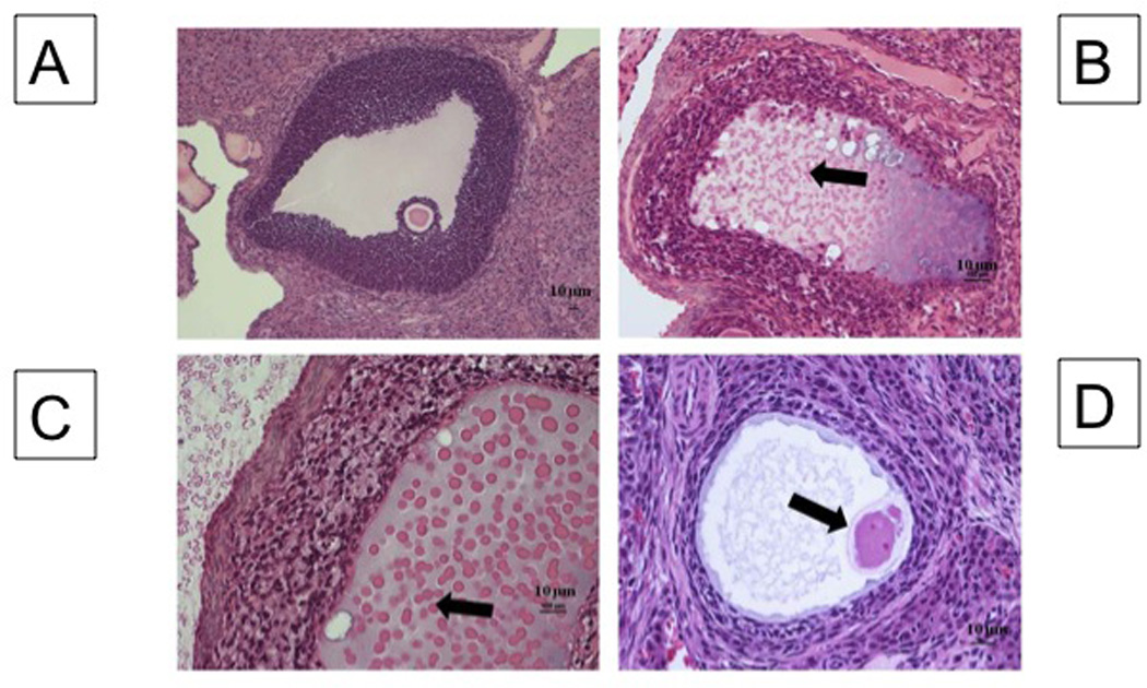
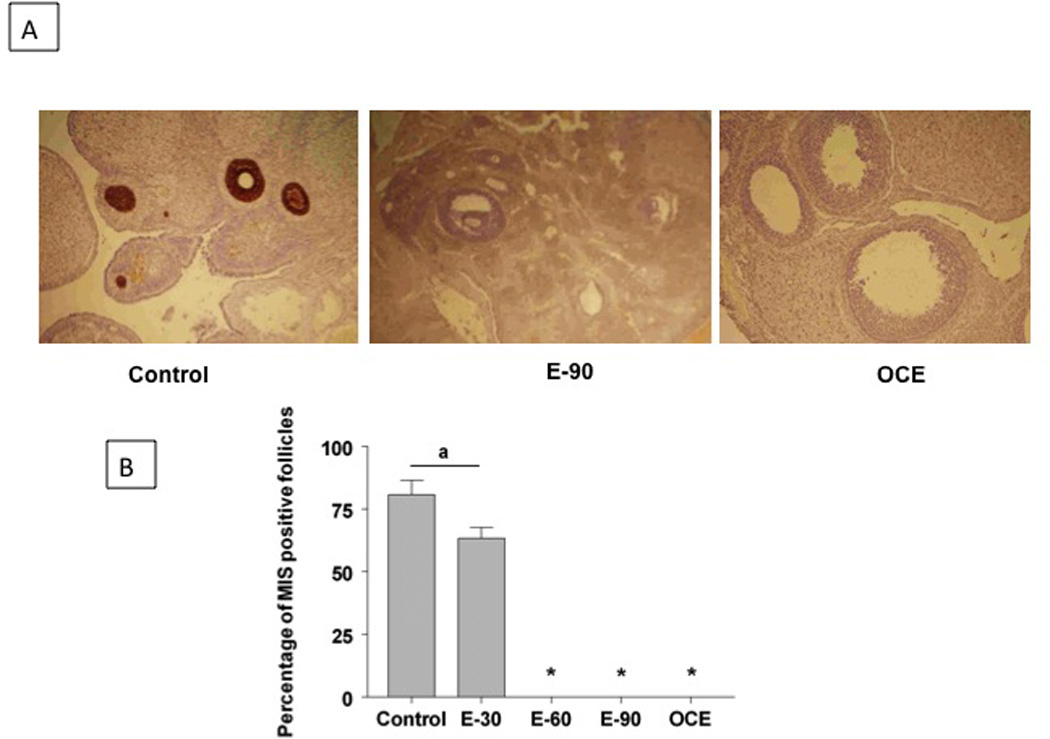
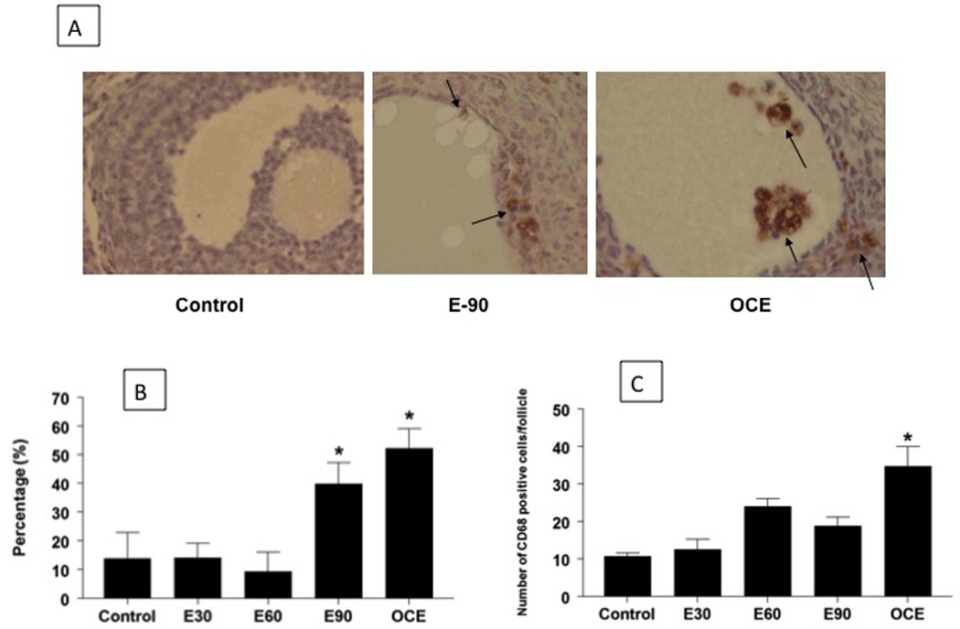
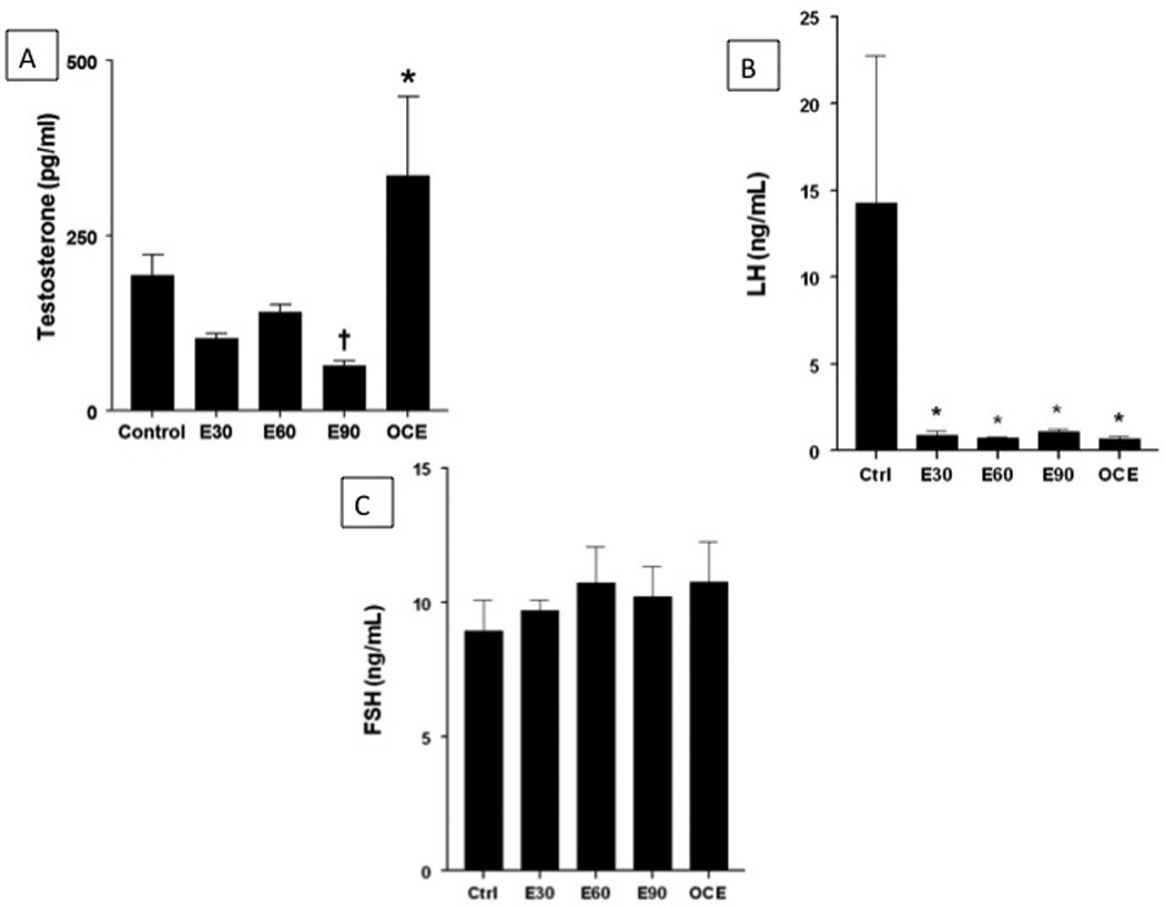
Aging in female rats is characterized by a state called "constant estrous" in which rats are unable to ovulate, have polycystic ovaries and moderately elevated estrogen levels. We hypothesized that chronic exposure of young animals to low levels of E2 can produce reproductive changes similar to that seen in aging animals. Adult female rats were sham-implanted (control) or implanted with slow-release E2 (20 ng/day) pellets for 30, 60, or 90 days. Old constant estrous (OCE) rats were used for comparison. Estrous cyclicity was monitored periodically. At the end of treatment, animals were sacrificed, trunk blood was collected for hormone measurements and ovaries for immunohistochemistry. Young animals became acyclic with increasing duration of E2 exposure while OCE rats were in a state of acyclicity. Ovaries became increasingly more cystic with E2 exposure, and were comparable to OCE rats; however, there was a marked reduction in interstitial tissue with exogenous E2 treatment. Exogenous E2 also decreased Mullerian inhibiting substance expression, increased infiltration of macrophages without much impact on apoptosis in the ovaries. Serum testosterone levels decreased in E2-treated young animals, while it increased significantly in OCE rats. There was a marked reduction in LH but not FSH levels with E2 exposure in both young and old animals. These results indicate that even very low doses of E2 are capable of inducing aging-like changes in young animals.
Keywords: CD68; Mullerian inhibiting substance; TUNEL; estrogen; follicular cysts; ovary.
Figures
Similar articles
-
Obligatory roles for follicle-stimulating hormone (FSH), estradiol and androgens in the induction of small polyfollicular ovarian cysts in hypophysectomized immature rats.Endocrine. 2007 Apr;31(2):179-92. doi: 10.1007/s12020-007-0028-5. Endocrine. 2007. PMID: 17873331
-
Effects of age on hormone levels and in vitro steroidogenesis by rat ovary and adrenal.Exp Aging Res. 1982 Fall-Winter;8(3-4):203-8. doi: 10.1080/03610738208260367. Exp Aging Res. 1982. PMID: 6820341
-
Effects of chronic exposure to estradiol on ovarian cyclicity in C57BL/6J mice: potentiation at low doses and only partial suppression at high doses.Biol Reprod. 1990 Aug;43(2):312-7. doi: 10.1095/biolreprod43.2.312. Biol Reprod. 1990. PMID: 2378943
-
Extremely high levels of estradiol and testosterone in a case of polycystic ovarian syndrome. Hormone and clinical similarities with the phenotype of the alpha estrogen receptor null mice.J Endocrinol Invest. 2000 Jul-Aug;23(7):467-72. doi: 10.1007/BF03343757. J Endocrinol Invest. 2000. PMID: 11005272
-
Induction of ovarian cysts in progesterone-synchronized immature rats: evidence that suppression of follicular aromatase activity is not a prerequisite for the induction of cystic follicles.Endocrinology. 1989 Apr;124(4):1646-53. doi: 10.1210/endo-124-4-1646. Endocrinology. 1989. PMID: 2924717
Cited by
-
Prenatal bisphenol A and/or diethylhexyl phthalate exposure followed by adult estradiol treatment affects behavior and brain monoamines in female rat offspring.Front Endocrinol (Lausanne). 2025 Jan 6;15:1479838. doi: 10.3389/fendo.2024.1479838. eCollection 2024. Front Endocrinol (Lausanne). 2025. PMID: 39839474 Free PMC article.
-
Chronic exposure to low doses of estradiol-17ß increases blood pressure in young female rats: A possible role for central Endothelin-1.Sci Rep. 2017 Mar 10;7(1):139. doi: 10.1038/s41598-017-00213-9. Sci Rep. 2017. PMID: 28273940 Free PMC article.
References
-
- Spencer AL, Bonnema R, McNamara MC. Helping women choose appropriate hormonal contraception: update on risks, benefits, and indications. Am J Med. 2009 Jun;122(6):497–506. - PubMed
-
- Brett KM, Reuben CA. Prevalence of estrogen or estrogen-progestin hormone therapy use. Obstet Gynecol. 2003 Dec;102(6):1240–1249. - PubMed
-
- Goodman A, Schorge J, Greene MF. The long-term effects of in utero exposures--the DES story. N Engl J Med. 2011 Jun 2;364(22):2083–2084. - PubMed
-
- Campbell CS, Schwartz NB, Firlit MG. The role of adrenal and ovarian steroids in the control of serum LH and FSH. Endocrinology. 1977 Jul;101(1):162–172. - PubMed
Grants and funding
LinkOut - more resources
Full Text Sources
Other Literature Sources