. 2016 Mar;12(3):167-73.
doi: 10.1038/nchembio.2001. Epub 2016 Jan 18.

A topological and conformational stability alphabet for multipass membrane proteins

Affiliations

A topological and conformational stability alphabet for multipass membrane proteins

Xiang Feng et al. Nat Chem Biol. 2016 Mar.

Abstract

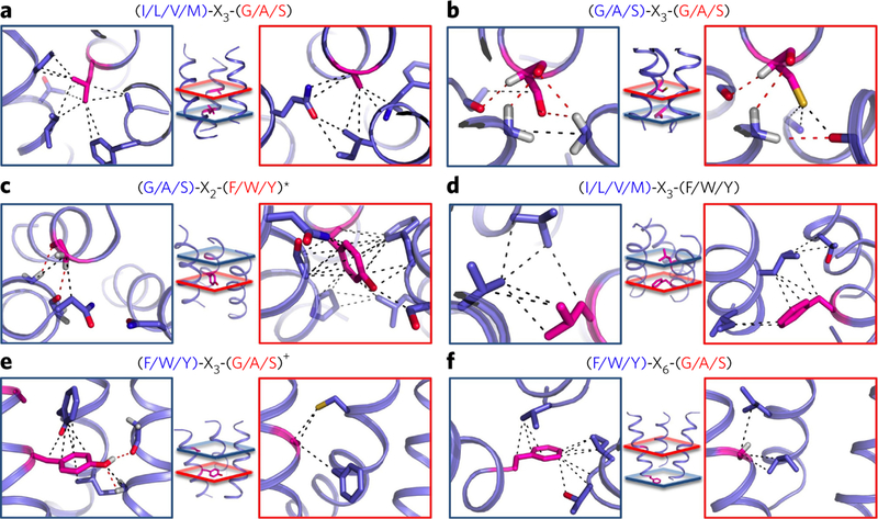
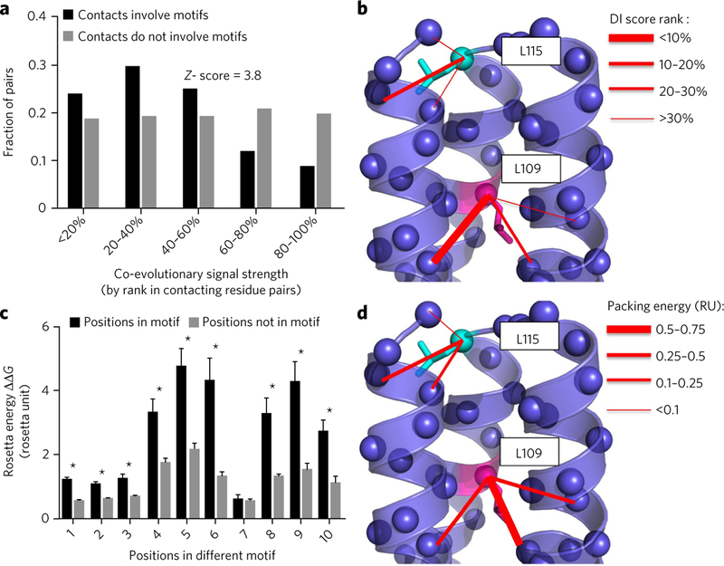
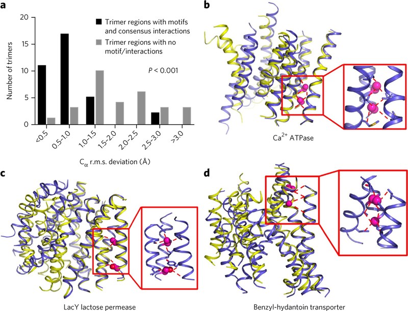
Multipass membrane proteins perform critical signal transduction and transport across membranes. How transmembrane helix (TMH) sequences encode the topology and conformational flexibility regulating these functions remains poorly understood. Here we describe a comprehensive analysis of the sequence-structure relationships at multiple interacting TMHs from all membrane proteins with structures in the Protein Data Bank (PDB). We found that membrane proteins can be deconstructed in interacting TMH trimer units, which mostly fold into six distinct structural classes of topologies and conformations. Each class is enriched in recurrent sequence motifs from functionally unrelated proteins, revealing unforeseen consensus and evolutionary conserved networks of stabilizing interhelical contacts. Interacting TMHs' topology and local protein conformational flexibility were remarkably well predicted in a blinded fashion from the identified binding-hotspot motifs. Our results reveal universal sequence-structure principles governing the complex anatomy and plasticity of multipass membrane proteins that may guide de novo structure prediction, design, and studies of folding and dynamics.


Conflict of interest statement

Competing financial interests

The authors declare no competing financial interests.

Figures

Figure 1
Figure 1. TMH trimers cluster in six major structure classes with enriched sequence motifs.
(a−f) Descriptions of six largest classes of TMH trimer structure with specific sequence motifs enriched at the interhelical interface. Sequence motifs are constituted by pairs of amino acids belonging to three chemical classes (G/A/S, small: glycine, alanine or serine; I/L/V/M, large: isoleucine, leucine, valine or methionine; F/W/Y or aromatic: phenylalanine, tryptophan or tyrosine). Residues in the motif are aligned along an ideal helix following their sequence separation (register). The helix number describes on which helix or helices of a reference trimer are found the motifs. For example, if a motif is found on helices 1 and 2 in a given trimer, the corresponding helix numbers are given as: ½. The topology and helix number of the reference trimer for each class are described in Supplementary Table 1. Each helix is colored using a blue to red spectrum from N to Cterminus. The reported motifs were found in at least 20% of trimer interacting regions in a given class and are ordered from left to right according to their frequency of occurrence in that class.
Figure 2
Figure 2. Recurrent sequence motifs create consensus interhelical trimer interactions across protein families.
(a−d) Consensus interaction networks created by each of the two sequence motif residues enriched at specific TMH trimer structure class labeled in the figure. A dashed red line indicates a consensus interhelical interaction between a motif residue (magenta) and a residue on an adjacent helix (cyan). Interaction networks are highlighted for a given sequence motif in three functionally unrelated membrane proteins.
Figure 3
Figure 3. Consensus patterns of contacts display unique combinations of atomic interactions.
(a−f) Enriched sequence motifs create unique trimer-specific interatomic van der Waals (black dotted line) and hydrogen bonding (red dotted line) interactions in local regions of the trimers defined by red and blue planes at the center of each panel. Interatomic interactions are defined for any pair of atoms distant by <5 Å belonging to one of the two motif residues (magenta) and a residue on an adjacent helix. The red and blue boxes highlight the atomic contacts from the corresponding colored planes created by each residue of the following motifs and trimer structure classes: (G/A/S)-X3-(G/A/S) motif specific to all right-handed trimers (a); (I/L/V/M)-X3-(G/A/S) motif specific to all left-handed trimers (b); (G/A/S)-X2-(F/W/Y/(M)) motif specific to parallel and left-handed trimers (c); (I/L/V/M)-X3-(F/W/Y) motif specific to parallel and right-handed trimers (d); (F/W/Y)-X3-(G/A/S) motif specific to left- and right-handed trimers (e); (F/W/Y)-X6-(G/A/S) motif specific to left- and right-handed IItrimers (f). *, methionine also found in the second position with similar contacts; +, histidine also found in the first position with similar contacts.
Figure 4
Figure 4. Sequence motifs create evolutionary conserved networks of interhelical stabilizing contacts.
(a) Distribution of residue pairs as a function of their coevolutionary strengths measured by the method EVfold23. The coevolutionary strength of a pair is reported relative to the distribution of coevolutionary scores for all residue pairs as a rank (the lower the rank, the higher the relative coevolutionary strength). Selected residue pairs are in contact across the trimer interhelical interface and involve motif residues (black) or do not involve motif residues (gray). The Z score describes the statistical significance of the difference in rank distribution between the two classes of residue pairs. (b) Example of coevolutionary scores (direct interaction (DI) score) for interresidue contacts involving either a residue in a motif (magenta) or the same residue type not in a motif (cyan) at a trimer interface. (c) Comparison between the energy contribution to the trimer stability of a residue in a motif (black) and that of the same residue type not in a motif (gray). The energy contribution was calculated by alanine scanning, and the comparison was performed for ten different motif residues and reported as mean values + s.d. *P < 0.01, comparison between motif and nonmotif residues’ energetic contributions (Welch’s two-sided t-test). (d) Example of interaction energy between contacts involving a motif residue (magenta) and contacts not involving motif residues (cyan) at a trimer interface.
Figure 5
Figure 5. Sequence−3D contact motifs are strong predictors of local conformational stability.
(a) Distribution of trimer unit structural changes (measured by Cα r.m.s. deviation in Å) in multipass membrane proteins crystallized in distinct conformations. The statistical significance in the distribution of Cα r.m.s. deviation between trimers bearing or not sequence−3Dcontact motifs was calculated using Welch’s two-sided t-test. (b−d) Examples of multipass membrane protein X-ray crystallography structures crystallized in two distinct conformations (superimposed backbone representations in blue and yellow). The trimer units containing a sequence-contact motif (red window) do not change conformations.

References

    1. von Heijne G Membrane-protein topology. Nat. Rev. Mol. Cell Biol 7, 909–918 (2006). - PubMed
    1. Matthews EE, Zoonens M & Engelman DM Dynamic helix interactions in transmembrane signaling. Cell 127, 447–450 (2006). - PubMed
    1. Krishnamurthy H, Piscitelli CL & Gouaux E Unlocking the molecular secrets of sodium-coupled transporters. Nature 459, 347–355 (2009). - PMC - PubMed
    1. Rakoczy EP, Kiel C, McKeone R, Stricher F & Serrano L Analysis of disease-linked rhodopsin mutations based on structure, function, and protein stability calculations. J. Mol. Biol 405, 584–606 (2011). - PubMed
    1. Partridge AW, Therien AG & Deber CM Missense mutations in transmembrane domains of proteins: phenotypic propensity of polar residues for human disease. Proteins 54, 648–656 (2004). - PubMed

Publication types

Substances