. 2016 May-Jul:52-54:246-259.
doi: 10.1016/j.matbio.2016.01.003. Epub 2016 Jan 15.

Accelerated enamel mineralization in Dspp mutant mice

Affiliations

Accelerated enamel mineralization in Dspp mutant mice

Kostas Verdelis et al. Matrix Biol. 2016 May-Jul.

Abstract

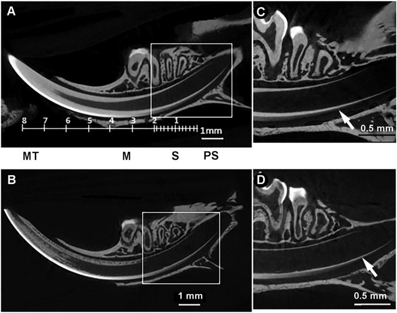
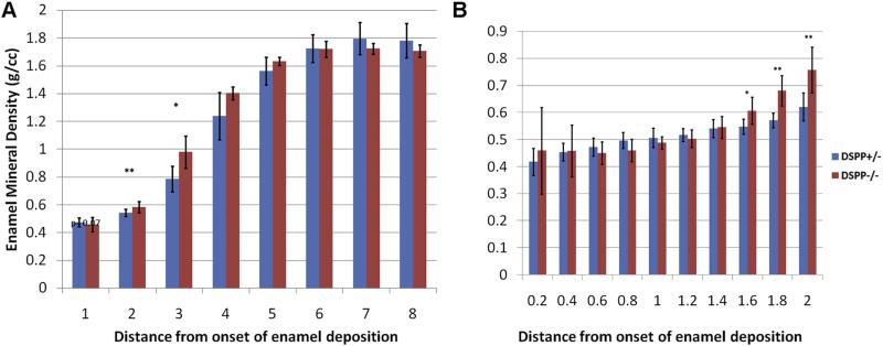
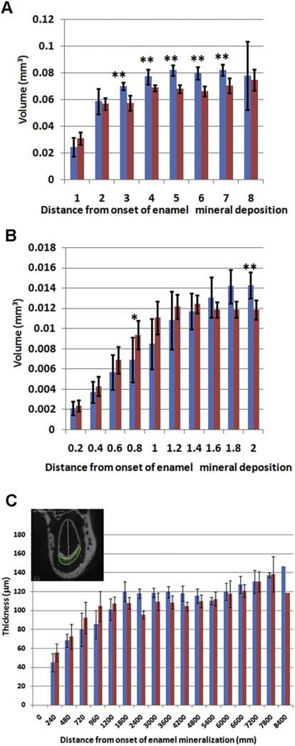
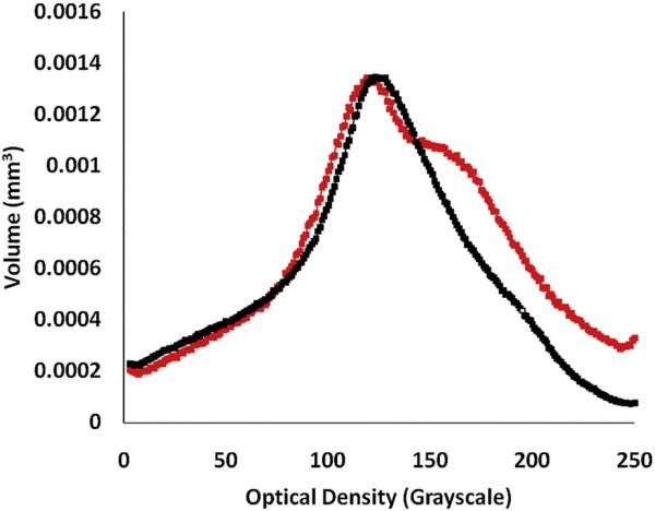
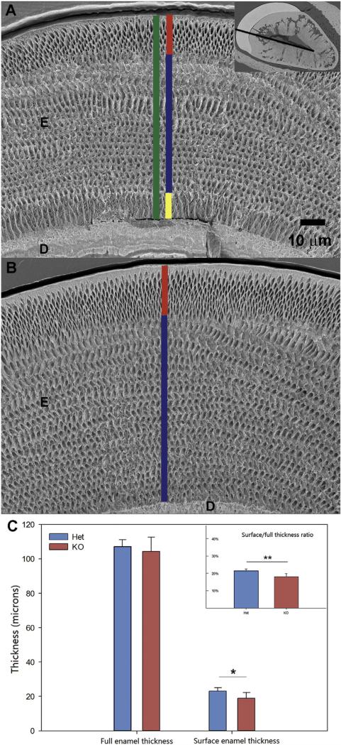
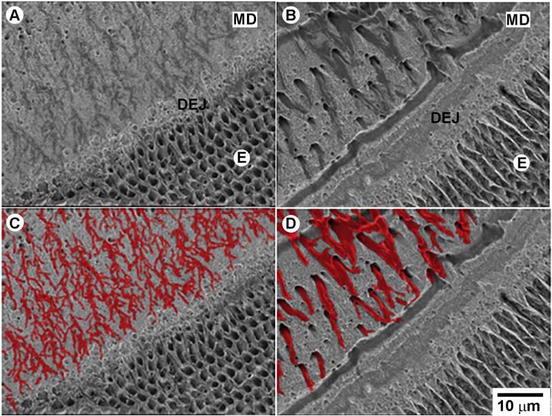
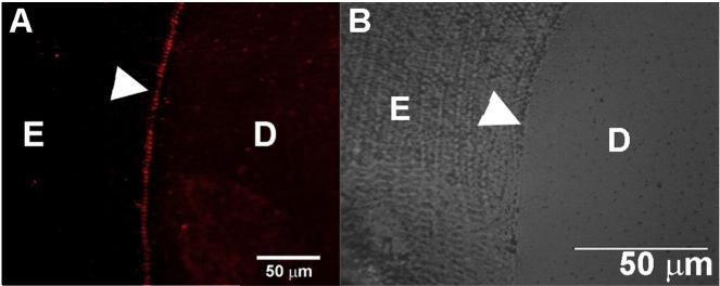
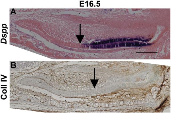
Dentin sialophosphoprotein (DSPP) is one of the major non-collagenous proteins present in dentin, cementum and alveolar bone; it is also transiently expressed by ameloblasts. In humans many mutations have been found in DSPP and are associated with two autosomal-dominant genetic diseases - dentinogenesis imperfecta II (DGI-II) and dentin dysplasia (DD). Both disorders result in the development of hypomineralized and mechanically compromised teeth. The erupted mature molars of Dspp(-/-) mice have a severe hypomineralized dentin phenotype. Since dentin and enamel formations are interdependent, we decided to investigate the process of enamel onset mineralization in young Dspp(-/-) animals. We focused our analysis on the constantly erupting mouse incisor, to capture all of the stages of odontogenesis in one tooth, and the unerupted first molars. Using high-resolution microCT, we revealed that the onset of enamel matrix deposition occurs closer to the cervical loop and both secretion and maturation of enamel are accelerated in Dspp(-/-) incisors compared to the Dspp(+/-) control. Importantly, these differences did not translate into major phenotypic differences in mature enamel in terms of the structural organization, mineral density or hardness. The only observable difference was the reduction in thickness of the outer enamel layer, while the total enamel thickness remained unchanged. We also observed a compromised dentin-enamel junction, leading to delamination between the dentin and enamel layers. The odontoblast processes were widened and lacked branching near the DEJ. Finally, for the first time we demonstrate expression of Dspp mRNA in secretory ameloblasts. In summary, our data show that DSPP is important for normal mineralization of both dentin and enamel.

Keywords: Dentin; Dentin sialophosphoprotein; Dentino-enamel junction; Enamel; Odontogenesis.


Figures

Fig. 1
Fig. 1
Enamel mineralization occurs precociously in the Dspp–/– incisor. MicroCT reconstructions of mandibles from one-month-old Dspp+/– (A, C) and Dspp–/– (B, D) mice. A and B represent sagittal views of whole jaw, note that incisor volumes were oriented to fit the whole incisor length. And C and D are close ups from the cervical parts of the incisors in the boxed areas of A and B; arrows pointing to the onset of amelogenesis. M — Maturation; MT — Mature; PS — Presecretory; S — Secretory.
Fig. 2
Fig. 2
Dspp–/– enamel has increased mineral density in the early secretory phase. Enamel mineral density profiles from one-month-old DSPP Dspp+/– (red) and Dspp–/– (blue) mouse A) whole incisor profile using 1 mm thick segments along the incisor axis and B) high resolution profile of first 2 mm of forming enamel using 0.2 mm thick segments along the incisor axis. The statistically significant differences are marked with asterisks: *–p < 0.05, **–p < 0.01. (For interpretation of the references to color in this figure legend, the reader is referred to the web version of this article.)
Fig. 3
Fig. 3
Dspp–/– enamel its maximum volume prematurely. A and B enamel volume profiles from one-month-old DSPP Dspp+/– (red) and Dspp–/– (blue) mouse — (A) whole incisor profile using 1 mm thick segments along the incisor axis and (B) high resolution profile of first 2 mm of forming enamel using 0.2 mm thick segments along the incisor axis. C. Enamel thickness profile. The inset marks the line along which the measurements were carried out. The statistically significant differences are marked with asterisks: *–p < 0.05, **–p < 0.01. (For interpretation of the references to color in this figure legend, the reader is referred to the web version of this article.)
Fig. 4
Fig. 4
Unerupted Dspp–/– molars have increased mineral density. Distribution of enamel mineral density in 1st molars from 10 day-old Dspp+/+ (black) and Dspp–/– (red) mice. (For interpretation of the references to color in this figure legend, the reader is referred to the web version of this article.)
Fig. 5
Fig. 5
The surface thickness of Dspp–/– enamel is similar to Dspp+/–, while the total enamel thickness is similar in both genotypes. Measurements of enamel thickness erupted portions of incisors in Dspp+/– and Dspp–/– one-month-old mice. A. SEM micrograph of the Dspp–/– erupted incisor showing total enamel thickness in green, surface enamel thickness in red, bulk enamel thickness in blue and inner enamel thickness in yellow. The inset shows the line along which the measurements were carried out, connecting labial and lingual corners of the pulp cavity; B. SEM micrograph of the Dspp+/– erupted incisor with the bulk enamel thickness shown in blue and the surface enamel thickness in yellow; C. The histograms show total enamel thickness, outer enamel thickness and outer to total enamel thickness ratio. One asterisk identifies p < 0.05, two asterisks identify p < 0.01. D—dentin, E—enamel. (For interpretation of the references to color in this figure legend, the reader is referred to the web version of this article.)
Fig. 6
Fig. 6
Dspp–/– teeth present with major defects at the DEJ, mantle dentin and inner enamel. BS SEM images of etched erupted portions of incisors mice, polished in the transverse plane and resin cast, from one-month-old mice. A. Dspp+/+ and B. Dspp–/–. C and D correspond to images A and B with the odontoblasts processes colored for a better visual perception. All micrographs are taken at the same magnification. E—enamel, MD—mantle dentin. (For interpretation of the references to color in this figure legend, the reader is referred to the web version of this article.)
Fig. 7
Fig. 7
DSP protein is present in the DEJ. Immunofluorescence image of a fully mineralized polished section of an erupted mouse incisor showing expression of DSPP. D—dentin, E—enamel.
Fig. 8
Fig. 8
Dspp mRNA is expressed in the secretory ameloblasts A) (mRNA) Dspp expression (purple) is found in the ameloblast and odontoblast layers of the incisor at E16.5. Arrow points to the onset of the DSPP expression. B) Collagen IV (brown stain) is found in the basal lamina which is digested during the onset of the secretory stage of amelogenesis (indicated by the arrow). Scale bars = 200 μm.

References

    1. Jernvall J, Thesleff I. Reiterative signaling and patterning during mammalian tooth morphogenesis. Mech. Dev. 2000;92:19–29. - PubMed
    1. Ten NA. Structure, and Function. Mosby; St. Luis: 2007. Cate's Oral Histology: Development.
    1. Takano Y, Sakai H, Baba O, Terashima T. Differential involvement of matrix vesicles during the initial and appositional mineralization processes in bone, dentin, and cementum. Bone. 2000;26:333–339. - PubMed
    1. Zaslansky P, Friesem AA, Weiner S. Structure and mechanical properties of the soft zone separating bulk dentin and enamel in crowns of human teeth: insight into tooth function. J. Struct. Biol. 2006;153:188–199. - PubMed
    1. Simmer JP, Hu JCC. Expression, structure, and function of enamel proteinases. Connect. Tissue Res. 2002;43:441–449. - PubMed

Publication types

LinkOut - more resources