. 2016 Feb;15(1):67-76.
doi: 10.1111/acel.12410. Epub 2015 Oct 8.

Reduction of fibroblast size/mechanical force down-regulates TGF-β type II receptor: implications for human skin aging

Affiliations

Reduction of fibroblast size/mechanical force down-regulates TGF-β type II receptor: implications for human skin aging

Gary J Fisher et al. Aging Cell. 2016 Feb.

Abstract

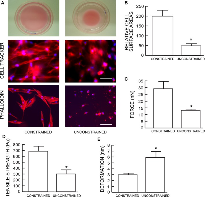
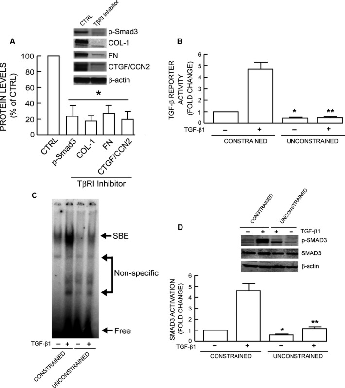
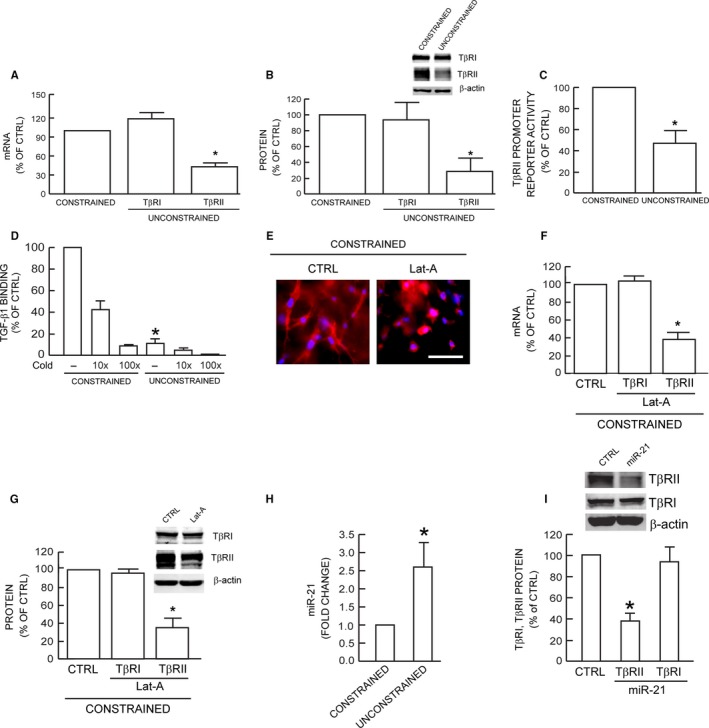
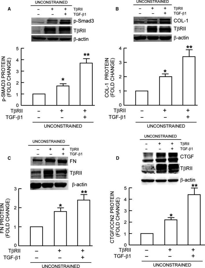
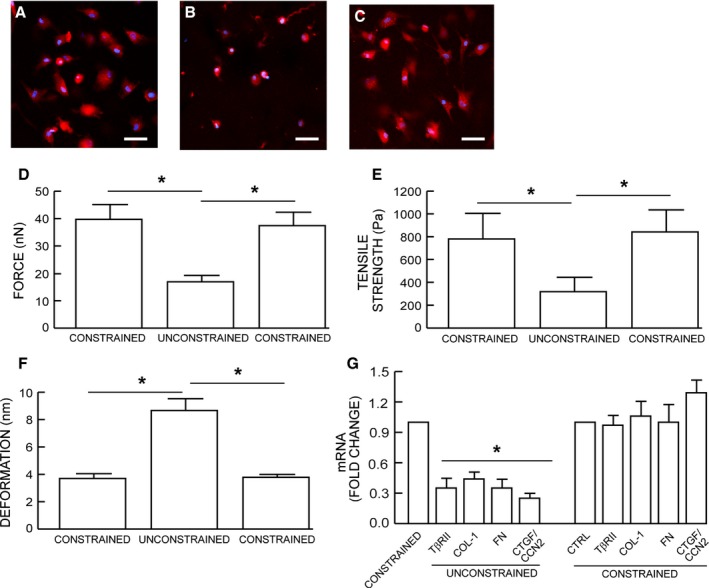
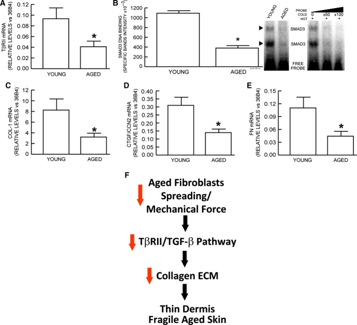
The structural integrity of human skin is largely dependent on the quality of the dermal extracellular matrix (ECM), which is produced, organized, and maintained by dermal fibroblasts. Normally, fibroblasts attach to the ECM and thereby achieve stretched, elongated morphology. A prominent characteristic of dermal fibroblasts in aged skin is reduced size, with decreased elongation and a more rounded, collapsed morphology. Here, we show that reduced size of fibroblasts in mechanically unrestrained three-dimensional collagen lattices coincides with reduced mechanical force, measured by atomic force microscopy. Reduced size/mechanical force specifically down-regulates TGF-β type II receptor (TβRII) and thus impairs TGF-β/Smad signaling pathway. Both TβRII mRNA and protein were decreased, resulting in 90% loss of TGF-β binding to fibroblasts. Down-regulation of TβRII was associated with significantly decreased phosphorylation, DNA-binding, and transcriptional activity of its key downstream effector Smad3 and reduced expression of Smad3-regulated essential ECM components type I collagen, fibronectin, and connective tissue growth factor (CTGF/CCN2). Restoration of TβRII significantly increased TGF-β induction of Smad3 phosphorylation and stimulated expression of ECM components. Reduced expression of TβRII and ECM components in response to reduced fibroblast size/mechanical force was fully reversed by restoring size/mechanical force. Reduced fibroblast size was associated with reduced expression of TβRII and diminished ECM production, in aged human skin. Taken together, these data reveal a novel mechanism that provides a molecular basis for loss of dermal ECM, with concomitant increased fragility, which is a prominent feature of human skin aging.

Keywords: TGF-β type II receptor, TGF-β/Smad; aging; cell size; extracellular matrix; mechanotransduction.


Figures

Figure 1
Figure 1
Dermal fibroblasts cultured in unconstrained collagen lattices display reduced size and mechanical force. (A) Dermal fibroblasts were cultured in mechanically constrained (upper left) or unconstrained (upper right) 3D type I collagen lattices. Cells were stained dye with CellTracker fluorescent, and nuclei were stained blue with DAPI (middle panels). Cellular actin filaments were stained red with phalloidin (lower panel) N = 8. (B) Relative cell surface areas were quantified by computerized image analysis (image‐pro plus software, version 4.1, Media Cybernetics, MD). Means ± SEM. N = 8, *P < 0.05. (C) traction forces, (D) tensile strength, and (E) deformation were determined by AFM PeakForce Quantitative NanoMechanics mode and nanoscope analysis software. Means ± SEM. N = 8, *< 0.05.
Figure 2
Figure 2
Reduced fibroblast size/mechanical force impairs TGF‐β signaling. (A) TGF‐β‐dependent ECM production. Constrained collagen lattices were treated with TβRI kinase inhibitor (SB431542, 10 μm) for 24 h. Smad3 phosphorylation and type I collagen, fibronectin, CTGF/CCN2 protein levels were determined by Western analysis and normalized to β‐actin (loading control). Inset shows representative Western blots. Mean ± SEM, N = 3, *< 0.05. (B) TGF‐β/Smad3 reporter activity. Cells were transfected with TGF‐β/Smad3‐dependent luciferase reporter construct (SBEX4). Twenty‐four hours after transfection, cells were harvested and cultured in collagen lattices for 1 day. Cultures were treated with TGF‐β1 (5 ng mL−1) for 16 h, and luciferase activity was determined. Mean±SEM, N = 3, *< 0.05 vs. control constrained, **< 0.05 vs. TGF‐β1 constrained. (C) Representative EMSA. Collagen lattice cultures were treated with TGF‐β1 (5 ng mL−1) for 4 h. Smad3 protein binding to target probe (containing the Smad3 binding element) was determined by EMSA, N = 3. (D) Smad3 phosphorylation. Collagen lattice cultures were treated with TGF‐β1 (5 ng mL−1) for 2 h. Smad3 phosphorylation was determined by Western analysis. Inset shows representative Western blots. Mean ± SEM, N = 3, *< 0.05 vs. control constrained, **< 0.05 vs. TGF‐β1 constrained.
Figure 3
Figure 3
Reduced fibroblast size/mechanical force specifically down‐regulates TβRII. TβRI and TβRII (A) mRNA and (B) protein levels were determined by real‐time RTPCR and Western analysis, respectively. mRNA and protein levels were normalized to 36B4 (internal housekeeping gene control) and β‐actin (loading control), respectively. Inset shows representative Western blots. Mean ± SEM, N = 8, *< 0.05. (C) TβRII promoter reporter activity. Fibroblasts were transfected with TβRII promoter reporter. Twenty‐four hours transfection, cells were harvested and cultured in collagen lattices for two days, prior to measurement of luciferase activity. Mean ± SEM, N = 3, *< 0.05. (D) Specific binding of [125I]TGF‐β1 to fibroblasts. Specificity of TGF‐β1 binding to receptor complex was confirmed by competition assay by addition of 10–100× excess of unlabeled TGF‐β1. Means ± SEM, N = 3, *< 0.05. (E) Fibroblasts were cultured in type I collagen lattices and treated with DMSO (CTRL, left) or Latrunculin‐A (Lat‐A, right, 100 nm) for 24 h. Fibroblasts were stained with CellTracker red fluorescent dye. TβRI and TβRII (F) mRNA and (G) protein levels were quantified by real‐time RTPCR and Western analysis, respectively. mRNA and protein levels were normalized by 36B4 (housekeeping gene internal control) and β‐actin (loading control), respectively. Inset shows representative Western blots. Mean ± SEM, N = 3, *< 0.05. (H) miR‐21 levels were determined by real‐time PCR and normalized to RNU6B, endogenous reference snRNA. Mean ± SEM, N = 3, *< 0.05. (I) Fibroblasts were treated with miR‐21 mimic for 24 h. Protein levels were determined by Western analysis and normalized to β‐actin (loading control). Inset shows representative Western blots. Mean ± SEM, N = 3, *< 0.05.
Figure 4
Figure 4
Expression of TβRII restores TGF‐β signaling and induction of ECM components. Fibroblasts were infected with control adenovirus virus or TβRII adenovirus. Twenty‐four hours after infection, cells were harvested and cultured in mechanically unconstrained collagen lattices for 1 day. Cultures were treated with TGF‐β1 (5 ng mL−1) for (A) one hour or (B, C, and D) 16 h. (A) Smad3 phosphorylation, (B) Type I procollagen, (C) Fibronectin, and (D) CTGF/CCN2 protein levels were determined by Western analysis. Protein levels were normalized by β‐actin (loading control). Insets show representative Western blots. Mean ± SEM, N = 3 for (A), N = 4 for (B, C, and D), *< 0.05 vs. control, **< 0.05 vs. no TGF‐β1.
Figure 5
Figure 5
Restoration of fibroblast size/mechanical force reverses impaired TGF‐β signaling and ECM component production. Fibroblasts were cultured in mechanically (A) constrained or (B) unconstrained 3D type I collagen lattices. Fibroblasts were harvested from mechanically (B) unconstrained collagen lattices and cultured in mechanically (C) constrained collagen lattices. Cell morphology and nuclei were visualized by staining cells with CellTracker red fluorescent dye, and DAPI (blue), respectively. Cell mechanical properties, (D) traction forces and (E) tensile strength, and (F) deformation, were determined by AFM PeakForce Quantitative NanoMechanics mode and nanoscope analysis software, as described in Method. Means ± SEM. N = 8, *< 0.05. (G) Type I procollagen, fibronectin, and CTGF/CCN2 mRNA levels were determined by real‐time RTPCR. mRNA levels were normalized by the housekeeping gene (36B4, internal control). Mean ± SEM, N = 4, *< 0.05.
Figure 6
Figure 6
Reduced gene expression of TβRII and ECM components in aged human skin. Dermal fibroblasts from buttocks skin of young (<21–30 years old) and aged (>80 years old) were obtained by laser capture microdissection. mRNA levels of indicated genes were quantified by real‐time RTPCR. mRNA levels were normalized to internal control, housekeeping gene 36B4. (A) type II TGF‐β receptor, (B) Nuclear extracts were prepared from young and aged human skin dermis. Smad3 protein binding to target probe, containing consensus Smad3 binding element, was determined by EMSA. Specificity of retarded complexes was determined by addition of excess unlabeled probe. Intensities of specific retarded complex bands were quantified by Storm Molecular Imager. Means ± SEM, N = 6, *< 0.05. (C) type I procollagen, (D) connective tissue growth factor/CCN2, (E) fibronectin. Means ± SEM, N = 12, *< 0.05. (F) Reduced fibroblast size/mechanical force contributes to skin aging by impairment of TGF‐β signaling and inhibition of ECM production. See details in Discussion.

Similar articles

Cited by

References

    1. Arora PD, Narani N, McCulloch CA (1999) The compliance of collagen gels regulates transforming growth factor‐beta induction of alpha‐smooth muscle actin in fibroblasts. Am. J. Pathol. 154, 871–882. - PMC - PubMed
    1. Assoian RK, Klein EA (2008) Growth control by intracellular tension and extracellular stiffness. Trends Cell Biol. 18, 347–352. - PMC - PubMed
    1. Bonnans C, Chou J, Werb Z (2014) Remodelling the extracellular matrix in development and disease. Nat. Rev. Mol. Cell Biol. 15, 786–801. - PMC - PubMed
    1. Butcher DT, Alliston T, Weaver VM (2009) A tense situation: forcing tumour progression. Nat. Rev. Cancer 9, 108–122. - PMC - PubMed
    1. Buxboim A, Swift J, Irianto J, Spinler KR, Dingal PC, Athirasala A, Kao YR, Cho S, Harada T, Shin JW, Discher DE (2014) Matrix elasticity regulates lamin‐A, C phosphorylation and turnover with feedback to actomyosin. Curr. Biol. 24, 1909–1917. - PMC - PubMed

Publication types

Substances