. 2016 Jan 19:6:19234.
doi: 10.1038/srep19234.

Plug-and-Display: decoration of Virus-Like Particles via isopeptide bonds for modular immunization

Affiliations

Plug-and-Display: decoration of Virus-Like Particles via isopeptide bonds for modular immunization

Karl D Brune et al. Sci Rep. .

Abstract

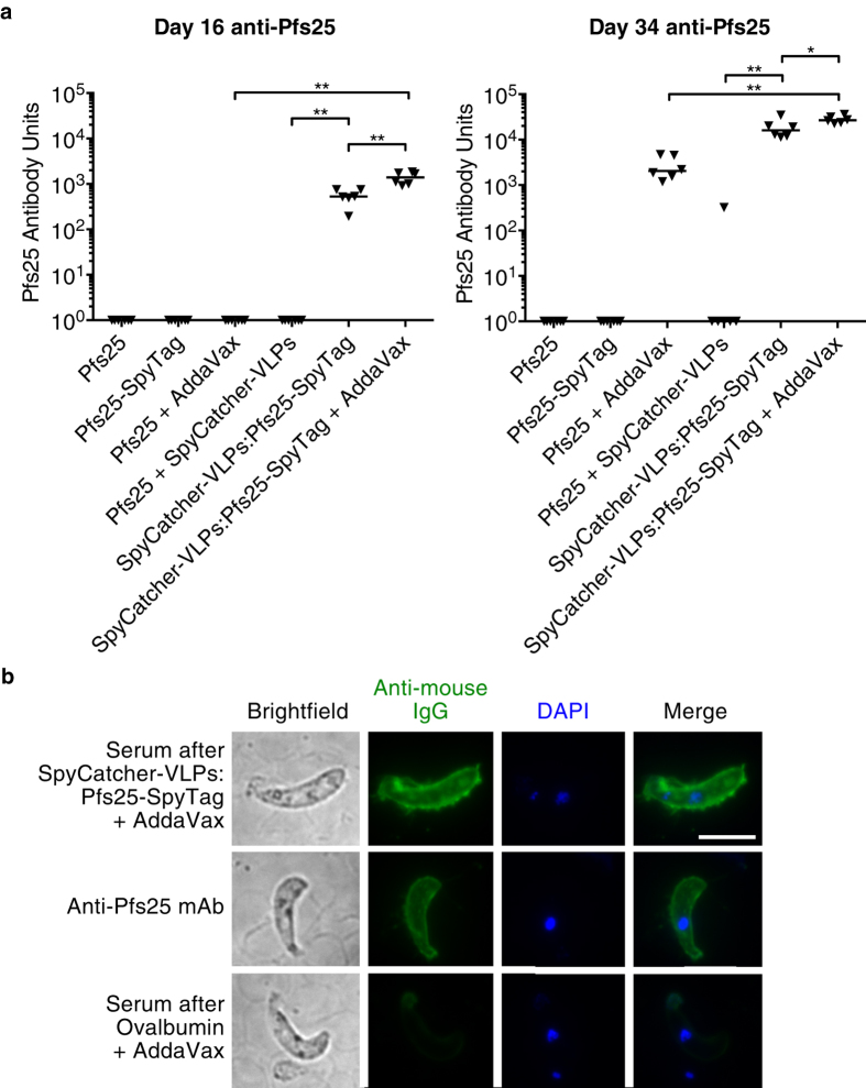
Virus-like particles (VLPs) are non-infectious self-assembling nanoparticles, useful in medicine and nanotechnology. Their repetitive molecularly-defined architecture is attractive for engineering multivalency, notably for vaccination. However, decorating VLPs with target-antigens by genetic fusion or chemical modification is time-consuming and often leads to capsid misassembly or antigen misfolding, hindering generation of protective immunity. Here we establish a platform for irreversibly decorating VLPs simply by mixing with protein antigen. SpyCatcher is a genetically-encoded protein designed to spontaneously form a covalent bond to its peptide-partner SpyTag. We expressed in E. coli VLPs from the bacteriophage AP205 genetically fused to SpyCatcher. We demonstrated quantitative covalent coupling to SpyCatcher-VLPs after mixing with SpyTag-linked to malaria antigens, including CIDR and Pfs25. In addition, we showed coupling to the VLPs for peptides relevant to cancer from epidermal growth factor receptor and telomerase. Injecting SpyCatcher-VLPs decorated with a malarial antigen efficiently induced antibody responses after only a single immunization. This simple, efficient and modular decoration of nanoparticles should accelerate vaccine development, as well as other applications of nanoparticle devices.


Conflict of interest statement

M.H. is an inventor on a patent regarding peptide targeting via spontaneous amide bond formation (EP2534484).

Figures

Figure 1
Figure 1. Overview of Plug-and-Display VLP assembly. SpyCatcher is genetically fused to the AP205 phage coat protein (AP205 CP3) and expressed in E. coli.
Self-assembly of monomers generates SpyCatcher-VLPs. Upon mixing, SpyTag-antigen forms a spontaneous isopeptide bond with SpyCatcher-VLPs, yielding decorated particles for immunization.
Figure 2
Figure 2. Characterization of SpyCatcher-VLP assembly.
(a) Size-exclusion chromatography showed SpyCatcher-CP3 assembly into VLPs, analyzed by absorbance at 260 and 280 nm and compared to Mw markers. (b) Negatively-stained TEM image of SpyCatcher-VLPs. Scale bar 100 nm. (c) Size distribution of SpyCatcher-VLPs from TEM (n = 100).
Figure 3
Figure 3. Decoration of VLPs by spontaneous isopeptide bond formation.
(a) Complete reaction of SpyCatcher-VLPs with SpyTag. SpyCatcher-VLPs were incubated with SpyTag-MBP or the negative control SpyTag DA-MBP, before boiling in SDS-loading buffer and analysis by SDS-PAGE with Coomassie staining. (b) SpyCatcher-VLP reacted with malarial protein antigens. As in (a), except with SpyTag-CIDR(HB3var03), SpyTag-CIDR(IT4var07) or Pfs25-SpyTag. (c) Native agarose gel electrophoresis of VLPs. The gel was stained with SybrSafe (nucleic acid) and then Coomassie dye (protein), showing unconjugated SpyCatcher-VLPs and SpyCatcher-VLPs conjugated with SpyTag-CIDR(IT4var07).
Figure 4
Figure 4. Plug-and-Display immunization against CIDR.
(a) Schematic of immunization route with SpyCatcher-VLPs covalently decorated with SpyTag-CIDR(IT4var07) or the negative controls: SpyCatcher-VLPs + untagged CIDR (no covalent association) or CIDR alone. (b) Time-course of immunization and sampling. (c) Antibody response to CIDR after Plug-and-Display immunization. 6 mice per condition were immunized on days 0 and 14 with CIDR, SpyCatcher-VLPs + untagged CIDR, or SpyCatcher-VLPs conjugated to SpyTag-CIDR. After 13 days (left) or 28 days (right), the total IgG anti-CIDR titer was determined by ELISA. Triangles represent the value for each mouse, while the horizontal bar represents the median. ns not significant, **p < 0.01, determined by Mann-Whitney test (n = 6).
Figure 5
Figure 5. Plug-and-Display immunization against Pfs25.
(a) Antibodies were raised to Pfs25 after SpyCatcher-VLP immunization. 6 mice per condition were immunized on day 0 and 17 with Pfs25, Pfs25-SpyTag, Pfs25-SpyTag conjugated to SpyCatcher-VLPs, or SpyCatcher-VLPs + untagged Pfs25. Where indicated, the adjuvant AddaVax was included. After prime (16 days, left) or boost (34 days, right), total anti-Pfs25 IgG was determined by ELISA (expressed as Pfs25 Antibody Units, by comparison to reference serum). Triangles represent the value for each mouse, while the horizontal bar represents the median. *p < 0.05, **p < 0.01, determined by Mann-Whitney test (n = 6). (b) Antibodies from SpyCatcher-VLPs:Pfs25-SpyTag immunization bound the ookinete surface. Ookinetes expressing Pfs25 were stained with day 34 serum from a mouse immunized with SpyCatcher-VLPs:Pfs25-SpyTag and AddaVax (top row). As a positive control, cells were stained with a monoclonal antibody against Pfs25 (middle row). As a negative control, cells were stained with serum from a mouse immunized with ovalbumin and AddaVax (bottom row). Cells were visualized in the fluorescence microscope, shown with brightfield imaging (grayscale, left), antibody staining (green, center left), DAPI DNA stain (blue, center right), and an overlay of DAPI and antibody staining (right). Scale bar = 5 μm.

References

    1. Sapsford K. E. et al. Functionalizing Nanoparticles with Biological Molecules: Developing Chemistries that Facilitate Nanotechnology. Chem. Rev. 113, 1904–2074 (2013). - PubMed
    1. De Gregorio E. & Rappuoli R. From empiricism to rational design: a personal perspective of the evolution of vaccine development. Nat. Rev. Immunol. 14, 505–514 (2014). - PMC - PubMed
    1. Draper S. J. & Heeney J. L. Viruses as vaccine vectors for infectious diseases and cancer. Nat. Rev. Microbiol. 8, 62–73 (2010). - PubMed
    1. Marston H. D., Folkers G. K., Morens D. M. & Fauci A. S. Emerging Viral Diseases: Confronting Threats with New Technologies. Sci. Transl. Med. 6, 1–6 (2014). - PubMed
    1. Smith D. M., Simon J. K. & Baker J. R. Jr Applications of nanotechnology for immunology. Nat. Rev. Immunol. 13, 592–605 (2013). - PMC - PubMed

Publication types