. 2016 Mar;34(3):601-13.
doi: 10.1002/stem.2298. Epub 2016 Feb 19.

Comprehensive Proteomic Analysis of Mesenchymal Stem Cell Exosomes Reveals Modulation of Angiogenesis via Nuclear Factor-KappaB Signaling

Affiliations

Comprehensive Proteomic Analysis of Mesenchymal Stem Cell Exosomes Reveals Modulation of Angiogenesis via Nuclear Factor-KappaB Signaling

Johnathon D Anderson et al. Stem Cells. 2016 Mar.

Abstract

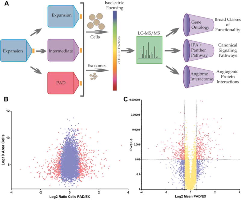
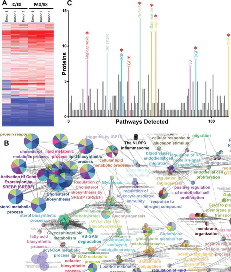
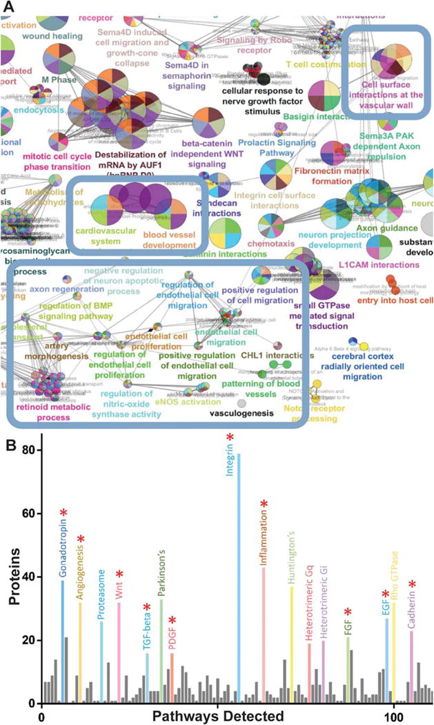
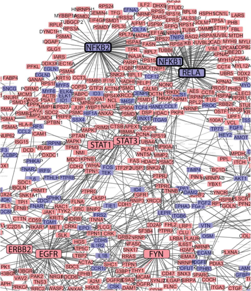
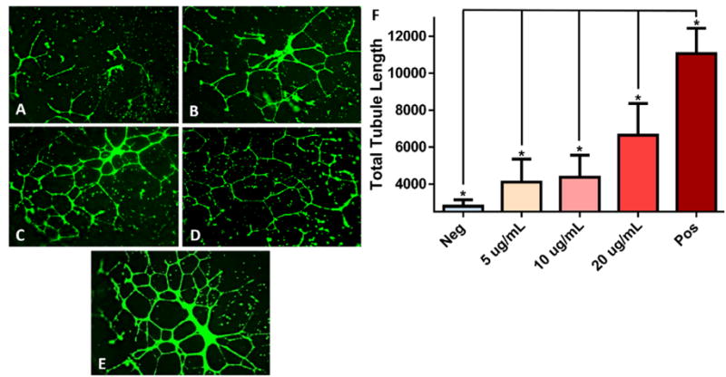
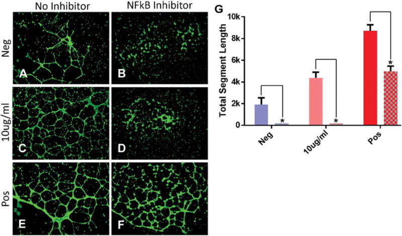
Mesenchymal stem cells (MSC) are known to facilitate healing of ischemic tissue related diseases through proangiogenic secretory proteins. Recent studies further show that MSC derived exosomes function as paracrine effectors of angiogenesis, however, the identity of which components of the exosome proteome responsible for this effect remains elusive. To address this we used high-resolution isoelectric focusing coupled liquid chromatography tandem mass spectrometry, an unbiased high throughput proteomics approach to comprehensively characterize the proteinaceous contents of MSCs and MSC derived exosomes. We probed the proteome of MSCs and MSC derived exosomes from cells cultured under expansion conditions and under ischemic tissue simulated conditions to elucidate key angiogenic paracrine effectors present and potentially differentially expressed in these conditions. In total, 6,342 proteins were identified in MSCs and 1,927 proteins in MSC derived exosomes, representing to our knowledge the first time these proteomes have been probed comprehensively. Multilayered analyses identified several putative paracrine effectors of angiogenesis present in MSC exosomes and increased in expression in MSCs exposed to ischemic tissue-simulated conditions; these include platelet derived growth factor, epidermal growth factor, fibroblast growth factor, and most notably nuclear factor-kappaB (NFkB) signaling pathway proteins. NFkB signaling was identified as a key mediator of MSC exosome induced angiogenesis in endothelial cells by functional in vitro validation using a specific inhibitor. Collectively, the results of our proteomic analysis show that MSC derived exosomes contain a robust profile of angiogenic paracrine effectors, which have potential for the treatment of ischemic tissue-related diseases.

Keywords: Exosomes; High-resolution isoelectric focusing; Liquid chromatography tandem mass spectrometry; Mesenchymal stem cells; Nuclear factor kappaB; Peripheral arterial disease; Proteomics.


Conflict of interest statement

Disclosure of Potential Conflicts of Interest

The authors indicate no potential conflicts of interest.

Figures

Figure 1
Figure 1
Experimental design workflow and ratio distribution of mesenchymal stem cell (MSC) proteomics. (A): Schematic representation of proteomics workflow. MSCs were isolated from human bone marrow and expanded to passage 6 using EX conditions. Cells were then washed three times with phosphate buffered saline and switched to either EX, IC, or PAD-like (PAD) conditions for 40 hours. Cells or exosomes were then lysed, trypsinized, and ran on high-resolution isoelectric focusing strips which were divided into 72 individual fractions and ran on LC-MS/MS. Identified proteins were analyzed using three different types of analysis software: gene ontology, canonical signaling pathways, and network analysis of the angiome interactome. ClueGO gene ontology analysis was used to characterize enrichment for proteins based on their functionalities. Panther and Ingenuity pathway analysis were used to characterize enrichment for proteins of specific canonical signaling pathways. CytoScape network analysis of the angiome interactome was used to visualize the physical interactions of known angiogenesis-mediating proteins (angiome) with proteins for which there is experimental evidence of physical interaction. (B): Plot of PAD/EX ratios (Log2, fold change) versus area (Log10, abundance) of MSC proteins; red dots represent significantly differentially expressed proteins (FDR1% [false discovery rate]), blue dots represent all nonsignificantly differentially expressed proteins. (C): PAD/EX ratios (Log2, fold change) versus p value; yellow dots represent differentially expressed proteins with mean fold changes < ±0.5 Log2, red dots represent > ±0.5 Log2 mean fold change with p value < .01 and blue dots with a p value of >.01. Abbreviations: EX, expansion condition; LC-MS/MS, liquid chromatography tandem mass spectrometry; PAD, peripheral arterial disease.
Figure 2
Figure 2
Analysis of high-resolution isoelectric focusing coupled liquid chromatography tandem mass spectrometry proteomics data from IC and PAD conditions compared to control condition EX. (A): Heatmap of mesenchymal stem cell (MSC) cluster analysis of differentially regulated proteins in IC and PAD conditions as compared to EX. (B): ClueGO gene ontology analysis of proteins upregulated in PAD MSCs shows enrichment for cholesterol biosynthesis, lipid biosynthesis, angiogenesis, and glycolysis associated proteins. (C): Panther pathway analysis of proteins upregulated in MSCs under PAD-like conditions show abundance of canonical angiogenesis related pathway proteins: epidermal growth factor, fibroblast growth factor, and platelet derived growth factor (red asterisk indicate angiogenesis associated pathways). Analysis of three different donors for each condition. For differential expression t tests with multiple testing correction with an false discovery rate of 1% was used. Circles are color coded according to their associated functionality. Number of circles and larger diameter of circles indicate greater over representation. Abbreviations: EX, expansion condition; IC, intermediate condition; PAD, peripheral arterial disease.
Figure 3
Figure 3
Mesenchymal stem cells (MSCs) increase secretion of exosomes upon exposure to PAD-like conditions. (A): Quantification of total protein content of vesicles derived from MSC under EX, IC, and PAD culture conditions using DC assay. (B): Scanning electron micrograph of MSCs cultured in EX culture conditions indicating microvesicle release (blue arrows) from the cell surface (scale bar = 5 μm, × 5k). (C): Scanning electron micrograph of MSCs cultured under PAD conditions (scale bar 2 μm, × 10k) indicating exosome adhesion to cell surface (red arrows). (D): Transmission electron micrograph of MSC derived exosomes with 2% uranyl acetate negative staining (scale bar 200 nm, × 25k). Abbreviations: EX, expansion condition; Exo, exosomes; IC, intermediate condition; MV, microvesicle; PAD, peripheral arterial disease.
Figure 4
Figure 4
Analysis of high-resolution isoelectric focusing coupled liquid chromatography tandem mass spectrometry proteomics data of mesenchymal stem cell (MSC) exosomes comparing peripheral arterial disease (PAD) to intermediate condition (IC). (A): ClueGO network analysis of 400 most abundant proteins in PAD exosomes shows enrichment for effector proteins (blue boxes). (B): Panther pathway analysis of PAD exosomes shows abundance of angiogenesis related pathway proteins: epidermal growth factor receptor, fibroblast growth factor, and platelet derived growth factor pathway associated proteins (red asterisk indicate angiogenesis associated pathways). Analysis of three different donors for each condition. For differential expression t tests with multiple testing correction with an false discovery rate of 1% was used. Circles are color coded according to their associated functionality. Number of circles and larger diameter of circles indicate greater over representation.
Figure 5
Figure 5
Network analysis of mesenchymal stem cell (MSC) exosome angiogenesis interactome. Network analysis using CytoScape of the MSC exosome angiogenesis interactome reveals clustering around nodes involved in nuclear factor kappaB signaling (emboldened boxes). Red boxes indicate presence of angiome interacting proteins in MSC exosomes, blue boxes indicate absence of these network proteins in MSC exosomes. Boxes of major clustering nodes of known effectors were enlarged for clarity. Edges connecting boxes indicates experimental evidence of physical contact (e.g., coimmunopreciptitation, yeast-2-hybrid).
Figure 6
Figure 6
Mesenchymal stem cell (MSC) exosome-induced in vitro tubule formation of human umbilical cord vein endothelial cells. (A): Basal media (Neg), (B) 5 μg/mL, (C) 10 μg/mL, (D) 20 μg/mL of MSC exosomes in basal media, (E) EndoGRO media positive control (Pos). Stained with Calcein AM and imaged at 14 hours poststimulation with × 4 objective. (F): Quantification of total segment length of tubule formation analyzed using ImageJ’s Angiogenesis plugin. EndoGRO positive control media contains 2% fetal bovine serum, epidermal growth factor 5 ng/mL and heparin sulfate 0.75 U/mL. (*) Indicates a p value < .05 using ANOVA, LSD post hoc analysis (n = 12).
Figure 7
Figure 7
NFkB inhibition abrogates mesenchymal stem cell (MSC) exosome-mediated tubule formation in human umbilical cord vein endothelial cells (HUVECs) in vitro. (A) Basal media, (B) basal media + NFkB inhibitor, (C) 10 μg/mL, (D) 10 μg/mL + NFkB inhibitor, (E) EndoGRO media, (F) EndoGRO media + NFkB inhibitor. HUVECs stained with Calcein AM and imaged 14 hours poststimulation with a ×4 objective. (G): Quantification of total segment length of tubule formation using ImageJ’s Angiogenesis plugin. EndoGRO media contains 2% fetal bovine serum, epidermal growth factor 5 ng/mL, and heparin sulfate 0.75 U/mL. (*) Indicates a p value < .01 using ANOVA, LSD post hoc analysis (n = 6). Abbreviation: NFkB, nuclear factor kappaB.

Similar articles

Cited by

References

    1. Milani RV, Lavie CJ. The role of exercise training in peripheral arterial disease. Vasc Med. 2007;12:351–358. - PubMed
    1. Katz G, Harchandani B, Shah B. Drug-eluting stents: The past, present, and future. Curr Atherosclerosis Rep. 2015;17:485. - PubMed
    1. Banfi A, von Degenfeld G, Gianni-Barrera R, et al. Therapeutic angiogenesis due to balanced single-vector delivery of VEGF and PDGF-BB. FASEB J. 2012;26:2486–2497. - PMC - PubMed
    1. Yla-Herttuala S, Rissanen TT, Vajanto I, et al. Vascular endothelial growth factors: Biology and current status of clinical applications in cardiovascular medicine. J Am Coll Cardiol. 2007;49:1015–1026. - PubMed
    1. Meyerrose T, Olson S, Pontow S, et al. Mesenchymal stem cells for the sustained in vivo delivery of bioactive factors. Adv Drug Deliv Rev. 2010;62:1167–1174. - PMC - PubMed

Publication types