

Near Infrared Light Scattering Changes Following Acute Brain Injury
- PMID: 26782205
- PMCID: PMC5973267
- DOI: 10.1007/978-1-4939-3023-4_17
Near Infrared Light Scattering Changes Following Acute Brain Injury
Erratum in
-
Erratum.Adv Exp Med Biol. 2016;876:E1-E2. doi: 10.1007/978-1-4939-3023-4_66. Adv Exp Med Biol. 2016. PMID: 27785776 Free PMC article. No abstract available.
Abstract
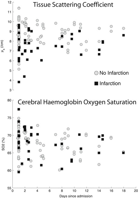
Acute brain injury (ABI) is associated with changes in near infrared light absorption reflecting haemodynamic and metabolic status via changes in cerebral oxygenation (haemoglobin oxygenation and cytochrome-c-oxidase oxidation). Light scattering has not been comprehensively investigated following ABI and may be an important confounding factor in the assessment of chromophore concentration changes, and/or a novel non-invasive optical marker of brain tissue morphology, cytostructure, hence metabolic status. The aim of this study is to characterize light scattering following adult ABI. Time resolved spectroscopy was performed as a component of multimodal neuromonitoring in critically ill brain injured patients. The scattering coefficient (μ's), absorption coefficient and cerebral haemoglobin oxygen saturation (SO2) were derived by fitting the time resolved data. Cerebral infarction was subsequently defined on routine clinical imaging. In total, 21 patients with ABI were studied. Ten patients suffered a unilateral frontal infarction, and mean μ' s was lower over infarcted compared to non-infarcted cortex (injured 6.9/cm, non-injured 8.2/cm p=0.002). SO2 did not differ significantly between the two sides (injured 69.3%, non-injured 69.0% p=0.7). Cerebral infarction is associated with changes in μ' s which might be a novel marker of cerebral injury and will interfere with quantification of haemoglobin/cytochrome c oxidase concentration. Although further work combining optical and physiological analysis is required to elucidate the significance of these results, μ' s may be uniquely placed as a non-invasive biomarker of cerebral energy failure as well as gross tissue changes.
Keywords: Brain injury; Cerebral ischaemia; Near infrared spectroscopy; Scattering.
Figures

References
Publication types
MeSH terms
Grants and funding
LinkOut - more resources
Full Text Sources
Other Literature Sources
Research Materials
