. 2016 Mar;48(3):1007-15.
doi: 10.3892/ijo.2016.3323. Epub 2016 Jan 5.

Insulin-like growth factor 2 mRNA-binding protein-3 as a marker for distinguishing between cutaneous squamous cell carcinoma and keratoacanthoma

Affiliations

Insulin-like growth factor 2 mRNA-binding protein-3 as a marker for distinguishing between cutaneous squamous cell carcinoma and keratoacanthoma

Akiko Kanzaki et al. Int J Oncol. 2016 Mar.

Abstract

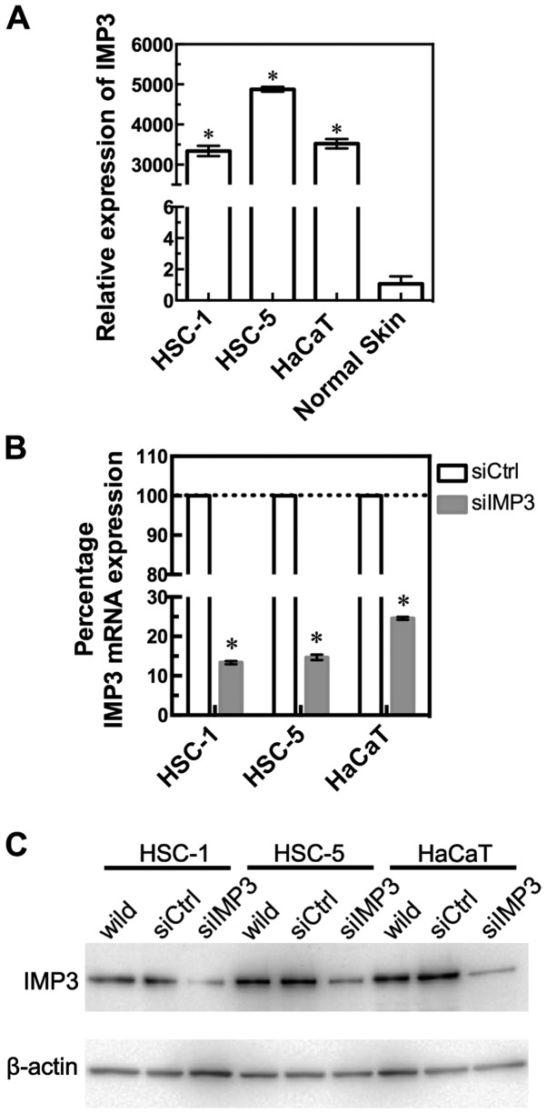
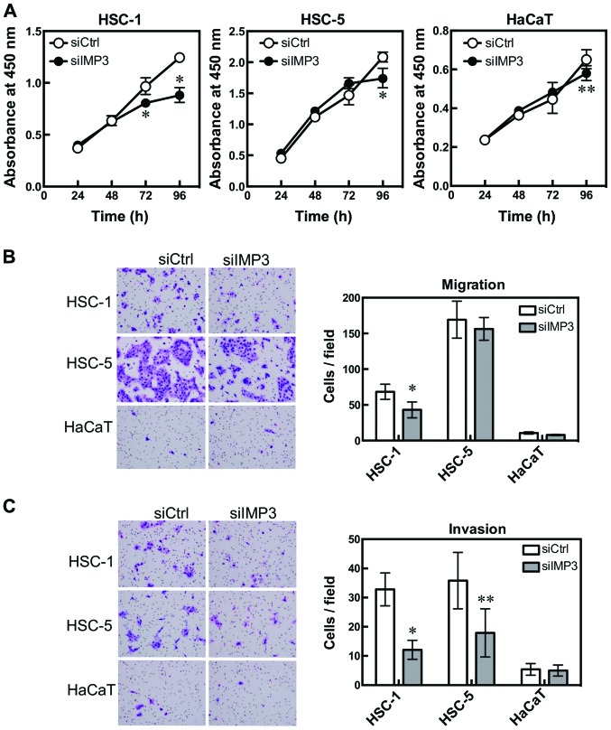
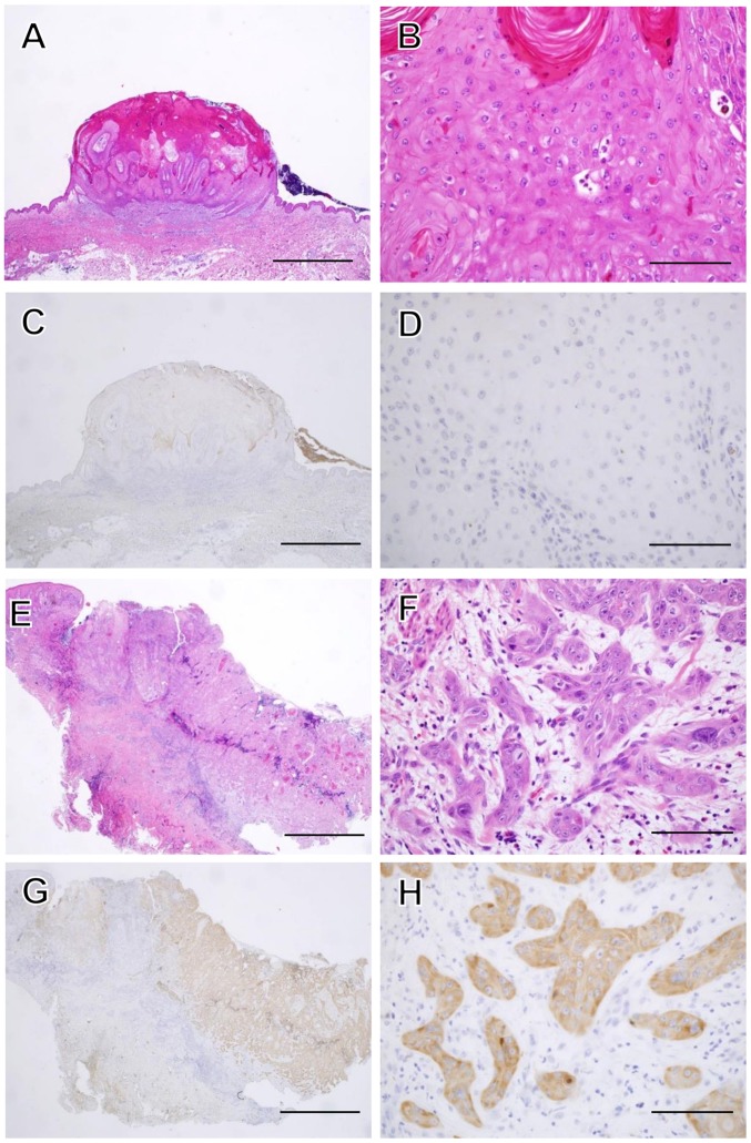
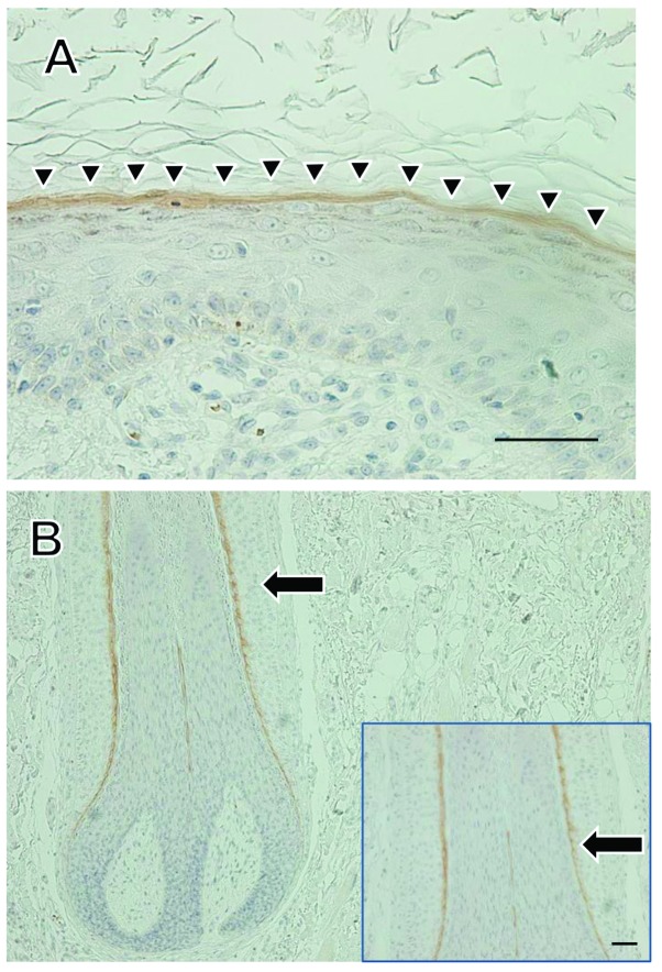
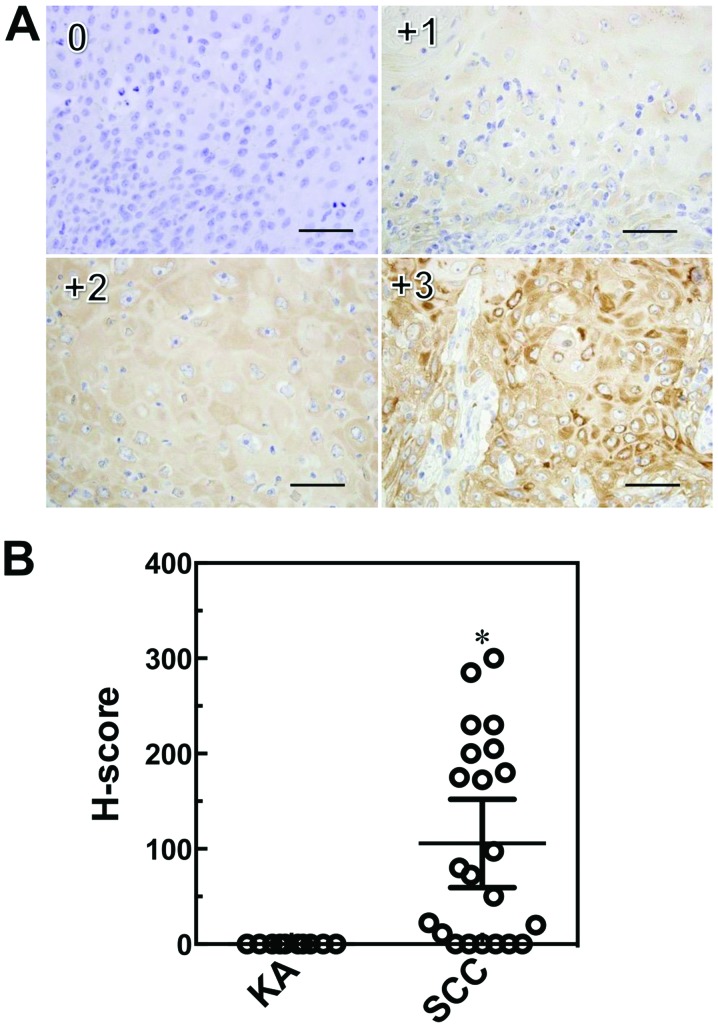
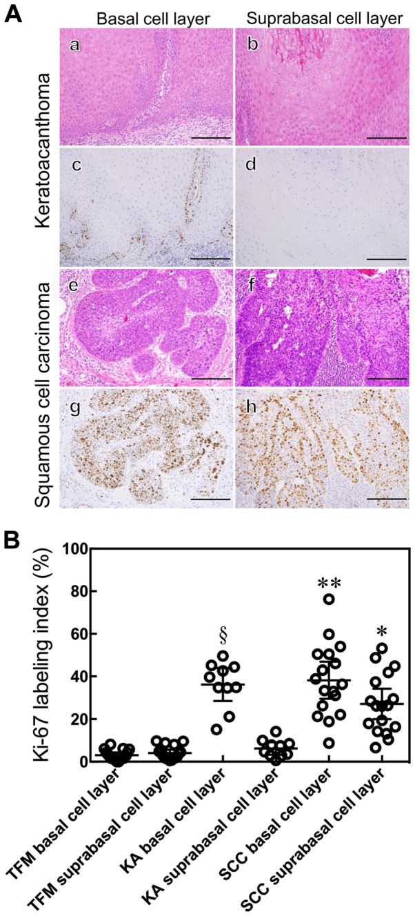
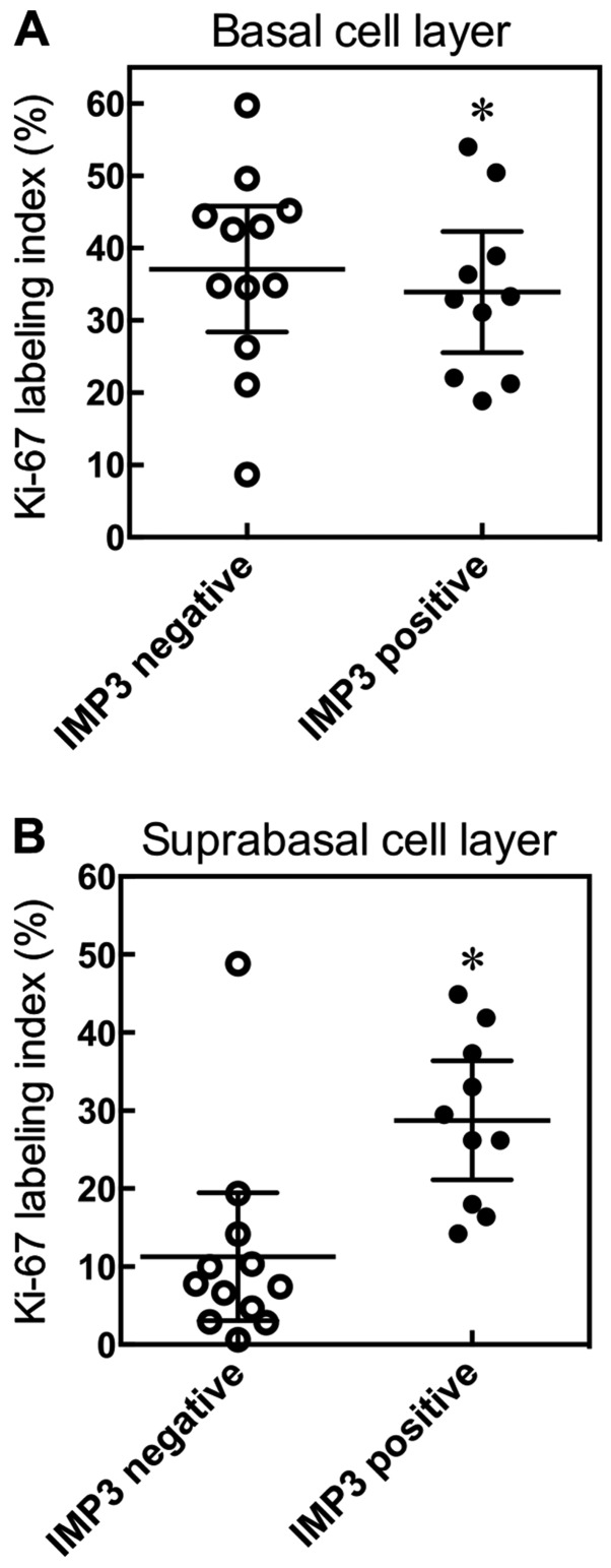
In the histopathological diagnosis of cutaneous tumors, the differential diagnosis of squamous cell carcinoma (SCC) with crateriform architecture and keratoacanthoma (KA) is often difficult so an accurate understanding of the biological features and the identification of reliable markers of SCC and KA are crucial issues. Insulin-like growth factor 2 mRNA-binding protein-3 (IGF2BP3, also known as IMP3) is thought of as a bona fide oncofetal protein, which is overexpressed and is involved in cell proliferation, migration, and invasion in several kinds of tumors. However, the role of IMP3 in cutaneous SCC and KA has not been well studied. Therefore, we focused on studying the biological functions of IMP3 in SCC and KA. In human skin SCC cell lines, HSC-1 and HSC-5, and the human keratinocyte cell line, HaCaT, IMP3 mRNA levels were significantly higher than that of normal human skin. The knockdown of IMP3 expression reduced the proliferation of HSC-1, and significantly reduced invasion by HSC-1 and HSC-5. In contrast, the knockdown of IMP3 did not significantly affect invasion by HaCaT cells. In immunohistochemical studies of SCC and KA tissues, the Ki-67 labeling index (LI) of the suprabasal cell layer was significantly higher in SCC, compared with KA tissues and the tumor-free margin (TFM) adjacent to SCC and KA. Most SCC tissues stained strongly positive for IMP3, but KA tissues and TFM were mostly negative for IMP3. The Ki-67 LI of the IMP3-positive group was significantly higher than that of the IMP3-negative group in the suprabasal cell layer of SCC. These results suggest that IMP3 plays an important role in proliferation and, more significantly, in the invasion of SCC, and may be a suitable marker for the histopathological diagnosis of SCC with a crateriform architecture and KA. Furthermore, IMP3 may potentially be a new therapeutic target for SCC.


Figures

Figure 1
Figure 1
IMP3 expression in HSC-1, HSC-5 and HaCaT cells, and normal human skin, and knockdown of IMP3 using siRNA. IMP3 mRNA expression was observed in HSC-1, HSC-5 and HaCaT cell lines and normal skin by RT-qPCR analysis. The expression of IMP3 relative to that in normal skin is shown (A). Knockout of IMP3 mRNA by siRNA in HSC-1, HSC-5 and HaCaT cell lines. IMP3 mRNA is expressed as a percentage of that in siRNA control transfected cells (100%) (B). For (A) and (B), results are expressed as mean ±95% confidence interval. *P<0.0001, compared to normal skin (A) and si-control (siCtrl)-transfected cells (B). Western blot of IMP3 protein expression after treatment with IMP3 siRNA in HSC-1, HSC-5 and HaCaT cell lines. IMP3 protein levels in untransfected wild-type, si control (siCtrl) or siIMP3 RNA-transfected cells are shown (C).
Figure 2
Figure 2
IMP3 is associated with cell proliferation, migration and invasion. HSC-1, HSC-5 and HaCaT cells were transfected with IMP3 siRNA and cell proliferation, migration and invasion were measured. Cell proliferation was determined by WST-8 cell counting reagent. HSC-1: *P<0.0001 for 72 and 96 h, Sidak's multiple comparison test, and two-way ANOVA; and HSC-5: *P<0.0001 for 96 h; HaCaT: **P=0.0008 for 96 h, Sidak's multiple comparison test, compared with si-control (siCtrl)-transfected cells (A). Cell migration was measured using BioCoat chambers. HSC-1: *P=0.0010, Mann-Whitney U-test, compared with siCtrl-transfected cells. HSC-5 and HaCaT cells were not affected (B). Cell invasion was determined using Matrigel-coated inserts in Boyden chambers. HSC-1: *P<0.0001; HSC-5: **P=0.0063, Mann-Whitney U-test compared with siCtrl-transfected cells (C). Results are expressed as mean ±95% confidence interval. (B and C) Diff-Quick-stained cells. Magnification, x200.
Figure 3
Figure 3
Histopathological features of KA and SCC. A magnified histological section showing a characteristic architectural pattern (an exo-endophytic lesion with a central keratotic horn) with the involvement of unclear epithelial lips on both sides by the lesions themselves (A). A close-up view of the KA of a lobule made up of large, pale pink cells with a glassy appearance without nuclear atypia (B). IMP3 was not expressed in the KA at all (C and D). A magnified histological section showed an elevated lesion with hyperkeratosis and acanthosis (E). In a close-up view of a SCC section, the neoplastic lobules consisted of squamoid cells, which showed nuclear atypia in the dermis (F). IMP3 was expressed diffusely in the neoplastic lobules of SCC (G and H). Hematoxylin and eosin (H&E) staining was used (A, B, E and F). (C, D, G and H) Sections were stained with anti-IMP3 antibody and peroxidase activity visualized by diaminobenzidine; sections were counterstained with Mayer's hematoxylin. The scale bar is 2 mm (A, C, E and G), and 100 μm (B, D, F and H).
Figure 4
Figure 4
Immunohistochemical staining for IMP3 in normal skin. Immunohistochemical staining for IMP3 in normal skin sections was observed. IMP3 staining was observed in the granular cell layer (A, arrow heads), and in the internal root sheath of hair follicles (B, arrow). Scale bars: 50 μm.
Figure 5
Figure 5
Immunohistochemical scoring system (H-score) for IMP3. Sections were stained with anti-IMP3 and peroxidase activity visualized by diaminobenzidine; sections were counterstained with Mayer's hematoxylin. Staining intensities for IMP3 are shown above (0, +1, +2, +3), with an example each of a membranous and cytoplasmic staining pattern. Scale bars: 50 μm. Immunohistochemical scoring system (H-score) values for IMP3 in SCC and KA tissues. Results are expressed as mean ±95% confidence interval. *P=0.0003, Mann-Whitney U-test compared with KA (B).
Figure 6
Figure 6
Histological and immunohistochemical staining of KA and SCC. KA (a–d) and SCC (e–h) were examined by either a histological hematoxylin and eosin (H&E) stain (a, b, e and f) or immunohistochemical staining with anti-Ki-67 antibody (c, d, g and h) and peroxidase activity visualized by diaminobenzidine; sections were counterstained with Mayer's hematoxylin. Ki-67 expression was localized in the basal cell layer of the tumor nest in KA (c and d) and in the whole layer of the tumor nest in SCC (g and h). Scale bars: 50 μm (A). The Ki-67 labeling index was determined. Results are expressed as mean ±95% confidence interval. *P<0.0001 compared with the suprabasal cell layer of KA and TFM, Mann-Whitney U-test. **P=0.0445 compared with the suprabasal cell layer of SCC, **P=0.8428 compared with the basal cell layer of KA, Mann-Whitney U-test. §P<0.0001 compared with the suprabasal of KA and basal cell layer of TFM, Mann-Whitney U-test (B).
Figure 7
Figure 7
Relationship between Ki-67 and IMP3. SCC cases were divided into two groups, an IMP3 positive group and an IMP3 negative group, and the relationship between ki-67 and IMP3 in basal and suprabasal layers examined. Results are expressed as means ±95% confidence interval. *P=0.4483 compared with IMP3 negative group, Mann-Whitney U-test (A). *P=0.0022 compared with IMP3 negative group, Mann-Whitney U-test (B).

References

    1. Schwartz RA. The keratoacanthoma: A review. J Surg Oncol. 1979;12:305–317. doi: 10.1002/jso.2930120404. - DOI - PubMed
    1. Misago N, Inoue T, Koba S, Narisawa Y. Keratoacanthoma and other types of squamous cell carcinoma with crateriform architecture: Classification and identification. J Dermatol. 2013;40:443–452. doi: 10.1111/1346-8138.12104. - DOI - PubMed
    1. Hodak E, Jones RE, Ackerman AB. Solitary keratoacanthoma is a squamous-cell carcinoma: Three examples with metastases. Am J Dermatopathol. 1993;15:332–342. doi: 10.1097/00000372-199308000-00007. discussion 343–352. - DOI - PubMed
    1. Weedon DD, Malo J, Brooks D, Williamson R. Squamous cell carcinoma arising in keratoacanthoma: A neglected phenomenon in the elderly. Am J Dermatopathol. 2010;32:423–426. doi: 10.1097/DAD.0b013e3181c4340a. - DOI - PubMed
    1. Kossard S, Tan KB, Choy C. Keratoacanthoma and infundibulocystic squamous cell carcinoma. Am J Dermatopathol. 2008;30:127–134. doi: 10.1097/DAD.0b013e318161310c. - DOI - PubMed

Publication types

MeSH terms