. 2016 Mar;13(3):2094-100.
doi: 10.3892/mmr.2016.4763. Epub 2016 Jan 12.

Myricetin induces apoptosis via endoplasmic reticulum stress and DNA double-strand breaks in human ovarian cancer cells

Affiliations

Myricetin induces apoptosis via endoplasmic reticulum stress and DNA double-strand breaks in human ovarian cancer cells

Ye Xu et al. Mol Med Rep. 2016 Mar.

Abstract

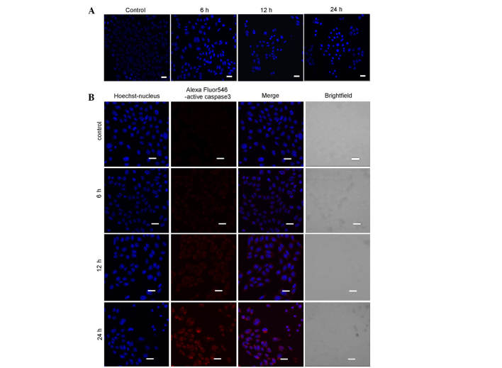
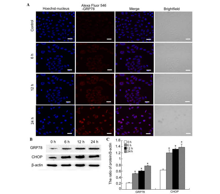
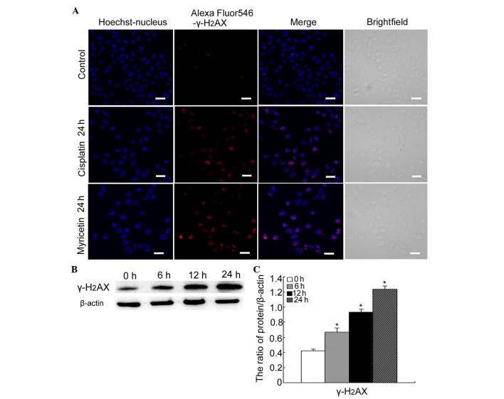
The mechanisms underlying myricetin-induced cancer cell apoptosis remain to be elucidated. Certain previous studies have shown that myricetin induces apoptosis through the mitochondrial pathway. Apoptosis, however, can also be induced by other classical pathways, including endoplasmic reticulum (ER) stress and DNA double‑strand breaks (DSBs). The aim of the present study was to assess whether these two apoptotic pathways are involved in myricetin‑induced cell death in SKOV3 ovarian cancer cells. The results revealed that treatment with myricetin inhibited viability of SKOV3 cells in a dose‑dependent manner. Myricetin induced nuclear chromatin condensation and fragmentation, and also upregulated the protein levels of active caspase 3 in a time‑dependent manner. In addition, myricetin upregulated ER stress‑associated proteins, glucose‑regulated protein‑78 and C/EBP homologous protein in SKOV3 cells. Phosphorylation of H2AX, a marker of DNA DSBs, was revealed to be upregulated in myricetin-treated cells. The data indicated that myricetin induces DNA DSBs and ER stress, which leads to apoptosis in SKOV3 cells.


Figures

Figure 1
Figure 1
Myricetin inhibits the viability of SKOV3 cells. SKOV3 cells were treated with different doses of myricetin for 24 h. (A) Cell viability was measured by a 3-(4,5-dimetrylthiazol-2-yl)-2,5-diphenyltetrazolium bromide assay and decreased in a dose-dependent manner. (B) Images were captured using an inverted phase contrast microscope at a magnification of ×100 (scale bar, 50 µm). Data are expressed as the mean ± standard deviation (n=3; *P<0.05, vs. control).
Figure 2
Figure 2
Myricetin induces apoptosis in SKOV3 cells. (A) The cells were treated with 40 µg/ml myricetin for 0, 6, 12 and 24 h, and were subsequently stained with Hoechst 33342. Confocal microscopy was used to observe cell morphology (scale bar, 20 mm). (B) The cells were treated with 40 µg/ml myricetin for 0, 6, 12 and 24 h. Nuclear staining and fluorescence of active Caspase 3 was observed using confocal microscopy (scale bar, 20 µm).
Figure 3
Figure 3
Myricetin induces ER stress-associated apoptosis in SKOV3 cells. (A) The cells were treated with 40 µg/ml myricetin for 0, 6, 12 and 24 h. The expression of GRP-78 was detected using confocal microscopy (scale bar, 20 µm). (B) Western blot analysis was performed to determine the expression levels of GRP-78 and CHOP in SKOV3 cells following treatment with 40 µg/ml myricetin for 0, 6, 12 and 24 h. (C) Quantification of the protein expression levels of GRP-78 and CHOP. The data were normalized against β-actin and are expressed as the mean ± standard deviation (n=3; *P<0.05, vs. control). GRP, glucose-regulated protein; CHOP, C/EBP homologous protein.
Figure 4
Figure 4
Myricetin facilitates DNA double-strand breaks in SKOV3 cells. (A) The cells were treated with cisplatin (6 µg/ml) or myricetin (40 µg/ml) for 0 and 24 h. The expression of γ-H2AX was observed using confocal microscopy (scale bar, 20 µm). (B) Western blot analysis was performed to detect the expression of γ-H2AX in SKOV3 cells following treatment with 40 µg/ml myricetin for 0, 6, 12 and 24 h. (C) Quantification of the protein expression of γ-H2AX. The data were normalized against β-actin and are expressed as the mean ± standard deviation (n=3). *P<0.05 vs. control.

References

    1. Siegel R, Naishadham D, Jemal A. Cancer statistics, 2013. CA Cancer J Clin. 2013;63:11–30. doi: 10.3322/caac.21166. - DOI - PubMed
    1. Tumanian SV, Iartseva DV. Effect of hepatic functional activity of the liver and endogenous intoxication in patients with ovarian cancer. Khirurgiia (Mosk) 2014:45–47. In Russian. - PubMed
    1. Shilpa V, Bhagat R, Premalata CS, Pallavi VR, Ramesh G, Krishnamoorthy L. Relationship between promoter methylation & tissue expression of MGMT gene in ovarian cancer. Indian J Med Res. 2014;140:616–623. - PMC - PubMed
    1. Shibata Y. Initial safety and efficacy of cisplatin and gemcitabine combination chemotherapy for unresectable biliary tract cancer. Gan To Kagaku Ryoho. 2014;41:2599–2602. - PubMed
    1. Yang J, Shi Y, He X, Dong M, Zhang C, Liu P, Zhou S, Qin Y, Gui L, Yang S, Sun Y. A pilot study of the safety and efficacy of dexamethasone, ifosfamide, methotrexate and gemcitabine chemotherapy for natural killer/T-cell lymphoma. Leuk Lymphoma. 2015;56:2218–2221. doi: 10.3109/10428194.2014.999323. - DOI - PubMed

Publication types