. 2016 Mar 18;44(5):2362-77.
doi: 10.1093/nar/gkw016. Epub 2016 Jan 17.

Attachment of ribosomal complexes and retrograde scanning during initiation on the Halastavi árva virus IRES

Affiliations

Attachment of ribosomal complexes and retrograde scanning during initiation on the Halastavi árva virus IRES

Irina S Abaeva et al. Nucleic Acids Res. .

Abstract

Halastavi árva virus (HalV) has a positive-sense RNA genome, with an 827 nt-long 5' UTR and an intergenic region separating two open reading frames. Whereas the encoded proteins are most homologous to Dicistrovirus polyproteins, its 5' UTR is distinct. Here, we report that the HalV 5' UTR comprises small stem-loop domains separated by long single-stranded areas and a large A-rich unstructured region surrounding the initiation codon AUG828, and possesses cross-kingdom internal ribosome entry site (IRES) activity. In contrast to most viral IRESs, it does not depend on structural integrity and specific interaction of a structured element with a translational component, and is instead determined by the unstructured region flanking AUG828. eIF2, eIF3, eIF1 and eIF1A promote efficient 48S initiation complex formation at AUG828, which is reduced ∼5-fold on omission of eIF1 and eIF1A. Initiation involves direct attachment of 43S preinitiation complexes within a short window at or immediately downstream of AUG828. 40S and eIF3 are sufficient for initial binding. After attachment, 43S complexes undergo retrograde scanning, strongly dependent on eIF1 and eIF1A. eIF4A/eIF4G stimulated initiation only at low temperatures or on mutants, in which areas surrounding AUG828 had been replaced by heterologous sequences. However, they strongly promoted initiation at AUG872, yielding a proline-rich oligopeptide.


Figures

Figure 1.
Figure 1.
The HalV 5-UTR possesses the IRES activity. (A) Model of the secondary structure of the HalV 5′ UTR, derived as described in the text, and indicating the positions of nucleotides modified by NMIA (blue circles). (B) Schematic representation of monocistronic and dicistronic mRNA constructs containing cyclin B2 (XL) and HalV cistrons. (C and D) The activity of the HalV 5′ UTR in monocistronic and dicistronic constructs assayed by translation in RRL, wheat germ and S. frugiperda cell-free systems.
Figure 2.
Figure 2.
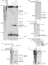
Factor requirements for initiation on HalV mRNA. (A–F) Toe-printing analysis of (A and B) 48S complex formation on MC Stem-HalV mRNA from mammalian 40S subunits, Met-tRNAiMet and indicated combinations of eIFs, (C) 48S complex formation on MC Stem-HalV mRNA from S. frugiperda 40S subunits, and mammalian Met-tRNAiMet and eIFs 2/3/1/1A, (D) elongation competence of 48S complexes assembled at AUG828, assayed using MC Stem-HalV-MTN-Stop mRNA containing a stop codon three triplets downstream of the initiation codon, (E) the influence of a short stem introduced after nt 805 on 48S complex formation on MC Stem-HalV mRNA, and (F) the influence of eIF1 and eIF1A on 40S/eIF3/HalV IRES ternary complex formation. The positions of 48S complexes (solid lines), pre-TCs (solid line), as well as 40S/HalV IRES binary and 40S/eIF3/HalV IRES ternary complexes (dashed lines), are indicated on the right.
Figure 3.
Figure 3.
40S subunits can attach to multiple regions in the HalV 5′ UTR. (AD) Interactions of 40S subunits with different regions of the HalV 5′ UTR and assembly of 48S complexes upstream of the AUG828, assayed by toe-printing. The positions of 48S complexes (solid lines), and 40S/IRES binary and 40S/eIF3/IRES ternary complexes (dashed lines) are indicated on the right. (E) Positions of toe-prints corresponding to 40S/IRES binary complexes (blue) and 40S/eIF3/IRES ternary complexes (red) mapped onto the secondary structure of the HalV 5′ UTR. (F) Association of the 32P-labeled MC Stem-HalV mRNA (nt 1–957) with 40S subunits, depending on the presence of eIF3. Ribosomal complexes were separated by centrifugation in 10–30% linear sucrose density gradients, and aliquots of gradient fractions were analyzed by scintillation counting. The position of 40S subunits determined by optical density is indicated. Sedimentation was from right to left. (G) The activity of Ligatin in 48S complex formation on MC Stem-HalV mRNA, assayed by toe-printing. The positions of 48S complexes (solid lines) and 40S/eIF3/IRES ternary complexes (dashed lines) are indicated on the right.
Figure 4.
Figure 4.
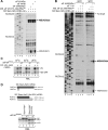
The influence of the group 4 eIFs on initiation on the HalV mRNA. (A, B) The influence of (A) eIF4A, eIF4B and eIF4Gm and (B) the reaction temperature on 48S complex formation at AUG828 and AUG872 of MC Stem-HalV mRNA, assayed by toe-printing. The positions of 48S complexes are indicated on the right. (C) The influence of the eIF4AR362Q mutant on translation of DC ΔXL-HalV mRNA in RRL at different temperatures. (D) Upper panel - schematic representation of the ORFs starting at AUG828 or AUG872 in the wt and (Δnt.934) mutant MC Stem-HalV mRNAs. Lower panel – the influence of the eIF4AR362Q mutant on translation in RRL from AUG828 and AUG872 of the wt and (Δnt.934) mutant MC Stem-HalV mRNAs.
Figure 5.
Figure 5.
The mechanism of selection of the HalV initiation codon AUG828. (A) Mutations in AUG828 and positions of new AUGs introduced into the DC Stem-ΔXL-HalV construct. (B) Toe-printing analysis of 48S complex formation on mutant DC Stem-ΔXL-HalV mRNAs with substitutions in AUG828, depending on the presence of different sets of eIFs. (CF) Toe-printing analysis of 48S complex formation on mutant DC Stem-ΔXL-HalV mRNAs containing additional AUGs introduced (C, D) upstream and (E, F) downstream of the AUG828, depending on the presence of different sets of eIFs. The positions of 48S complexes are indicated on the right (B–F).
Figure 6.
Figure 6.
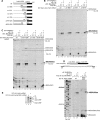
Mutational analysis of the HalV 5′ UTR. (A) Schematic representation of deletions introduced into the HalV 5′ UTR in the context of the DC Stem-ΔXL-HalV construct. (B and C) Toe-printing analysis of 48S complex formation on mutant DC Stem-ΔXL-HalV mRNAs containing deletions in the HalV 5′ UTR, depending on the presence of different sets of eIFs. The positions of 48S complexes (solid lines) and 40S/eIF3/HalV IRES ternary complexes (dashed lines) are indicated on the right. (D) Upper panel - schematic representation of the second cistron of DC XL-HalV-Aichi(ORF) mRNA, showing the sequence of the first 43 coding nucleotides in the insert. Lower panel – toe-printing analysis of 48S complex formation on DC ΔXL-HalV and DC XL-HalV-Aichi(ORF) mRNAs, depending on the presence of different sets of eIFs. The positions of 48S complexes are indicated on the right. (E) Relative activities of DC ΔXL-HalV and DC XL-HalV-Aichi(ORF) mRNAs assayed by translation in RRL.

References

    1. Jackson R.J., Hellen C.U., Pestova T.V. The mechanism of eukaryotic translation initiation and principles of its regulation. Nat. Rev. Mol. Cell. Biol. 2010;11:113–127. - PMC - PubMed
    1. Hinnebusch A.G. The scanning mechanism of eukaryotic translation initiation. Annu. Rev. Biochem. 2014;83:779–812. - PubMed
    1. Pisareva V.P., Pisarev A.V., Komar A.A., Hellen C.U., Pestova T.V. Translation initiation on mammalian mRNAs with structured 5′ UTRs requires DExH-box protein DHX29. Cell. 2008;135:1237–1250. - PMC - PubMed
    1. Abaeva I.S., Marintchev A., Pisareva V.P., Hellen C.U., Pestova T.V. Bypassing of stems versus linear base-by-base inspection of mammalian mRNAs during ribosomal scanning. EMBO J. 2011;30:115–129. - PMC - PubMed
    1. Hashem Y., des Georges A., Dhote V., Langlois R., Liao H.Y., Grassucci R.A., Hellen C.U., Pestova T.V., Frank J. Structure of the mammalian ribosomal 43S preinitiation complex bound to the scanning factor DHX29. Cell. 2013;153:1108–1119. - PMC - PubMed

Publication types

MeSH terms