. 2016 Apr 8;291(15):8130-9.
doi: 10.1074/jbc.M115.707380. Epub 2016 Jan 18.

Crystal Structure of Manganese Lipoxygenase of the Rice Blast Fungus Magnaporthe oryzae

Affiliations

Crystal Structure of Manganese Lipoxygenase of the Rice Blast Fungus Magnaporthe oryzae

Anneli Wennman et al. J Biol Chem. .

Abstract

Lipoxygenases (LOX) are non-heme metal enzymes, which oxidize polyunsaturated fatty acids to hydroperoxides. All LOX belong to the same gene family, and they are widely distributed. LOX of animals, plants, and prokaryotes contain iron as the catalytic metal, whereas fungi express LOX with iron or with manganese. Little is known about metal selection by LOX and the adjustment of the redox potentials of their protein-bound catalytic metals. Thirteen three-dimensional structures of animal, plant, and prokaryotic FeLOX are available, but none of MnLOX. The MnLOX of the most important plant pathogen, the rice blast fungusMagnaporthe oryzae(Mo), was expressed inPichia pastoris.Mo-MnLOX was deglycosylated, purified to homogeneity, and subjected to crystal screening and x-ray diffraction. The structure was solved by sulfur and manganese single wavelength anomalous dispersion to a resolution of 2.0 Å. The manganese coordinating sphere is similar to iron ligands of coral 8R-LOX and soybean LOX-1 but is not overlapping. The Asn-473 is positioned on a short loop (Asn-Gln-Gly-Glu-Pro) instead of an α-helix and forms hydrogen bonds with Gln-281. Comparison with FeLOX suggests that Phe-332 and Phe-525 might contribute to the unique suprafacial hydrogen abstraction and oxygenation mechanism of Mo-MnLOX by controlling oxygen access to the pentadiene radical. Modeling suggests that Arg-525 is positioned close to Arg-182 of 8R-LOX, and both residues likely tether the carboxylate group of the substrate. An oxygen channel could not be identified. We conclude that Mo-MnLOX illustrates a partly unique variation of the structural theme of FeLOX.

Keywords: crystal structure; enzyme mechanism; fatty acid oxidation; lipoxygenase pathway; metalloenzyme.


Figures

FIGURE 1.
FIGURE 1.
Overview of LOX with catalytic iron or manganese. A, phylogenetic tree of MnLOX from filamentous fungi and a selection of FeLOX. The GenBankTM numbers are for the MnLOX enzymes are as follows: G. graminis (AAK81882.2); Magnaporthe salvinii (CAD61974); M. oryzae (ALE27899) (27); F. oxysporum (EGU80482.1); Colletotrichum gloeosporioides (EQB45907.1); and Aspergillus fumigatus MnLOX (EDP47436.1). The listed FeLOX are as follows: P. aeruginosa (Q8RNT4.2); Plexaura homomalla (PDB code 4QWT); Glycine max FeLOX (P08170.2); Homo sapiens 5-LOX (P09917.2); F. oxysporum (EXK38530.1); G. graminis FeLOX (EJT77580.1), and A. fumigatus (EAL84806). The tree was generated by MEGA6 (25) as described (38). B, overview of the oxidation of linoleic acid to hydroperoxides by FeLOX and MnLOX. Both enzymes catalyze the abstraction of the pro-11S hydrogen. The formed radical is delocalized over the pentadiene, and oxygen is typically inserted in an antarafacial way at the 13S or 9R positions by FeLOX, and in a suprafacial way at the 13R, 9S and 11S positions by MnLOX. Note that if the fatty acid enters in the reverse orientation in the catalytic channel, FeLOX can abstract the pro-11R hydrogen and form hydroperoxides with 9S and 13R configuration.
FIGURE 2.
FIGURE 2.
Crystal structure of Mo-MnLOX. A, schematic illustrations in two directions of the overall structure, colored in rainbow spectrum with the N terminus in blue, the C terminus in red, and the catalytic manganese in purple. The broken helix covering the active site is colored in light green. B, variation of the α2 helix in different LOX structures as follows: Mo-MnLOX (blue, PDB code 5FNO); 5-LOX (red, PDB code 3O8Y); sLOX-1 (green, PDB code 1YGE); and P. aeruginosa 15S-LOX (yellow, PDB code 4G32). The α2 helix of Mo-MnLOX is an 11-turn-long helix (blue), which runs along the whole length of the protein and leaves an open access to the substrate channel and the catalytic metal (light orange).
FIGURE 3.
FIGURE 3.
Overview of the metal ligands of Mo-MnLOX and FeLOX. A, catalytic manganese (orange sphere) of Mo-MnLOX (gray) is coordinated by the His-284, His-289, His-469, Asn-473, and Val-605 in a distorted octahedral configuration. The coral 8R-LOX (PDB code 4QWT, pink) is superimposed for comparison with an r.m.s. deviation of 0.57 Å. The largest differences are between the Asn residues and the C-terminal Ile/Val residues. B, metal ligands to the catalytic iron of coral 8R-LOX (PDB code 4QWT, pink), sLOX-1 (PDB code 1YGE, green), and 15S-LOX of P. aeruginosa (PDB code 4G32, yellow) are superimposed with an r.m.s. deviation between 0.23 and 0.29 Å.
FIGURE 4.
FIGURE 4.
Factors influencing the metal coordination of Mo-MnLOX. A, unbiased 2FoFc electron density map is shown at contour level of one σ. The metal-coordinating Asn-473 residue is situated on a loop and might provide the increased flexibility necessary for the use of manganese as catalytic metal. Gln-474 and Ser-604 are in close positions. B, comparison of the structure of the loop with Asn-473 (light blue) with the corresponding part of 8R-LOX (gray). C, hydrogen bond network close to the active site of MnLOX and Asn-473, which forms three hydrogen bonds as follows: with the conserved Gln-281, the main chain oxygen of the metal coordinating His-469, and the main chain of Glu-476. Gln-474 is forming a hydrogen bond network with Ser-604 proximate to the C-terminal Val-605; it also forms a weak interaction with a coordinated water molecule that also interacts with the main chain of Arg-528 and the side chain of Asn-527.
FIGURE 5.
FIGURE 5.
Substrate channel entrance of Mo-MnLOX and a comparison with 8R-LOX. A, proposed substrate channel entrance of Mo-MnLOX (PDB code 5FNO) is illustrated in surface rendering (gray), superimposed with the structure of 8R-LOX (PDB code 4QWT chain C), showing arachidonic acid as substrate. The Arg-525 is positioned in the opening to the channel in suitable distance for ionic interaction between the Arg-525 side chain and the carboxyl of the fatty acid substrate. B, Mo-MnLOX (blue) and 8R-LOX (pink) are superimposed. The Arg-182 of 8R-LOX has been found to tether the carboxylate of the substrate. The Arg-525 of MnLOX is provided by a helix closer to the C terminus, but these two Arg residues seem nevertheless to play similar roles in the tethering of the carboxyl group.
FIGURE 6.
FIGURE 6.
Possible oxygen access routes in the U-shaped substrate channel of Mo-MnLOX. Leu-331 from the arched helix is defining the upper wall of the channel at the bottom of the U-shaped substrate channel in analogy with Leu-431 of 8R-LOX. Phe-332 may shield the pentadiene for oxygen insertion in an antarafacial way so that oxygen may reach the pentadiene radical from the other side as indicated in by the arrows in the two side pockets. Phe-526 is likely to bend the substrate to allow oxygen access from the same side as the catalytic metal. Arachidonic acid, bound to coral 8R-LOX (PDB code 4QWT, chain C), is included for clarity; the natural substrates of Mo-MnLOX are linoleic and α-linolenic acids, but 20:2n-6, 20:3n-3, and 22:5n-6 are also oxidized by the enzyme (supplemental Fig. S4).
FIGURE 7.
FIGURE 7.
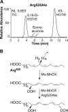
RP-HPLC-MS/MS analysis of the biosynthesis of 9S,16S-DiHPOTrE from 9S-HPOTrE by the R525A mutant and an overview of the sequential biosynthesis of 9,16-DiHPOTrE. A, RP-HPLC-MS/MS analysis of the lipoxygenation of 9S-HPOTrE by the R525A mutant of Mo-MnLOX after reduction of hydroperoxides to alcohols with triphenylphosphine. B, overview of the biosynthesis of 9S,16S-DiHPOTrE by Mo-MnLOX and the R525A mutant. NL, normalized to 100%. TIC, total ion current.
FIGURE 8.
FIGURE 8.
Overview of the active site of Mo-MnLOX. Arachidonic acid, bound in the substrate channel of coral 8R-LOX (PDB code 4QWT, chain C), is included in the U-shaped active site of Mo-MnLOX for clarity. The carboxyl group of arachidonic acid is likely tethered by Arg-525 and the ω end by Phe-342. Leu-332 clamps the substrate in position, and Phe-332 and Phe-526 may position pentadiene for suprafacial hydrogen abstraction and oxygenation. Three His residues, Asn-473, Val-605, and the catalytic water are coordinating manganese (pink). Hydrogen bonds are likely formed between Gln-281 and Asn-473 and between Val-605 and the catalytic water (red).

References

    1. Brash A. R. (1999) Lipoxygenases: occurrence, functions, catalysis, and acquisition of substrate. J. Biol. Chem. 274, 23679–23682 - PubMed
    1. Newcomer M. E., and Brash A. R. (2015) The structural basis for specificity in lipoxygenase catalysis. Protein Sci. 24, 298–309 - PMC - PubMed
    1. Haeggström J. Z., and Funk C. D. (2011) Lipoxygenase and leukotriene pathways: biochemistry, biology, and roles in disease. Chem. Rev. 111, 5866–5898 - PubMed
    1. Brodhun F., and Feussner I. (2011) Oxylipins in fungi. FEBS J. 278, 1047–1063 - PubMed
    1. Heshof R., Jylhä S., Haarmann T., Jørgensen A. L., Dalsgaard T. K., and de Graaff L. H. (2014) A novel class of fungal lipoxygenases. Appl. Microbiol. Biotechnol. 98, 1261–1270 - PubMed

Publication types

LinkOut - more resources