. 2016 Jan 19;11(1):e0146618.
doi: 10.1371/journal.pone.0146618. eCollection 2016.

Ceramide Synthase 6 Is a Novel Target of Methotrexate Mediating Its Antiproliferative Effect in a p53-Dependent Manner

Affiliations

Ceramide Synthase 6 Is a Novel Target of Methotrexate Mediating Its Antiproliferative Effect in a p53-Dependent Manner

Baharan Fekry et al. PLoS One. .

Abstract

We previously reported that ceramide synthase 6 (CerS6) is elevated in response to folate stress in cancer cells, leading to enhanced production of C16-ceramide and apoptosis. Antifolate methotrexate (MTX), a drug commonly used in chemotherapy of several types of cancer, is a strong inhibitor of folate metabolism. Here we investigated whether this drug targets CerS6. We observed that CerS6 protein was markedly elevated in several cancer cell lines treated with MTX. In agreement with the enzyme elevation, its product C16-ceramide was also strongly elevated, so as several other ceramide species. The increase in C16-ceramide, however, was eliminated in MTX-treated cells lacking CerS6 through siRNA silencing, while the increase in other ceramides sustained. Furthermore, the siRNA silencing of CerS6 robustly protected A549 lung adenocarcinoma cells from MTX toxicity, while the silencing of another ceramide synthase, CerS4, which was also responsive to folate stress in our previous study, did not interfere with the MTX effect. The rescue effect of CerS6 silencing upon MTX treatment was further confirmed in HCT116 and HepG2 cell lines. Interestingly, CerS6 itself, but not CerS4, induced strong antiproliferative effect in several cancer cell lines if elevated by transient transfection. The effect of MTX on CerS6 elevation was likely p53 dependent, which is in agreement with the hypothesis that the protein is a transcriptional target of p53. In line with this notion, lometrexol, the antifolate inducing cytotoxicity through the p53-independent mechanism, did not affect CerS6 levels. We have also found that MTX induces the formation of ER aggregates, enriched with CerS6 protein. We further demonstrated that such aggregation requires CerS6 and suggests that it is an indication of ER stress. Overall, our study identified CerS6 and ceramide pathways as a novel MTX target.


Conflict of interest statement

Competing Interests: The authors have declared that no competing interests exist.

Figures

Fig 1
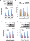
Fig 1. Effects of MTX on CerS6, ceramide levels and cellular proliferation.
(A) Levels of CerS6 (Western blot) in cancer cells with and without MTX treatment. Cells were collected 48 h after MTX was added. (B) Changes in CerS6 levels after treatment with 10 nM MTX for 48 h (calculated from A). The intensity of bands was quantified using ImageJ software. In all cases, normalization for levels of actin was performed. (C) Changes in levels of selected ceramides in A549 cells after treatment with 10 nM MTX; Pi, total phospholipid inorganic phosphate present in cell extracts. Student’s t-test was performed for statistical analysis of the changes in ceramide levels and asterisks indicate statistically significant differences (p<0.05). (D) Antiproliferative effect of MTX (10 and 100 nM) in a panel of cancer cell lines. Live cells were assessed by MTT assay. Error bars show SD of four measurements. Differences between control and drug treated groups were statistically significant (calculated by one-way ANOVA) with p value below 0.0007 in all cases with the exception of A549 p53-/- cells (p = 0.0357). (E) Scatter plot of CerS6 protein levels (from panel B) versus cell survival after MTX treatment (from panel D) for ten cell lines indicated in the above panels. Pearson’s coefficient (r = −0.83; n = 10; p<0.01) indicates strong negative correlation between changes in CerS6 levels and cell survival upon MTX treatment.
Fig 2
Fig 2. The effect of CerS6 transient expression on cancer cell lines highly sensitive to MTX.
(A) Transient expression of CerS6 in A549 cells inhibits proliferation. (B) Antiproliferative effect of C16-ceramide on A549 cells. Live cells were assessed by MTT assay after exposure to C16-ceramide for indicated time; stock solution of ceramide (10 mM) was prepared in 50:50 mixture of DMSO/methanol and was added directly to cultured cells; control cells were treated with the same volume of DMSO/methanol mixture. Error bars show SD of four measurements. Differences between control and drug treated groups were statistically significant (calculated by one-way ANOVA) with p value below 0.0009. (C and D) Transient expression of CerS6 in HepG2 or HCT116 cells inhibits proliferation. For panels A, C, and D cells were transiently transfected with the CerS6 expression vector and proliferation was assessed by MTT assay at indicated time points. Data from MTT assay are plotted as fold change of live cells compared to control (number of cells at 0 h). Data show average of a representative experiment, six measurements for each data point, with error bars indicating SD; Student’s t test was performed for statistical analysis (in all data sets the difference between control and CerS6-transfected cells were statistically significant with p<0.005). Insets show levels of CerS6; actin was used as a loading control.
Fig 3
Fig 3. CerS6 is a target of MTX in mediation of antiproliferative effect.
(A) Silencing CerS6 by siRNA rescues A549 cells from cytotoxic effect of MTX while silencing CerS4 does not change the drug toxicity. Cells were transfected with scrambled siRNA, CerS6 siRNA or CerS4 siRNA (25 pmol), MTX was added 6 h later and live cells were assessed by MTT assay 48 h after the beginning of MTX treatment. Data represent an average of two independent experiments, each performed in quadruplicates with error bars representing SD. Student’s t test was performed for statistical analysis (difference in cell number between control and CerS6-silenced cells were statistically significant with p<0.005; there were no significant differences in cell number between control and CerS4-silenced cells). Panels on the right show the efficiency of CerS6 and CerS4 silencing (evaluated at 48 h of MTX treatment). (B) Levels of ceramide in control A549 cells (Cntr, transfected with scrambled siRNA), MTX-treated cells transfected with scrambled siRNA (MTX) and MTX-treated cells after CerS6 silencing (CerS6 siRNA/MTX, transfected with CerS6 siRNA). Differences between the groups were analyzed by one-way ANOVA and asterisks indicate statistically significant changes (p<0.05). (C) Silencing of CerS6 by siRNA protects cancer cells from MTX cytotoxic effect. Student’s t-test was performed and the differences between MTX effect in control and CerS6-silenced cells were statistically significant (p<0.01). (D) MTX (10 nM) leads to elevation of CerS6 but not of CerS4 in A549 cells.
Fig 4
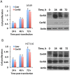
Fig 4. The effect of CerS 4 transient expression on cancer cell lines highly sensitive to MTX.
Transient expression of CerS4 in A549 or HCT116 cells does not inhibit cellular proliferation (MTT assay). Data are average of two independent experiments, each done in quadruplicates with error bars representing SD. Differences between control and CerS4-expressing cells were not statistically significant (p>0.1) in both panel A and panel B. Insets show levels of CerS4 and CerS6 in the cells after transient expression of CerS4.
Fig 5
Fig 5. Targeting of CerS6 by MTX is p53-dependent.
(A) MTX (10 nM) treatment strongly elevates CerS6 in p53+/+ but not p53-/- cells. (B) Antifolate lometrexol (LTX) bypasses p53 and does not affect CerS6: left panel, LTX inhibits both p53+/+ and p53-/- A549 cells (Experiments were performed three times with six wells per concentration point in each experiment; error bars, SD) no statistically significant differences between p53+/+ and p53-/- cells were observed (p>0.1); right panel, levels of p53 and CerS6 (Western blot) in the cells after LTX treatment. Cells were exposed to LTX for 48 h.
Fig 6
Fig 6. Methotrexate induces CerS6-dependent formation of ER membrane aggregates in A549 non-small cell lung carcinoma cells.
(A) Treatment with MTX (10 nM) resulted in the increase of autophagosome number, but CerS6 was not associated with these organelles. (B) MTX (10 nM) induced aggregation of ER membranes and the accumulation of CerS6 in the aggregates. Arrows indicate ER membrane aggregates. Note the lack of such aggregates after lometrexol (1.0 μM) treatment. (C) MTX (10 nM) induced ER aggregation (detected by monitoring CALR-RFP) in the absence of exogenous CerS6. (D) A549 cells lacking CerS6 (siRNA silencing) did not form ER aggregates in response to MTX. Cells were co-transfected with GFP-CerS6 and LC3-RFP (panel A) or CALR-RFP (panel B), MTX was added 6 h after transfection and live cell images were captured 48 h later. In experiments with endogenous CerS6, cells were co-transfected with GFP and CALR-RFP. Six hours before co-transfection, cells were transfected with scrambled siRNA (panel C) or CerS6 siRNA (panel D). MTX was added 6 h after the second transfection and images were captured 48 h later. Pearson’s coefficient for co-localization (calculated using Fiji software) is indicated for each panel.
Fig 7
Fig 7. Proposed two-step mechanism for CerS6-dependent MTX cytotoxicity.
Step 1: MTX induces p53, which functions as the transcriptional activator of CerS6 and increases levels of the protein in ER. Step 2: MTX induces ER stress leading to aggregation of CerS6-containing ER membranes. Enhanced generation of C16-ceramide by CerS6 further promotes cytotoxic effect.

Similar articles

Cited by

References

    1. Visentin M, Zhao R, Goldman ID. The antifolates. Hematol Oncol Clin North Am. 2012;26(3):629–48, ix 10.1016/j.hoc.2012.02.002 - DOI - PMC - PubMed
    1. Goldman ID, Chattopadhyay S, Zhao R, Moran R. The antifolates: evolution, new agents in the clinic, and how targeting delivery via specific membrane transporters is driving the development of a next generation of folate analogs. Curr Opin Investig Drugs. 2010;11(12):1409–23. Epub 2010/12/15. . - PubMed
    1. Wilson PM, Danenberg PV, Johnston PG, Lenz HJ, Ladner RD. Standing the test of time: targeting thymidylate biosynthesis in cancer therapy. Nat Rev Clin Oncol. 2014;11(5):282–98. 10.1038/nrclinonc.2014.51 . - DOI - PubMed
    1. Jaffe N. Historical perspective on the introduction and use of chemotherapy for the treatment of osteosarcoma. Adv Exp Med Biol. 2014;804:1–30. 10.1007/978-3-319-04843-7_1 . - DOI - PubMed
    1. Paci A, Veal G, Bardin C, Leveque D, Widmer N, Beijnen J, et al. Review of therapeutic drug monitoring of anticancer drugs part 1—cytotoxics. Eur J Cancer. 2014;50(12):2010–9. 10.1016/j.ejca.2014.04.014 . - DOI - PubMed

Publication types

MeSH terms