. 2016 Jan 19;11(1):e0146540.
doi: 10.1371/journal.pone.0146540. eCollection 2016.

Chronic Exposure to Androgenic-Anabolic Steroids Exacerbates Axonal Injury and Microgliosis in the CHIMERA Mouse Model of Repetitive Concussion

Affiliations

Chronic Exposure to Androgenic-Anabolic Steroids Exacerbates Axonal Injury and Microgliosis in the CHIMERA Mouse Model of Repetitive Concussion

Dhananjay R Namjoshi et al. PLoS One. .

Abstract

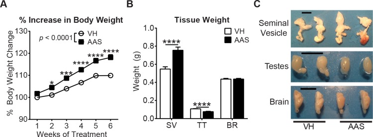
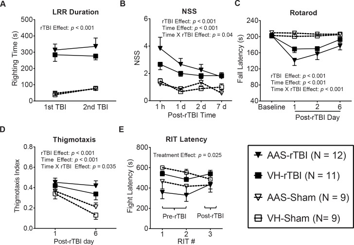
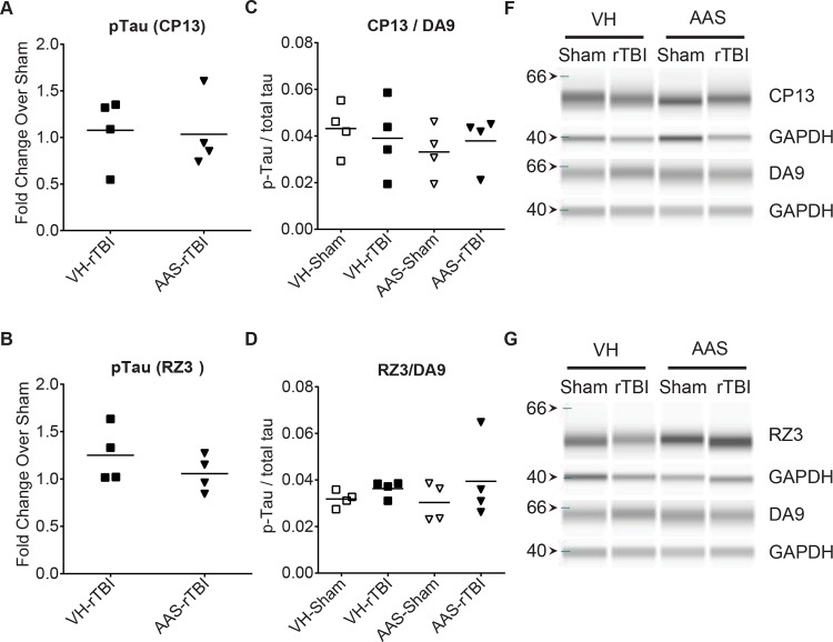
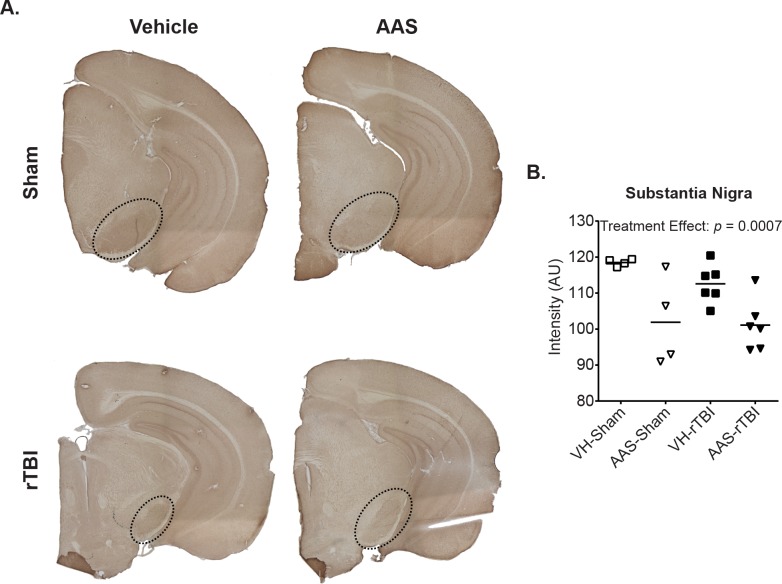
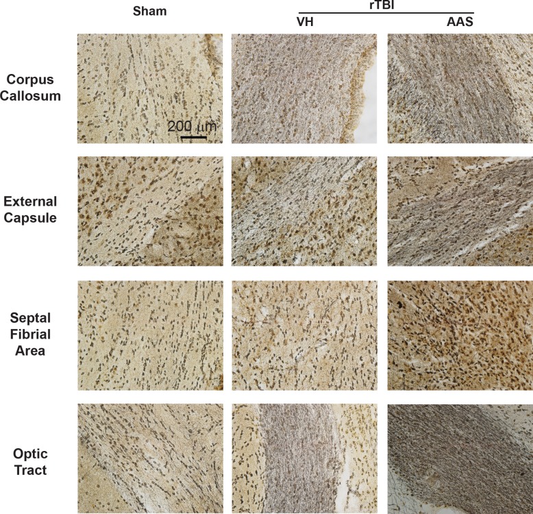
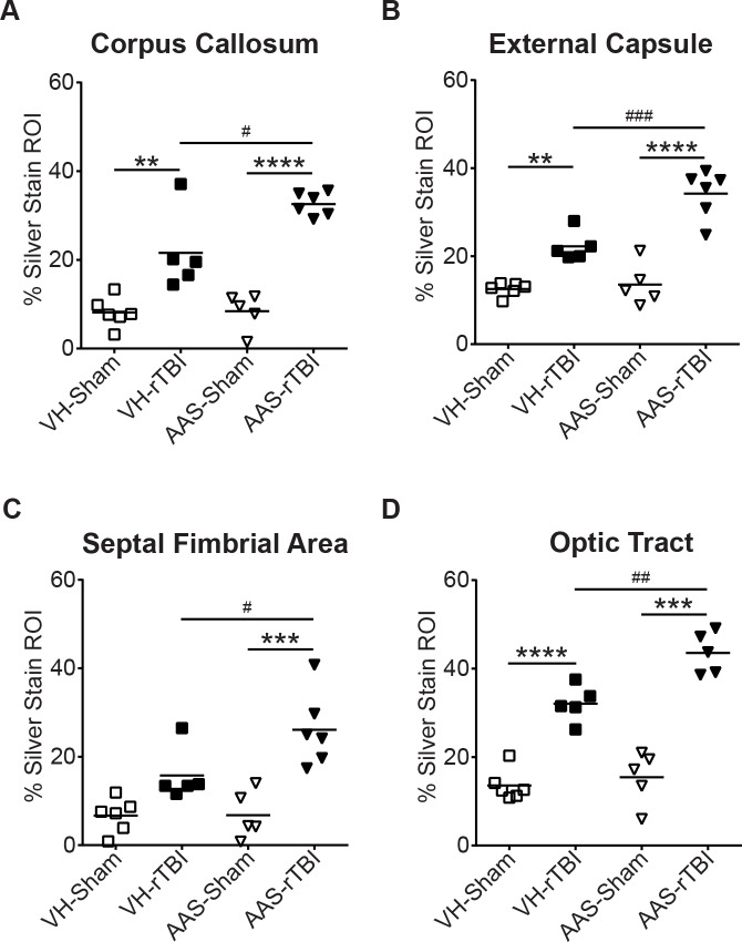
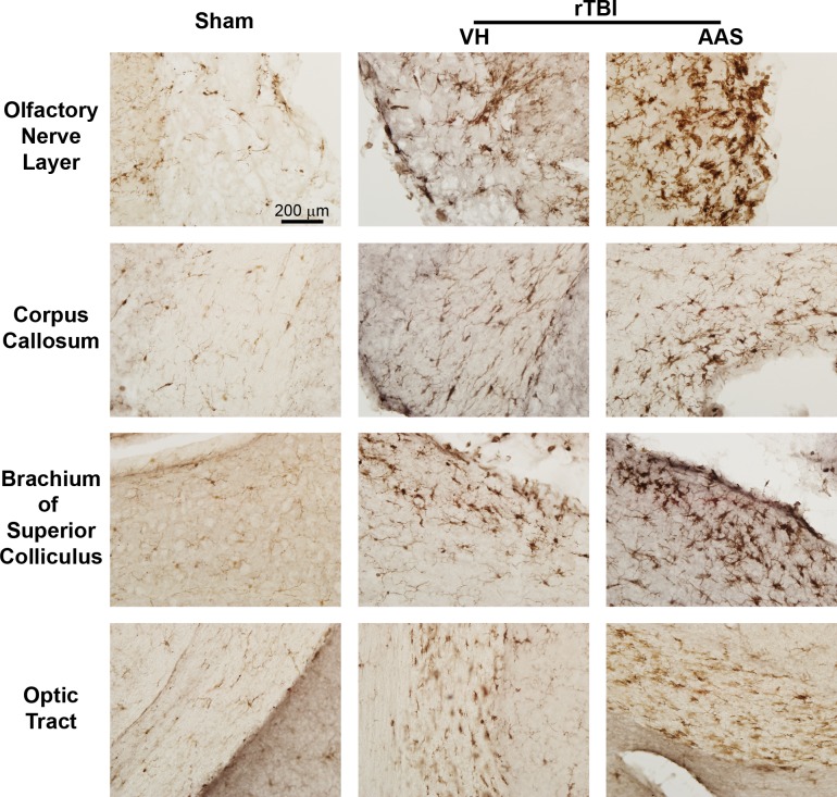
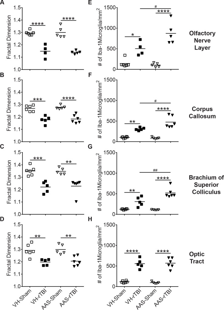
Concussion is a serious health concern. Concussion in athletes is of particular interest with respect to the relationship of concussion exposure to risk of chronic traumatic encephalopathy (CTE), a neurodegenerative condition associated with altered cognitive and psychiatric functions and profound tauopathy. However, much remains to be learned about factors other than cumulative exposure that could influence concussion pathogenesis. Approximately 20% of CTE cases report a history of substance use including androgenic-anabolic steroids (AAS). How acute, chronic, or historical AAS use may affect the vulnerability of the brain to concussion is unknown. We therefore tested whether antecedent AAS exposure in young, male C57Bl/6 mice affects acute behavioral and neuropathological responses to mild traumatic brain injury (TBI) induced with the CHIMERA (Closed Head Impact Model of Engineered Rotational Acceleration) platform. Male C57Bl/6 mice received either vehicle or a cocktail of three AAS (testosterone, nandrolone and 17α-methyltestosterone) from 8-16 weeks of age. At the end of the 7th week of treatment, mice underwent two closed-head TBI or sham procedures spaced 24 h apart using CHIMERA. Post-repetitive TBI (rTBI) behavior was assessed for 7 d followed by tissue collection. AAS treatment induced the expected physiological changes including increased body weight, testicular atrophy, aggression and downregulation of brain 5-HT1B receptor expression. rTBI induced behavioral deficits, widespread axonal injury and white matter microgliosis. While AAS treatment did not worsen post-rTBI behavioral changes, AAS-treated mice exhibited significantly exacerbated axonal injury and microgliosis, indicating that AAS exposure can alter neuronal and innate immune responses to concussive TBI.


Conflict of interest statement

Competing Interests: The authors have declared that no competing interests exist.

Figures

Fig 1
Fig 1. Chronic AAS treatment in mice induces physiological changes.
Prior to rTBI, mice were treated 5 d per week for 7 weeks with a cocktail of androgenic-anabolic steroids (AAS) or sesame oil vehicle (VH). Cohort size: VH: N = 22, AAS: N = 23. (A) Percent increase in body weight in AAS and VH-treated mice over time. (B) Comparison of weights of seminal vesicles (SV), testes (TT), and brain (BR) collected at 7 d post-rTBI. (C) Macroscopic size comparison of seminal vesicle, testes and brain. Scale bar = 1cm. In all graphs, data are presented as mean ± SEM values. Body weight data are analyzed by two-way repeated measures ANOVA followed by a Bonferroni post hoc test, tissue weight data were analyzed by two-tailed unpaired t test. For all graphs, *: p < 0.05, ***: p < 0.001, ****: p < 0.0001.
Fig 2
Fig 2. Chronic AAS treatment does not exacerbate acute post-rTBI behavioral deficits.
(A) Duration of loss of righting reflex (LRR) was assessed immediately following the sham or TBI procedure. (B) Composite neurological severity score (NSS) was assessed at 1 h and at 1, 2 and 7 d post-rTBI. (C) Motor performance was assessed on an accelerating rotarod at 1, 2, and 7 d post-rTBI. The graph depicts fall latency in seconds at baseline before rTBI and at three post-rTBI time points. (D) Thigmotaxis was quantified at 1 and 6 d post-rTBI and is represented as thigmotaxis index. (E) Aggressive behavior was assessed with the RIT at the 5th (RIT # 1) and 6th (RIT # 2) week following initiation of AAS treatment (pre-rTBI) as well as at 5 d (RIT # 3) post-rTBI. Graphs represent latency to initiate fighting by the resident mouse. Data in all graphs are presented as mean ± SEM. Data are analyzed by repeated measures general linear model. Legends and cohort sizes are consistent across all graphs.
Fig 3
Fig 3. CHIMERA rTBI and AAS treatment do not affect endogenous tau phosphorylation at 7 days post injury.
Tau phosphorylation was assessed using the Simple Western system (Protein Simple). Graphs in the left column (A and B) show fold change in endogenous phosphorylated tau levels in half-brain homogenates collected at 7 d rTBI compared to sham brains using antibodies CP13 (pSer202 and pThr205, Panel A) and RZ3 (pThr231, Panel B), respectively. Graphs in the middle column (C and D) depict quantitation of phosphorylated tau as a proportion of total tau (DA9). Representative digital immunoblots of phosphorylated and corresponding total tau are depicted in the right column (E and F).
Fig 4
Fig 4. Chronic AAS treatment downregulates 5-HT1B receptor expression in the substantia nigra.
Immunohistochemistry was used to assess 5-HT1B receptor expression levels. Representative images of whole-mount sections for AAS-treated and control brains are depicted in Panel A. 5-HT1B receptor expression in the substantia nigra (dashed circles) was quantified by measuring the mean grey intensity of the selected brain area on an 8-bit grayscale image. Graph in Panel B depicts mean staining intensity in arbitrary units.
Fig 5
Fig 5. Chronic AAS treatment in mice exacerbates post-rTBI axonal injury.
Post-rTBI axonal damage was assessed with silver staining. Representative 40X-magnified images of corpus callosum, external capsule, septal-fimbrial area and optic tract of sham (left column) and VH- (middle column) and AAS-treated (right column) rTBI brains are depicted.
Fig 6
Fig 6. Quantitative assessment of silver stained images.
Silver stained images were quantified by calculating the percent of region of interest (ROI) in the white matter tract area that was stained with silver. Graphs indicate percent of the ROI showing positive signal in the respective white matter regions. For all graphs, * indicates a significant rTBI effect compared to the respective sham values and # indicates a significant treatment effect between rTBI groups. Data are analyzed by two-way ANOVA followed by a Tukey post-hoc test. For all graphs **: p < 0.01, ***: p < 0.001, ****: p < 0.0001, #: p < 0.05, ##: p < 0.01, ###: p < 0.001.
Fig 7
Fig 7. Chronic AAS treatment in mice augments post-rTBI microgliosis.
Post-rTBI microglial activation was assessed with Iba1 immunohistochemistry at 7 d. Representative 40X-magnified images of white matter regions show resting microglia in sham brains (left column) and activated microglia in injured brains (second and third columns).
Fig 8
Fig 8. Quantitative analysis of the microglial response to rTBI.
Microglial morphology was quantitatively assessed using fractal analysis. Graphs in the left column represent fractal dimension for microglial morphology in (A) olfactory nerve layer, (B) corpus callosum, (C) brachium of superior colliculus and (D) optic tract. Graphs in the right column (E-H) show number of Iba1-positive cells per mm2 in the same white matter regions. For all graphs, * indicates a significant rTBI effect compared to the respective sham values and # indicates a significant treatment effect between rTBI groups. Data are analyzed by two-way ANOVA followed by a Tukey post-hoc test. For all graphs *: p < 0.05, **: p < 0.01, ***: p < 0.001, ****: p < 0.0001, #: p < 0.05, ##: p < 0.01.

References

    1. Reilly P. The impact of neurotrauma on society: an international perspective. Prog Brain Res. 2007;161:3–9. Epub 2007/07/10. 10.1016/s0079-6123(06)61001-7 . - DOI - PubMed
    1. Gerberding JL, Binder S. The report to congress on mild traumatic brain injury in the United States: Steps to prevent a serious public health problem Atlanta, GA: Centers for Disease Control and Prevention, 2003.
    1. Langlois JA, Rutland-Brown W, Wald MM. The Epidemiology and Impact of Traumatic Brain Injury: A Brief Overview. J Head Trauma Rehabil. 2006;21(5):375–8. - PubMed
    1. Faul MM, Xu L, Wald MM, Coronado VG. Traumatic brain injury in the united states: emergency department visits, hospitalizations and death 2002–2006 Atlanta, GA: Centers for Disease Control and Prevention, National Center for Injury Prevention and Control, 2010.
    1. Centers for Disease Control and Prevention. Nonfatal traumatic brain injuries related to sports and recreation activities among persons aged <19 years—United States, 2001–2009. MMWR Morb Mortal Wkly Rep. 2011;30(39):1337–76. Epub 2007/07/28. doi: mm5629a2 [pii]. - PubMed

Publication types

MeSH terms