. 2016 Jan 19;11(1):e0146952.
doi: 10.1371/journal.pone.0146952. eCollection 2016.

Depletion of Paraspeckle Protein 1 Enhances Methyl Methanesulfonate-Induced Apoptosis through Mitotic Catastrophe

Affiliations

Depletion of Paraspeckle Protein 1 Enhances Methyl Methanesulfonate-Induced Apoptosis through Mitotic Catastrophe

Xiangjing Gao et al. PLoS One. .

Abstract

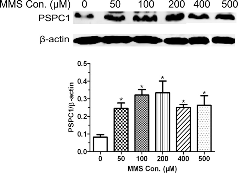
Previously, we have shown that paraspeckle protein 1 (PSPC1), a protein component of paraspeckles that was involved in cisplatin-induced DNA damage response (DDR), probably functions at the G1/S checkpoint. In the current study, we further examined the role of PSPC1 in another DNA-damaging agent, methyl methanesulfonate (MMS)-induced DDR, in particular, focusing on MMS-induced apoptosis in HeLa cells. First, it was found that MMS treatment induced the expression of PSPC1. While MMS treatment alone can induce apoptosis, depletion of PSPC1 expression using siRNA significantly increased the level of apoptosis following MMS exposure. In contrast, overexpressing PSPC1 decreased the number of apoptotic cells. Interestingly, morphological observation revealed that many of the MMS-treated PSPC1-knockdown cells contained two or more nuclei, indicating the occurrence of mitotic catastrophe. Cell cycle analysis further showed that depletion of PSPC1 caused more cells entering the G2/M phase, a prerequisite of mitosis catastrophe. On the other hand, over-expressing PSPC1 led to more cells accumulating in the G1/S phase. Taken together, these observations suggest an important role for PSPC1 in MMS-induced DDR, and in particular, depletion of PSPC1 can enhance MMS-induced apoptosis through mitotic catastrophe.


Conflict of interest statement

Competing Interests: The authors have declared that no competing interests exist. Although one author is employed by a commercial company "Zhejiang CONBA Pharmaceutical Co. Ltd.", it does not alter the authors' adherence to PLOS ONE policies on sharing data and materials.

Figures

Fig 1
Fig 1. Cell viability and DNA damage were determined after methyl methanesulfonate (MMS) treatment.
(A) HeLa cells were treated with the indicated concentrations of MMS for 12, 24, 36, and 48 h. Cell viability was measured by MTT assay. (B) HeLa cells were treated with different concentrations of MMS for 12 h, then detected the level of DNA damage by immunofluorescence (a) and Western blot (b). Scale bar, 50 μm. The results are shown as the mean ± SD of three independent experiments. *p < 0.05, **p < 0.01, as compared with the solvent control group.
Fig 2
Fig 2. Paraspeckle protein 1 (PSPC1) is induced by MMS.
HeLa cells were treated with indicated concentrations of MMS for 12 h. The levels of PSPC1 were examined by Western blot. The results are shown as the mean ± SD of three independent experiments. *p < 0.05, as compared with the control group.
Fig 3
Fig 3. Modulation of PSPC1 influences MMS-induced cell cycle redistribution.
(A) HeLa cells were transfected with siPSPC1 or siControl for 24 h, then treated with 0 μM and 100 μM of MMS for 12 h. The expression of PSPC1 was examined by Western blot and cell cycle was analyzed by flow cytometry. (B) HeLa cells were transfected with pPSPC1 or pCON to overexpress PSPC1 for 24 h, then treated with 0 μM and 100 μM of MMS for 12 h. The expression of PSPC1 was examined by Western blot and cell cycle was analyzed by flow cytometry. The values represent averages of three independent experiments, mean ± SD.*p < 0.05, **p < 0.01, as compared with the control group.
Fig 4
Fig 4. Effect of PSPC1 on MMS-induced apoptosis.
(A) HeLa cells were transfected with siPSPC1, siControl, pPSPC1, or pControl for 24 h, followed by 200 or 400 μM of MMS treatment for 12 h prior to analysis of PSPC1 and PARP expression by Western blot. For the detection of apoptosis, HeLa cells transfected with siRNAs (B) or overexpression plasmids (C) were treated with 200 or 400 μM of MMS for 12 h, and then analyzed by dual-parameter flow cytometry utilizing Annexin V-FITC and PI double staining. Representative dot plot data from three independent experiments are shown in the left panel, and the histogram graph in the right panel represents the percentage of dual-parameter positive cells pooled from three independent experiments. Data are presented as the mean ± SD of three independent experiments. **p < 0.01, as compared with the control group.
Fig 5
Fig 5. Knockdown of PSPC1 induces mitotic catastrophe.
HeLa cells were transfected with siPSPC1 or siControl. 24 h post-transfection, cells were harvested and the expression of tubulin and the nucleolus were examined by immunofluorescence microscopy (A). The percentage of multinuclear cells were shown as a histogram, and densitometry data of three independent experiments were presented, mean ± SD (n = 200 cells). (B). (C) Using the same treated cells, the levels of representative mitotic catastrophe proteins (phospho-histone H3 [Ser10], Cdc2, cyclinB, and Chk2) were examined by western blot. **p < 0.01, as compared with the control group.

References

    1. Molz L, Booher R, Young P, Beach D. cdc2 and the regulation of mitosis: six interacting mcs genes. Genetics. 1989;122(4):773–82. - PMC - PubMed
    1. Ayscough K, Hayles J, MacNeill SA, Nurse P. Cold-sensitive mutants of p34cdc2 that suppress a mitotic catastrophe phenotype in fission yeast. Molecular & general genetics: MGG. 1992;232(3):344–50. . - PubMed
    1. Andreassen PR, Lohez OD, Lacroix FB, Margolis RL. Tetraploid state induces p53-dependent arrest of nontransformed mammalian cells in G1. Molecular biology of the cell. 2001;12(5):1315–28. - PMC - PubMed
    1. Margottin-Goguet F, Hsu JY, Loktev A, Hsieh HM, Reimann JD, Jackson PK. Prophase destruction of Emi1 by the SCF(betaTrCP/Slimb) ubiquitin ligase activates the anaphase promoting complex to allow progression beyond prometaphase. Developmental cell. 2003;4(6):813–26. . - PubMed
    1. Lock RB, Stribinskiene L. Dual modes of death induced by etoposide in human epithelial tumor cells allow Bcl-2 to inhibit apoptosis without affecting clonogenic survival. Cancer research. 1996;56(17):4006–12. . - PubMed

Publication types

MeSH terms