. 2016 Mar 11;291(11):5512-5526.
doi: 10.1074/jbc.M115.694877. Epub 2016 Jan 19.

A Signaling Network Controlling Androgenic Repression of c-Fos Protein in Prostate Adenocarcinoma Cells

Affiliations

A Signaling Network Controlling Androgenic Repression of c-Fos Protein in Prostate Adenocarcinoma Cells

Eswar Shankar et al. J Biol Chem. .

Abstract

The transcription factor c-Fos controls many important cellular processes, including cell growth and apoptosis. c-Fos expression is rapidly elevated in the prostate upon castration-mediated androgen withdrawal through an undefined mechanism. Here we show that androgens (5α-dihydrotestosterone and R1881) suppress c-Fos protein and mRNA expression induced by 12-O-tetradecanoylphorbol-13-acetate (TPA) or EGF in human prostate cancer (PCa) cell lines. Such suppression transpires through a transcriptional mechanism, predominantly at the proximal serum response element of the c-fos promoter. We show that androgen signaling suppresses TPA-induced c-Fos expression through repressing a PKC/MEK/ERK/ELK-1 signaling pathway. Moreover, our results support the hypothesis that p38(MAPK), PI3K, and PKCδ are involved in the androgenic regulation of c-Fos through controlling MEK/ERK. Stable silencing of c-Fos and PKCδ with shRNAs suggests that R1881 promotes cell death induced by low-dose TPA through a mechanism that is dependent on both PKCδ and loss of c-Fos expression. Reciprocally, loss of either PKCδ or c-Fos activates p38(MAPK) while suppressing the activation of ERK1/2. We also provide the first demonstration that R1881 permits cell death induced by low-dose TPA in the LNCaP androgen-dependent PCa cell line and that TPA-induced cell death is independent of exogenous androgen in the castration-resistant variants of LNCaP, C4-2 and C4-2B. Acquisition of androgen-independent killing by TPA correlates with activation of p38(MAPK), suppression of ERK1/2, and loss of c-Fos. These results provide new insights into androgenic control of c-Fos and use of PKC inhibitors in PCa therapy.

Keywords: Akt PKB; MAPK; PKC; androgen; androgen receptor; c-Fos; c-Jun transcription factor; p38 MAPK; phorbol ester; prostate cancer.


Figures

FIGURE 1.
FIGURE 1.
Androgen down-regulates the protein level of c-Fos in prostate epithelial cells. A, LNCaP, C4-2B, MDA-PCa-2b, and VCaP cells were incubated with either 10 nm R1881 or DHT 48 h prior to a 2-h treatment with 10 ng/ml TPA, and expression of c-Fos and β-actin in whole cell lysates were analyzed by Western blotting. B, comparison of 24 h of pretreatment with R1881 versus co-treatment with R1881 on the induction of c-Fos by TPA in LNCaP cells as assessed by Western blotting. C, Western blotting of the effective dose of R1881 that suppresses c-Fos induction by TPA in LNCaP cells. D, R1881 (48 h) suppresses EGF-induced (10 ng/ml, 30 min) c-Fos expression. E, Western blotting of the expression of c-Fos in CWR22Rv1 cells following 22-h treatment with 1 nm R1881 before a 2-h treatment with 10 ng/ml TPA or a 1-h treatment with 10 ng/ml EGF. F, R1881 suppression of TPA-induced c-Fos expression in LNCaP cells is not reversed by the proteasomal inhibitor MG132, as determined by Western blotting. Cells were treated for 20 h with 10 nm R1881 or vehicle prior to 30-min pretreatment with 5 μm MG132 or vehicle, followed by 2 h of treatment with 10 ng/ml TPA or vehicle. Results are representative of two to three independent experiments.
FIGURE 2.
FIGURE 2.
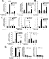
Suppression of c-Fos mRNA by R1881. A and B, LNCaP cells were pretreated with R1881 (10 nm) for 48 h and incubated with TPA (10 ng/ml) for the indicated times as c-Fos expression was measured by Northern blotting analysis (A) and semiquantitative RT-PCR with total RNA (10 μg) (B). Data were quantified by a PhosphorImager (A, right panel) or by ImageJ (B, right panel). The data in B are shown normalized to β-actin. C and D, real-time quantitative RT-PCR analysis of TPA-induced (2 h) or EGF-induced (1 h) c-fos expression (normalized to GAPDH) in LNCaP cells pretreated with R1881 (n = 4 replicates). Error bars represent the mean ± S.E. of triplicate to quadruplicate determinations. p Values were calculated by Student's t test (two-tailed). Data are representative of two to three different experiments/treatment.
FIGURE 3.
FIGURE 3.
Androgen controls c-fos expression at the promoter level. A, LNCaP cells were co-transfected with either pGL3-basic-luc (control) or c-Fos promoter-luc (p-c-Fos-luc) along with CMV-Renilla, followed by treatment with R1881 (10 nm) or DHT (10 nm) for 48 h before TPA (10 ng/ml) treatment for 2 h. B, separately for EGF treatment, cells were preincubated with R1881 for 24 h prior to EGF treatment (10 ng/ml) for 24 h. C, cells were first transfected with p-c-Fos-luc and CMV-Renilla, and then incubated with R1881 (10 nm) for 48 h prior to TPA treatment (10 ng/ml, 2 h). U0216 (10 μm), PD98059 (5 μm), SP600125 (10 μm), SB202190 (10 μm), and GF109203X (25 μm) were added 1 h before R1881. Data shown are relative values of firefly luciferase normalized to Renilla luciferase. D, LNCaP cells were co-transfected with SRE-luc, SRF-Luc, or CRE-Luc along with CMV-Renilla, followed by treatment with R1881 (10 nm) for 48 h before TPA treatment (10 ng/ml) for 2 h. Data are relative values of firefly luciferase normalized to Renilla luciferase. Error bars represent the mean ± S.E. of triplicate determinations. p Values were calculated by Student's t test (two-tailed). Data are representative of two to three different experiments/treatment.
FIGURE 4.
FIGURE 4.
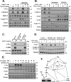
Androgenic control of the PKC/MEK/ERK pathway in PCa cells. A, LNCaP and C4-2B cells were pretreated with 10 nm R1881 48 h prior to 2-h treatment with 10 ng/ml TPA, and cell lysates were analyzed by Western blotting for expression of total and phosphorylated MEK1/2, ERK1/2 p38 MAPK, and SAPK/JNK. B and C, the effect of R1881 (10 nm, 48 h) on c-Fos promoter activity (assayed as in Fig. 3) by enforced expression of wild-type (B) MEK1 and constitutively active (Act) MEK1 (C) constructs transfected in LNCaP cells. D, LNCaP cells treated as in A were analyzed for expression of total and phospho-PKCs. E, LNCaP cells treated as in A were analyzed for expression of total and phospho-PKCs following treatment with TPA and R1881 as indicated. Data are representative of two to three experiments.
FIGURE 5.
FIGURE 5.
R1881 promotes TPA induced cell death through a mechanism that involves the suppression of c-Fos expression and activation of PKCδ. c-Fos was stably silenced by pLKO.1 lentivirus-mediated transduction of LNCaP cells, and silencing was assessed by Western blotting of TPA-treated (2 h, 10 ng/ml) cells (A). Silencing of c-Fos enhanced and suppressed TPA-induced P-p38MAPK and P-ERK1/2, respectively (A), and was permissive to cell death induced by low-dose TPA treatment (B). PKCδ was stably silenced by pLKO.1 lentivirus-mediated transduction of LNCaP cells, as assessed by Western blotting of TPA-treated (2 h, 10 ng/ml) cells (C). Silencing of PKCδ suppressed and enhanced TPA-induced P-p38MAPK and P-ERK1/2, respectively (C and D) and repressed cell death induced by low-dose TPA + R1881 treatment (E). Relative changes in cell density were assessed by crystal violet staining of adherent cells following 5 days of treatment (B and E). F, dose response of TPA on changes in the expression of c-Fos, P-p38, and P-ERK1/2 in vehicle versus R1881 pretreatment LNCaP cells (48 h, 1 nm).
FIGURE 6.
FIGURE 6.
Cross-talk of MEK1/2, p38MAPK, PI3K and AR in TPA-induced signals controlling c-Fos expression. A and B, LNCaP cells were pretreated with vehicle or an inhibitor of p38MAPK (SB203580, 10 μm), MEK1/2 (selumetinib, 1 μm) or PI3K (ZSTK474, 1 μm) for 1 h, followed by vehicle or 1 nm R1881 (22 h) before 2-h pretreatment with 2.5 ng/ml TPA or vehicle. C, effect of selumetinib and ZSTK474 on EGF-induced (10 ng/ml, 30 min) c-Fos and P-ERK. D, effect of MK2206 versus ZSTK474 on c-Fos expression induced by TPA (2 h) with or without R1881 (24 h). E, dose dependence of R1881 on alterations in the expression of c-Fos and prostate-specific antigen (PSA) and the activation of ERK1/2 and p-38MAPK. Cell lysates were analyzed by Western blotting for the expression of the indicated proteins. F, schematic of our model showing pathways involved in the control of c-Fos expression by AR and how AR signaling may promote TPA-induced death.
FIGURE 7.
FIGURE 7.
Androgenic control of the c-Fos promoter. A, effect of R1881 on nuclear localization of P-ERK following treatment with TPA in LNCaP cells, as assessed by Western blotting of cytosolic and nuclear fractions. LNCaP cells were treated with or without 1 nm R1881 (22 h), followed by TPA (2.5 ng/ml, 2 h) or no TPA. B, effect of R1881 on expression and TPA-induced phosphorylation of ELK-1 and SRF expression in LNCaP (treated as in A), as assessed by Western blotting analysis of whole cell lysates. C, immunoprecipitation of ELK-1 by AR in TPA- and R1881-treated LNCaP cells (treated as in A). Cell lysates were immunoprecipitated with rabbit anti-AR (catalog no. sc-816) or control rabbit IgG and immunoblotted with mouse anti-AR (catalog no. sc-7305), anti-ELK-1 (catalog no. sc-56896), and anti-ERK1/2 (catalog no. 9107) antibodies. D, nuclear lysates of LNCaP cells (pretreated with or without 10 nm R1881 for 46 h, followed by 2 h with or without 10 ng/ml TPA) were subjected to EMSA using radiolabeled WT SRE and mutant SRE containing WT TCF-dimerized oligonucleotides. The DNA-protein complex (complex 1) was supershifted by treatment of the dimerized oligos with 1 μg of anti-SRF IgG (catalog no. sc-335) 30 min prior to electrophoresis. exp, exposure.
FIGURE 8.
FIGURE 8.
Androgen-dependent and -independent killing of PCa cells by TPA. A—C, R1881 permits TPA-induced death of LNCaP cells (A) but not CRPC variants of LNCaP, C4-2 (B), and C4-2B (C), which are killed by low-dose TPA in the absence of androgen, as shown in Fig. 6, B and E. D, androgen-independent killing of C4-2 and C4-2B cells by low-dose TPA correlates with androgen-independent activation of p38MAPK and reduced activation of ERK1/2 by TPA, as assessed by Western blotting and growth conditions described in Fig. 6A. E, TPA failed to kill RWPE-1 and DU145 cells but suppressed R1881-induced growth in VCaP, MDA-PCa-2b, and CWR22Rv1 cells assayed as in A. F, Western blotting analysis of the expression of AR in CWR22Rv1 cells treated with or without R1881, TPA, or EGF for 3 days. Error bars represent the mean ± S.E. of triplicate to six replicates. p Values were calculated by Student's t test (two-tailed) and two-way analysis of variance). Data are representative of at least two independent experiments. #, p < 0.05; *, p < 0.01; **, p < 0.001.

Similar articles

Cited by

References

    1. Cooke P. S., Young P., and Cunha G. R. (1991) Androgen receptor expression in developing male reproductive organs. Endocrinology 128, 2867–2873 - PubMed
    1. Heinlein C. A., and Chang C. (2004) Androgen receptor in prostate cancer. Endocr. Rev. 25, 276–308 - PubMed
    1. Huggins C., and Hodges C. V. (1972) Studies on prostatic cancer: I: the effect of castration, of estrogen and androgen injection on serum phosphatases in metastatic carcinoma of the prostate. CA Cancer J. Clin. 22, 232–240 - PubMed
    1. Veldscholte J., Berrevoets C. A., Zegers N. D., van der Kwast T. H., Grootegoed J. A., and Mulder E. (1992) Hormone-induced dissociation of the androgen receptor-heat-shock protein complex: use of a new monoclonal antibody to distinguish transformed from nontransformed receptors. Biochemistry 31, 7422–7430 - PubMed
    1. Chen C. D., Welsbie D. S., Tran C., Baek S. H., Chen R., Vessella R., Rosenfeld M. G., and Sawyers C. L. (2004) Molecular determinants of resistance to antiandrogen therapy. Nat. Med. 10, 33–39 - PubMed

Publication types

MeSH terms