

Whole-exome Sequencing Analysis Identifies Mutations in the EYS Gene in Retinitis Pigmentosa in the Indian Population
- PMID: 26787102
- PMCID: PMC4726297
- DOI: 10.1038/srep19432
Whole-exome Sequencing Analysis Identifies Mutations in the EYS Gene in Retinitis Pigmentosa in the Indian Population
Abstract
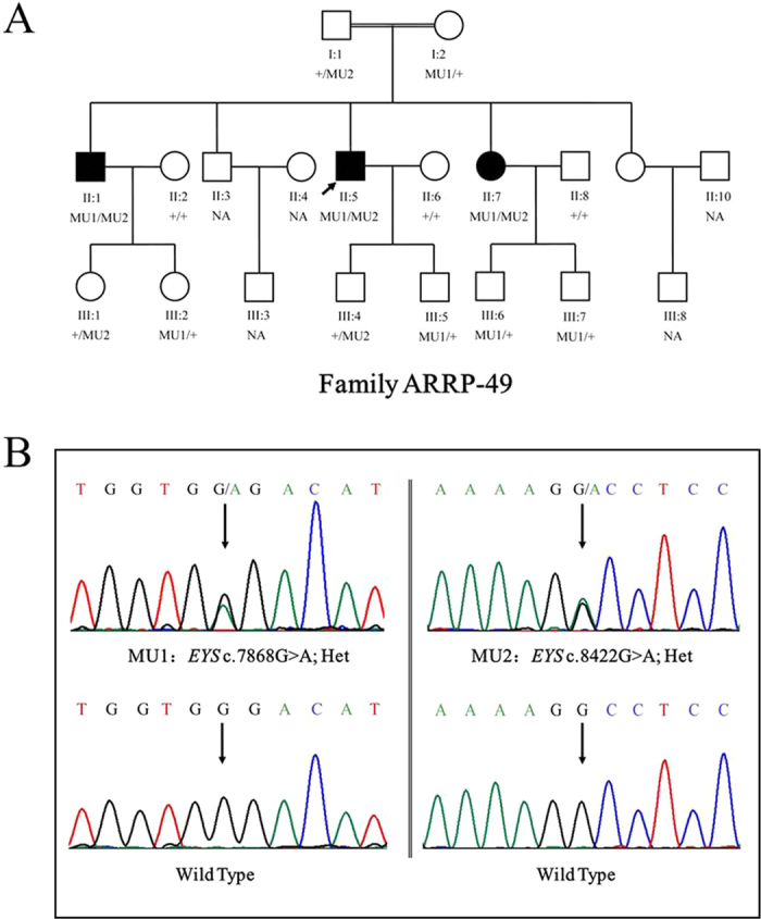
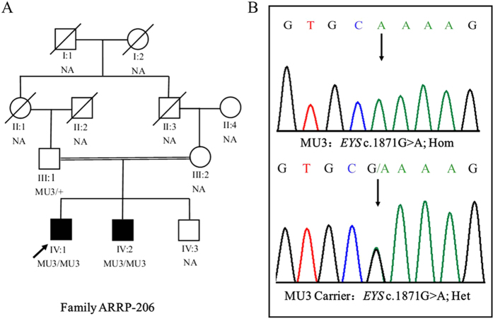
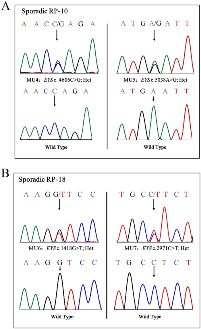
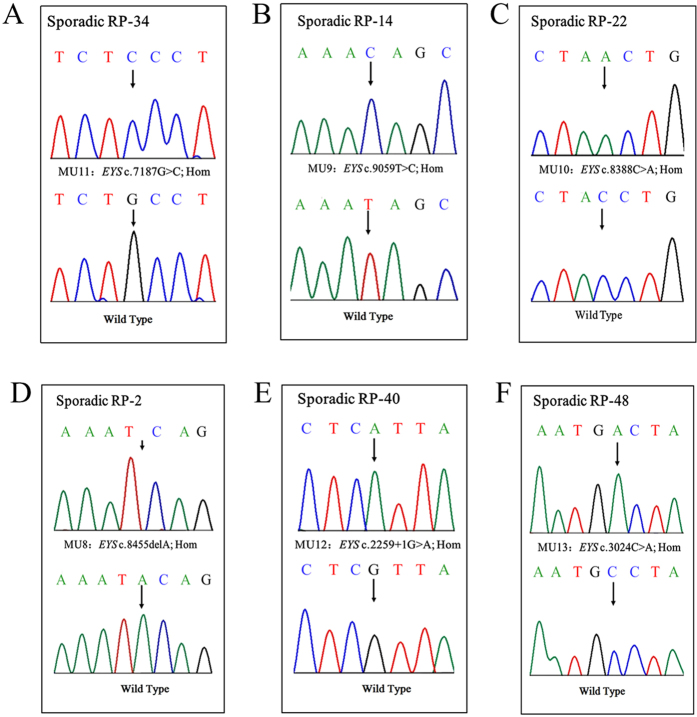
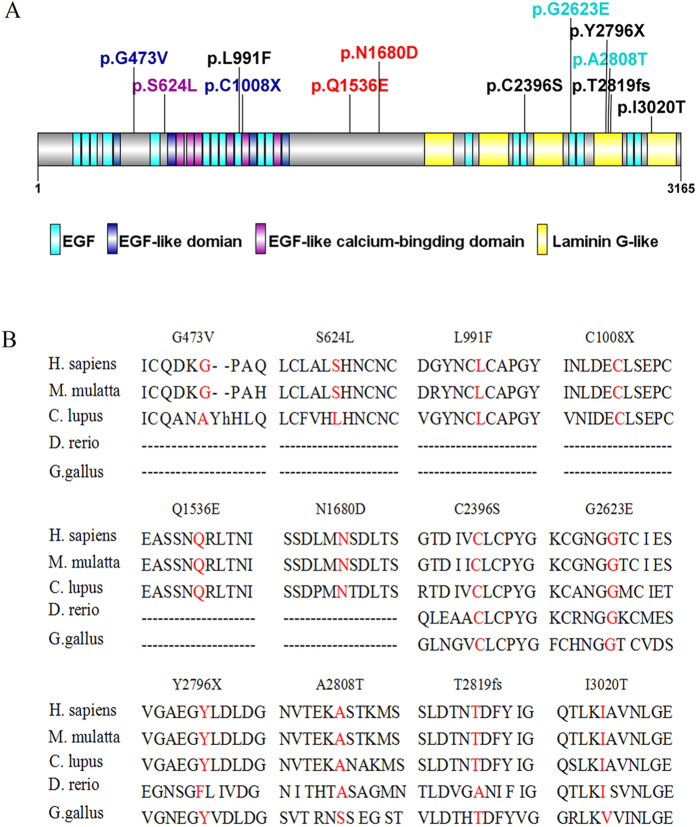
Retinitis pigmentosa (RP) is a rare heterogeneous genetic retinal dystrophy disease, and despite years of research, known genetic mutations can explain only approximately 60% of RP cases. We sought to identify the underlying genetic mutations in a cohort of fourteen Indian autosomal recessive retinitis pigmentosa (arRP) families and 100 Indian sporadic RP cases. Whole-exome sequencing (WES) was performed on the probands of the arRP families and sporadic RP patients, and direct Sanger sequencing was used to confirm the causal mutations identified by WES. We found that the mutations of EYS are likely pathogenic mutations in two arRP families and eight sporadic patients. Specifically, we found a novel pair of compound heterozygous mutations and a novel homozygous mutation in two separate arRP families, and found two novel heterozygous mutations in two sporadic RP patients, whereas we found six novel homozygous mutations in six sporadic RP patients. Of these, one was a frameshift mutation, two were stop-gain mutations, one was a splicing mutation, and the others were missense mutations. In conclusion, our findings expand the spectrum of EYS mutations in RP in the Indian population and provide further support for the role of EYS in the pathogenesis and clinical diagnosis of RP.
Figures
Similar articles
-
Whole exome sequencing reveals novel EYS mutations in Chinese patients with autosomal recessive retinitis pigmentosa.Mol Vis. 2019 Jan 20;25:35-46. eCollection 2019. Mol Vis. 2019. PMID: 30804660 Free PMC article.
-
Whole-exome sequencing reveals a novel frameshift mutation in the FAM161A gene causing autosomal recessive retinitis pigmentosa in the Indian population.J Hum Genet. 2015 Oct;60(10):625-30. doi: 10.1038/jhg.2015.92. Epub 2015 Aug 6. J Hum Genet. 2015. PMID: 26246154
-
Identification of Novel EYS Mutations by Targeted Sequencing Analysis.Genet Test Mol Biomarkers. 2020 Nov;24(11):745-753. doi: 10.1089/gtmb.2020.0186. Epub 2020 Oct 15. Genet Test Mol Biomarkers. 2020. PMID: 33058741
-
Genes and mutations causing retinitis pigmentosa.Clin Genet. 2013 Aug;84(2):132-41. doi: 10.1111/cge.12203. Epub 2013 Jun 19. Clin Genet. 2013. PMID: 23701314 Free PMC article. Review.
-
Characterizing the genotypic spectrum of retinitis pigmentosa in East Asian populations: a systematic review.Ophthalmic Genet. 2023 Apr;44(2):109-118. doi: 10.1080/13816810.2023.2182329. Epub 2023 Mar 1. Ophthalmic Genet. 2023. PMID: 36856324
Cited by
-
A Review on the Challenges in Indian Genomics Research for Variant Identification and Interpretation.Front Genet. 2020 Jul 22;11:753. doi: 10.3389/fgene.2020.00753. eCollection 2020. Front Genet. 2020. PMID: 32793285 Free PMC article. Review.
-
Genotypic spectrum and phenotype correlations of ABCA4-associated disease in patients of south Asian descent.Eur J Hum Genet. 2017 Jun;25(6):735-743. doi: 10.1038/ejhg.2017.13. Epub 2017 Mar 22. Eur J Hum Genet. 2017. PMID: 28327576 Free PMC article.
-
Whole genome sequencing and in vitro splice assays reveal genetic causes for inherited retinal diseases.NPJ Genom Med. 2021 Nov 18;6(1):97. doi: 10.1038/s41525-021-00261-1. NPJ Genom Med. 2021. PMID: 34795310 Free PMC article.
-
Ablation of EYS in zebrafish causes mislocalisation of outer segment proteins, F-actin disruption and cone-rod dystrophy.Sci Rep. 2017 Apr 5;7:46098. doi: 10.1038/srep46098. Sci Rep. 2017. PMID: 28378834 Free PMC article.
-
Compound pathogenic mutation in the USH2A gene in Chinese RP families detected by whole‑exome sequencing.Mol Med Rep. 2018 Dec;18(6):5016-5022. doi: 10.3892/mmr.2018.9530. Epub 2018 Oct 2. Mol Med Rep. 2018. PMID: 30280194 Free PMC article.
References
-
- Hartong D. T., Berson E. L. & Dryja T. P. Retinitis pigmentosa. Lancet 368, 1795–809 (2006). - PubMed
Publication types
MeSH terms
Substances
LinkOut - more resources
Full Text Sources
Other Literature Sources
