. 2016 Mar;139(Pt 3):937-52.
doi: 10.1093/brain/awv385. Epub 2016 Jan 19.

Improved proteostasis in the secretory pathway rescues Alzheimer's disease in the mouse

Affiliations

Improved proteostasis in the secretory pathway rescues Alzheimer's disease in the mouse

Yajing Peng et al. Brain. 2016 Mar.

Abstract

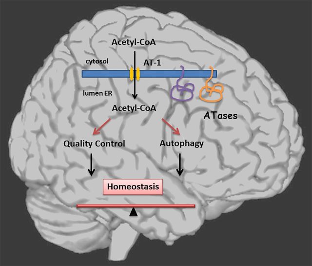
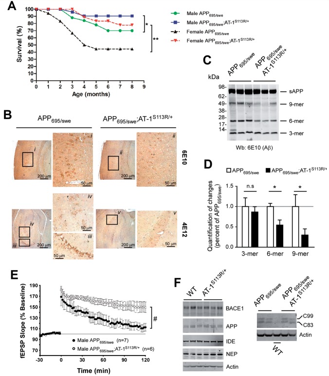
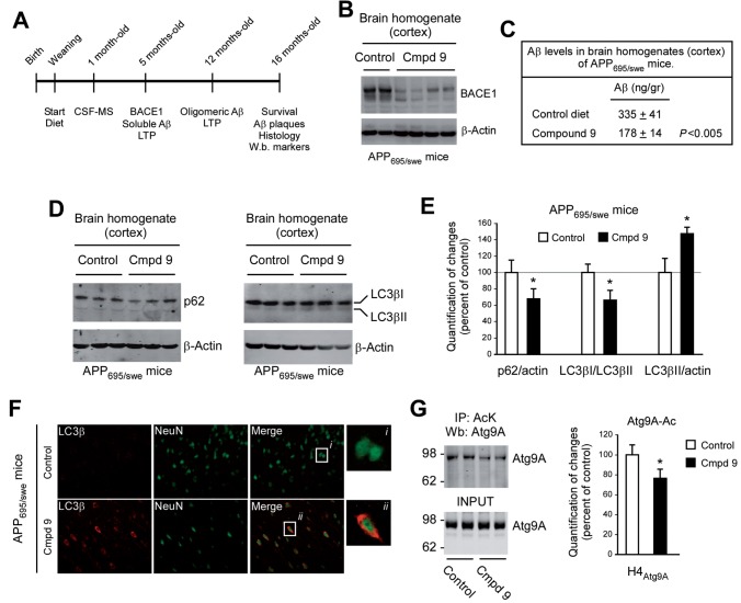
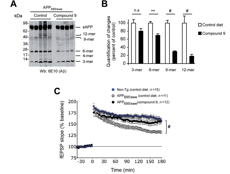
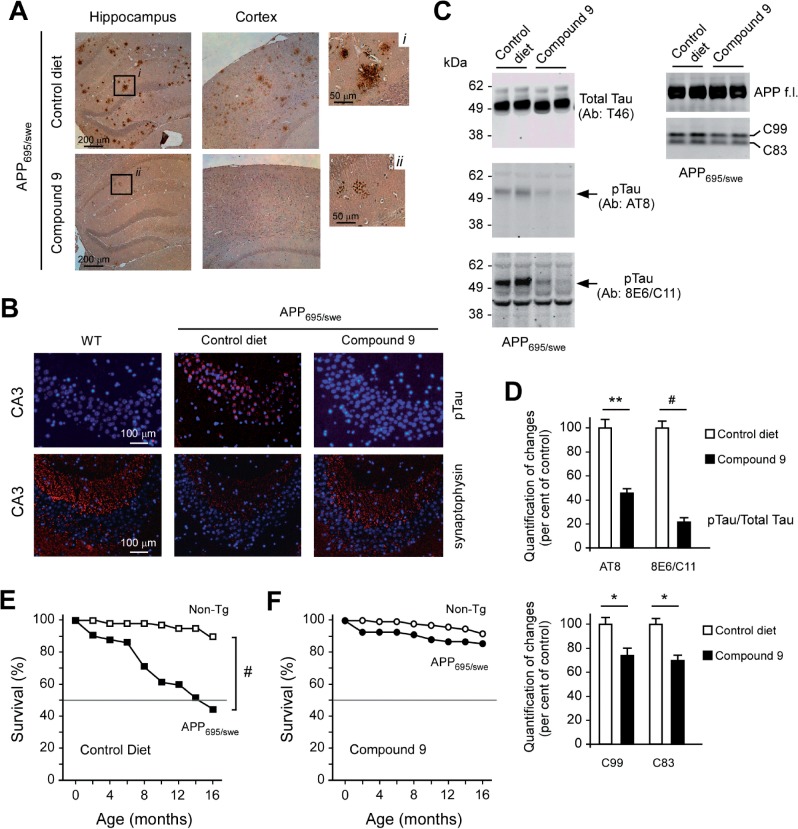
The aberrant accumulation of toxic protein aggregates is a key feature of many neurodegenerative diseases, including Huntington's disease, amyotrophic lateral sclerosis and Alzheimer's disease. As such, improving normal proteostatic mechanisms is an active target for biomedical research. Although they share common pathological features, protein aggregates form in different subcellular locations. Nε-lysine acetylation in the lumen of the endoplasmic reticulum has recently emerged as a new mechanism to regulate the induction of autophagy. The endoplasmic reticulum acetylation machinery includes AT-1/SLC33A1, a membrane transporter that translocates acetyl-CoA from the cytosol into the endoplasmic reticulum lumen, and ATase1 and ATase2, two acetyltransferases that acetylate endoplasmic reticulum cargo proteins. Here, we used a mutant form of α-synuclein to show that inhibition of the endoplasmic reticulum acetylation machinery specifically improves autophagy-mediated disposal of toxic protein aggregates that form within the secretory pathway, but not those that form in the cytosol. Consequently, haploinsufficiency of AT-1/SLC33A1 in the mouse rescued Alzheimer's disease, but not Huntington's disease or amyotrophic lateral sclerosis. In fact, intracellular toxic protein aggregates in Alzheimer's disease form within the secretory pathway while in Huntington's disease and amyotrophic lateral sclerosis they form in different cellular compartments. Furthermore, biochemical inhibition of ATase1 and ATase2 was also able to rescue the Alzheimer's disease phenotype in a mouse model of the disease. Specifically, we observed reduced levels of soluble amyloid-β aggregates, reduced amyloid-β pathology, reduced phosphorylation of tau, improved synaptic plasticity, and increased lifespan of the animals. In conclusion, our results indicate that Nε-lysine acetylation in the endoplasmic reticulum lumen regulates normal proteostasis of the secretory pathway; they also support therapies targeting endoplasmic reticulum acetyltransferases, ATase1 and ATase2, for a subset of chronic degenerative diseases.

Keywords: AT-1/SLC33A1; ATase; Alzheimer’s disease; autophagy; lysine acetylation; proteostasis.


Figures

None
See Duran-Aniotz et al . (doi: 10.1093/brain/awv401 ) for a scientific commentary on this article. Many neurodegenerative diseases are characterized by accumulation of toxic protein aggregates in specific subcellular locations. Using mouse models, Peng et al. show that inhibition of the endoplasmic reticulum acetylation machinery enhances autophagy-mediated disposal of aggregates that form within the secretory pathway and rescues Alzheimer’s disease.
Figure 1
Figure 1
Increased autophagy in AT-1 S113R/+ mice targets protein aggregates in the secretory pathway. ( A ) Western blot showing the migration profile of M-A53T syn and SP-A53T syn. ( B–F ) MEFs from wild-type (WT) and AT-1 S113R/+ mice were transfected with mutant α-synuclein (A53T syn). Levels of soluble (Triton™ X-100) and insoluble/aggregated (SDS) A53T syn were detected by western blotting. M-A53T syn, α-synuclein with an initiator methionine; SP-A53T syn, α-synuclein with a signal peptide at the N-terminus. Selected images are shown in B–E , while quantification of changes is shown in F . Values are mean ± SD * P < 0.05. Loading controls are shown in Supplementary Fig. 1 . ( G ) Media from SP-A53T syn transfected MEFs were used to immunoprecipitate α-synuclein prior to western blotting. Total cell lysate served as positive control. ( H ) Lactated dehydrogenase (LDH) activity was assayed in the media of SP-A53T syn transfected MEFs. Values are mean ( n = 4) ± SD. ** P < 0.005. ( I ) Immunolabelling showing co-localization of SP-A53T syn with LC3β puncta in AT-1 S113R/+ MEFs. As expected, LC3β displayed a diffuse cytosolic distribution in wild-type MEFs; puncta were only visible in AT-1 S113R/+ MEFs ( Peng et al. , 2014 ). No co-localization of α-synuclein with LC3β puncta was observed in AT-1 S113R/+ MEFs transfected with M-A53T syn. ( J ) Western blot showing increased levels of SP-A53T syn aggregates following BPE treatment to arrest the autophagy flux. Increased levels of p62, an autophagy-cargo protein that is normally degraded as part of the autophagy process, served as a marker of successful blockage of autophagy. Representative images are in the left panel; quantitative changes of α-syn/SDS are in the right panel.
Figure 2
Figure 2
Increased autophagy in AT-1 S113R/+ mice did not rescue the phenotype of mHtt Q160 mice. ( A–C ) Lifespan ( A ), body weight ( B ) and clasping ( C ) of indicated animals. Values in B are mean ± SD. Total numbers: females mHtt Q160 , n = 17; females mHtt Q160 ;AT-1 S113R/+ , n = 14; males mHtt Q160 , n = 18; males mHtt Q160 ;AT-1 S113R/+ , n = 18. ( D ) Western blot assessment of Htt aggregates in the striatum (Sm), cortex (Cx), and cerebellum (Cb). Lane 1: mHtt Q160 ;AT-1 S113R/+ mice; Lane 2: mHtt Q160 mice. Animals (males) were 2 months old when analysed.
Figure 3
Figure 3
Increased autophagy in AT-1 S113R/+ mice did not rescue the phenotype of hSOD1 G43A mice. ( A ) Median survival in hSOD1 G93A (163 days; n = 21) and hSOD1 G93A ;AT-1 S113R/+ (167 days; n = 18) mice. ( B ) Median onset of symptoms in hSOD1 G93A (121 days; n = 20) and hSOD1 G93A ;AT-1 S113R/+ (121 days; n = 17) mice. ( C ) Hindlimb grip-strength in wild-type ( n = 15), hSOD1 G93A ( n = 20), and hSOD1 G93A ;AT-1 S113R/+ ( n = 16) at 60 and 120 days. No significant difference was observed between hSOD1 G93A and hSOD1 G93A ;AT-1 S113R/+ mice. Average grip-strength in wild-type mice was 100.0 ± 9.8 gf for males and 93.1 ± 16.2 gf for females at 60 days; and 103.2 ± 11.2 gf for males and 101.9 ± 9.1 gf for females at 120 days. Data are mean ± SD. #P < 0.0005 (from wild-type). ( D ) Western blot against hSOD1 in the spinal cord of early symptomatic hSOD1 G93A and hSOD1 G93A ;AT-1 S113R/+ mice after differential detergent extraction. Non-ionic detergent insoluble hSOD1 (insoluble; P2 pellet) is shown in the upper panel. The lower panel shows hSOD1 in the NP-40 soluble fraction (soluble; S1 supernatant).
Figure 4
Figure 4
Increased autophagy in AT-1 S113R/+ mice rescued the phenotype of APP 695/swe mice. ( A ) Lifespan of indicated mice (Kaplan-Meier analysis). Numbers used: females APP 695/swe , n = 18; females APP 695/swe ;AT-1 S113R/+ , n = 18; males APP 695/swe , n = 25; males APP 695/swe ;AT-1 S113R/+ , n = 26. * P < 0.05; ** P < 0.005. Lifespan of APP 695/swe mice was similar to already published data (reviewed in Lalonde et al. , 2012 ). ( B ) Immunohistochemistry for intracellular amyloid-β aggregates. Two anti-amyloid-β antibodies were used (6E10 and 4E12). High magnification of indicated areas is shown. Animals (males) were 8 months old when analysed. ( C ) Western blot of extracellular amyloid-β oligomers in brain homogenate. Indicated bands correspond to already characterized amyloid-β oligomers ( Lesne et al. , 2006 ; Pehar et al. , 2010 ). soluble APP (sAPP) is also indicated. Animals (males) were 8 months old when analysed. ( D ) Quantification of major amyloid-β reactive species shown in C . Values are mean ( n = 3) ± SD. * P < 0.05. ( E ) Long-term potentiation induction in hippocampal slices. APP 695/swe mice lack the late component of long-term potentiation; these deficits were rescued by the AT-1 haploinsufficiency. Typical long-term potentiation of wild-type/non-transgenic animals is shown in Fig. 6 C. Values are mean ± SD. #P < 0.0005. ( F ) Western blot showing levels of BACE1, APP, NEP, IDE, C99 and C83.
Figure 5
Figure 5
Biochemical inhibition of ATase1 and ATase2 rescued the phenotype of APP 695/swe mice (males) displayed at 5 months of age. ( A ) Schematic view of the study plan. Description is in the text. ( B ) Western blot analysis of BACE1 levels. Control = control diet; Cmpd 9 = Compound 9. ( C ) ELISA determination of amyloid-β levels. Values are mean ( n = 7) ± SD. ( D ) Western blot assessment of commonly-used autophagy markers. ( E ) Quantification of results shown in D . Values are mean ( n = 6) ± SD. * P < 0.05. ( F ) Immunolabelling showing LC3β puncta in Compound 9-treated animals (brain cortex). LC3β positive labelling co-localizes with NeuN staining. ( G ) Western blot showing the acetylation status of ATG9A following Compound 9 treatment (10 µM; 3 days; n = 4) of ATG9A overexpressing H4 cells (H4 Atg9A ). Immunoprecipitation (IP) was performed with an anti-acetylated lysine antibody (AcK). INPUT is also shown. Levels of acetylated ATG9A (Atg9A-Ac) were normalized to the INPUT.
Figure 6
Figure 6
Biochemical inhibition of ATase1 and ATase2 rescued the phenotype of APP 695/swe mice (males) displayed at 12 months of age. ( A ) Western blot of extracellular amyloid-β oligomers in brain homogenate. Indicated bands correspond to already characterized amyloid-β oligomers ( Lesne et al. , 2006 ; Pehar et al. , 2010 ). Band specificity and loading controls are shown in Supplementary Fig. 4A and Supplementary Data . ( B ) Quantification of major amyloid-β reactive species shown in ( A ). ** P < 0.005; #P < 0.0005. ( C ) Long-term potentiation induction in hippocampal slices of indicated animals. Values are mean ± SD. #P < 0.0005.
Figure 7
Figure 7
Biochemical inhibition of ATase1 and ATase2 rescued the phenotype of APP 695/swe mice (males) displayed at 16 months of age. ( A ) Immunohistochemistry to visualize amyloid plaques. High magnification of indicated areas is shown. ( B ) Immunolabelling for phosphorylated tau (pTau) and synaptophysin in the CA3 region of the hippocampus. ( C ) Brain homogenate (cortex) of indicated animals were analysed by western blot. Levels of phosphorylated tau (pTau) were determined with two different phospho-specific antibodies (AT8 and 8EC6/C11). Total Tau and full-length APP (APP f.l.) served as internal loading controls. Only levels of pTau and C99/C83 showed significant changes ( D ). ( D ) Quantification of pTau, C99, and C83 levels shown in C . Values are mean ± SD. * P < 0.05; ** P < 0.005; #P < 0.0005. ( E and F ) Lifespan of wild-type/non-transgenic (Non-Tg) and APP 695/swe mice (males) fed a control diet ( E ) or a diet containing Compound 9 ( F ). Kaplan-Meier analysis is shown. Numbers used were as follows: Non-Tg, control diet, n = 30; Compound 9, n = 39); APP 695/swe (control diet, n = 26; Compound 9, n = 30). #P < 0.0005.

Comment in

References

    1. Axe EL, Walker SA, Manifava M, Chandra P, Roderick HL, Habermann A, et al. . Autophagosome formation from membrane compartments enriched in phosphatidylinositol 3-phosphate and dynamically connected to the endoplasmic reticulum . J Cell Biol 2008. ; 182 : 685 – 701 . - PMC - PubMed
    1. Bhattacharya A, Bokov A, Muller FL, Jernigan AL, Maslin K, Diaz V, et al. . Dietary restriction but not rapamycin extends disease onset and survival of the H46R/H48Q mouse model of ALS . Neurobiol Aging 2012. ; 33 : 1829 – 32 . - PubMed
    1. Bejarano E, Yuste A, Patel B, Stout RF, Jr., Spray DC, Cuervo AM . Connexins modulate autophagosome biogenesis . Nat Cell Biol 2014. ; 16 : 401 – 14 . - PMC - PubMed
    1. Bernales S, McDonald KL, Walter P . Autophagy counterbalances endoplasmic reticulum expansion during the unfolded protein response . PLoS Biol 2006. ; 4 : e423 . - PMC - PubMed
    1. Bhuiyan MS, Pattison JS, Osinska H, James J, Gulick J, McLendon PM, et al. . Enhanced autophagy ameliorates cardiac proteinopathy . J Clin Invest 2013. ; 123 : 5284 – 97 . - PMC - PubMed

Publication types

Substances