

Nanopore sequencing detects structural variants in cancer
- PMID: 26787508
- PMCID: PMC4848001
- DOI: 10.1080/15384047.2016.1139236
Nanopore sequencing detects structural variants in cancer
Abstract
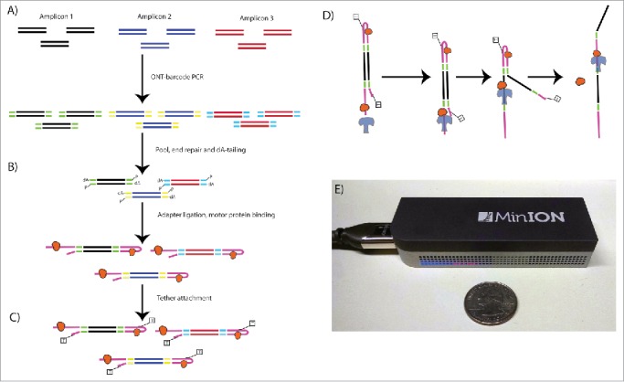
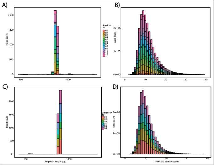
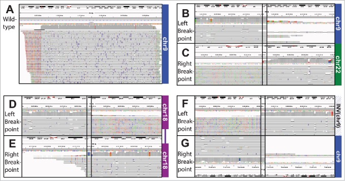
Despite advances in sequencing, structural variants (SVs) remain difficult to reliably detect due to the short read length (<300 bp) of 2nd generation sequencing. Not only do the reads (or paired-end reads) need to straddle a breakpoint, but repetitive elements often lead to ambiguities in the alignment of short reads. We propose to use the long-reads (up to 20 kb) possible with 3rd generation sequencing, specifically nanopore sequencing on the MinION. Nanopore sequencing relies on a similar concept to a Coulter counter, reading the DNA sequence from the change in electrical current resulting from a DNA strand being forced through a nanometer-sized pore embedded in a membrane. Though nanopore sequencing currently has a relatively high mismatch rate that precludes base substitution and small frameshift mutation detection, its accuracy is sufficient for SV detection because of its long reads. In fact, long reads in some cases may improve SV detection efficiency. We have tested nanopore sequencing to detect a series of well-characterized SVs, including large deletions, inversions, and translocations that inactivate the CDKN2A/p16 and SMAD4/DPC4 tumor suppressor genes in pancreatic cancer. Using PCR amplicon mixes, we have demonstrated that nanopore sequencing can detect large deletions, translocations and inversions at dilutions as low as 1:100, with as few as 500 reads per sample. Given the speed, small footprint, and low capital cost, nanopore sequencing could become the ideal tool for the low-level detection of cancer-associated SVs needed for molecular relapse, early detection, or therapeutic monitoring.
Keywords: 3rd generation sequencing; DNA sequencing; Deletions; cancer diagnostics; inversions; nanopore sequencing; next generation sequencing; structural variation; translocations; tumor suppressor gene.
Figures
References
-
- Nowell PC, Hungerford DA. Chromosome studies on normal and leukemic human leukocytes. J Natl Cancer Inst 1960; 25:85-109; PMID:14427847 - PubMed
-
- Dryja TP, Rapaport JM, Joyce JM, Petersen RA. Molecular detection of deletions involving band q14 of chromosome 13 in retinoblastomas. Proc Natl Acad Sci U S A 1986; 83:7391-4; PMID:2876425; http://dx.doi.org/10.1073/pnas.83.19.7391 - DOI - PMC - PubMed
-
- Slamon DJ, Clark GM, Wong SG, Levin WJ, Ullrich A, McGuire WL. Human breast cancer: correlation of relapse and survival with amplification of the HER-2/neu oncogene. Science 1987; 235:177-82; PMID:3798106; http://dx.doi.org/10.1126/science.3798106 - DOI - PubMed
-
- Jones S, Zhang X, Parsons DW, Lin JC, Leary RJ, Angenendt P, Mankoo P, Carter H, Kamiyama H, Jimeno A, et al.. Core signaling pathways in human pancreatic cancers revealed by global genomic analyses. Science 2008; 321:1801-6; PMID:18772397; http://dx.doi.org/10.1126/science.1164368 - DOI - PMC - PubMed
-
- Biankin AV, Waddell N, Kassahn KS, Gingras MC, Muthuswamy LB, Johns AL, Miller DK, Wilson PJ, Patch AM, Wu J, et al.. Pancreatic cancer genomes reveal aberrations in axon guidance pathway genes. Nature 2012; 491:399-405; PMID:23103869; http://dx.doi.org/10.1038/nature11547 - DOI - PMC - PubMed
Publication types
MeSH terms
Substances
Grants and funding
LinkOut - more resources
Full Text Sources
Other Literature Sources
Medical
Miscellaneous