. 2016 Jan 19;198(7):1044-57.
doi: 10.1128/JB.00679-15.

Insights into the Function of a Second, Nonclassical Ahp Peroxidase, AhpA, in Oxidative Stress Resistance in Bacillus subtilis

Affiliations

Insights into the Function of a Second, Nonclassical Ahp Peroxidase, AhpA, in Oxidative Stress Resistance in Bacillus subtilis

Nicole J Broden et al. J Bacteriol. .

Abstract

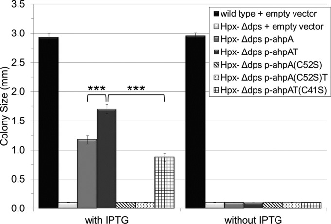
Organisms growing aerobically generate reactive oxygen-containing molecules, such as hydrogen peroxide (H2O2). These reactive oxygen molecules damage enzymes and DNA and may even cause cell death. In response, Bacillus subtilis produces at least nine potential peroxide-scavenging enzymes, two of which appear to be the primary enzymes responsible for detoxifying peroxides during vegetative growth: a catalase (encoded by katA) and an alkylhydroperoxide reductase (Ahp, encoded by ahpC). AhpC uses two redox-active cysteine residues to reduce peroxides to nontoxic molecules. A specialized thioredoxin-like protein, AhpF, is then required to restore oxidized AhpC back to its reduced state. Curiously, B. subtilis has two genes encoding Ahp: ahpC and ahpA. Although AhpC is well characterized, very little is known about AhpA. In fact, numerous bacterial species have multiple ahp genes; however, these additional Ahp proteins are generally uncharacterized. We seek to understand the role of AhpA in the bacterium's defense against toxic peroxide molecules in relation to the roles previously assigned to AhpC and catalase. Our results demonstrate that AhpA has catalytic activity similar to that of the primary enzyme, AhpC. Furthermore, our results suggest that a unique thioredoxin redox protein, AhpT, may reduce AhpA upon its oxidation by peroxides. However, unlike AhpC, which is expressed well during vegetative growth, our results suggest that AhpA is expressed primarily during postexponential growth.

Importance: B. subtilis appears to produce nine enzymes designed to protect cells against peroxides; two belong to the Ahp class of peroxidases. These studies provide an initial characterization of one of these Ahp homologs and demonstrate that the two Ahp enzymes are not simply replicates of each other, suggesting that they instead are expressed at different times during growth of the cells. These results highlight the need to further study the Ahp homologs to better understand how they differ from one another and to identify their function, if any, in protection against oxidative stress. Through these studies, we may better understand why bacteria have multiple enzymes designed to scavenge peroxides and thus have a more accurate understanding of oxidative stress resistance.


Figures

FIG 1
FIG 1
AhpA shares substantial sequence similarity to AhpC. (A) ClustalX alignment of the sequences of classical AhpC and nonclassical AhpA from B. subtilis. Arrows, the redox-active cysteine residues in AhpC. Symbols mark conserved positions: asterisks, identical residues; colons, strong similarity; periods, weak similarity. (B) Phylogenetic tree of representative Ahp proteins generated with ClustalX protein sequence alignments and the TreeView program. B. subtilis AhpC and AhpA are in bold. (C) Phylogenetic tree of representative thioredoxin family proteins generated with ClustalX protein sequence alignments and the TreeView program. B. subtilis AhpT is in bold.
FIG 2
FIG 2
B. subtilis ahpA complements E. coli ahpC mutants for peroxide sensitivity. (A) Western blot of crude lysates from E. coli strains expressing ahpA probed with anti-AhpC antibody developed against Salmonella Typhimurium AhpC. Lanes: 1, wild type with empty vector; 2, ΔahpCF mutant with empty vector; 3, ΔahpCF mutant with pTrc99a-ahpA; 4, ΔahpCF mutant with pTrc99a-ahpAT; 5, ΔahpCF mutant with pTrc99a-ahpA(C52S); 6, ΔahpCF mutant with pTrc99a-ahpA(C52S)T; 7, ΔahpCF mutant with pTrc99a-ahpAT(C41S). (B to E) Peroxide sensitivity of E. coli strains expressing B. subtilis ahpC (B, D) or ahpA (C, E) from pTrc99a (depicted as p-ahpC, p-ahpCF, p-ahpA, or p-ahpAT), as measured by disc diffusion assays. Assays were performed on LB agar containing either 10 μM (for ahpC) or 500 μM (for ahpA) IPTG at 37°C. Filter discs contained 3 μl of 270 mM CHP (B, C), 7 μl of 980 mM H2O2 (D), or 3 μl of 980 mM H2O2 (E). Error bars indicate the standard errors for three independent cultures. Significance was determined using Student's t test, comparing the results for the indicated samples to those for each other (B, C, E) or to those for the ΔkatG ΔahpCF strain (D). †, P < 0.1; *, P < 0.05; **, P < 0.01.
FIG 3
FIG 3
B. subtilis ahpA complements the aerobic growth defect of an E. coli Hpx Δdps strain. Shown is the aerobic growth of the E. coli Hpx Δdps strain expressing B. subtilis ahpA from pTrc99a (depicted as p-ahpA or p-ahpAT). Strains were grown aerobically on LB agar containing 500 μM IPTG, as indicated, at 37°C for 24 h. Shown is the average colony size of 20 isolated colonies of each strain, with error bars indicating the standard errors. Strains for which bars are barely visible on the graph did not grow aerobically. All strains grew anaerobically. Significance was determined using Student's t test, comparing the results for the indicated samples. ***, P < 0.001.
FIG 4
FIG 4
B. subtilis ahpA complements mutations conferring aerobic growth defects in B. subtilis strains. (A) Aerobic growth of B. subtilis strains containing mutations in catalase and peroxidases. Strains were grown on minimal medium at 37°C for 48 h. Shown is the average colony size of 25 isolated colonies of each strain, with error bars indicating the standard errors. (B) Growth of B. subtilis ΔkatA ΔahpCF strains overexpressing ahpA from a Pspac promoter (depicted as p-ahpA or p-ahpAT). Strains were grown aerobically on minimal medium containing 500 μM IPTG, as indicated, at 37°C for 48 h. Shown is the average colony size of 20 isolated colonies of each strain, with error bars indicating the standard errors. Significance was determined using Student's t test, comparing the results for all samples to those for the wild type (A) or the ΔkatA ΔahpCF strain (B). ***, P < 0.001. No significant differences were measured when strains lacking ahpA were compared to their isogenic strains containing wild-type ahpA (A); however, P was <0.1 when the result for the ΔkatA ΔahpCF ΔahpA strain was compared to that for the ΔkatA ΔahpCF strain.
FIG 5
FIG 5
Peroxide sensitivity of B. subtilis strains containing mutations in genes encoding catalase and peroxidases, as measured by disc diffusion assays. Disc diffusion assays were performed on minimal medium at 37°C. Filter discs contained 3 μl of either 1.96 M H2O2 (A) or 270 mM CHP (B, C). Error bars indicate the standard errors for three independent cultures. Significance was determined using Student's t test, comparing the results for all samples to those for the wild type. †, P < 0.1; *, P < 0.05; **, P < 0.01; ***, P < 0.001. No significant differences were measured when the results for strains lacking ahpA were compared to those for their isogenic strains containing wild-type ahpA.
FIG 6
FIG 6
B. subtilis ahpA is regulated by the transition-state regulator AbrB. (A) Basal expression levels of ahpC, katA, and ahpA during vegetative growth on the basis of β-galactosidase activity from transcriptional fusions of promoters of the indicated genes to lacZ. Strains were grown in LB broth at 37°C to an OD600 of 0.8. Similar results were obtained when strains were grown on minimal medium. (−), a wild-type strain lacking any lacZ fusion. (B to D) Expression level of ahpA based on β-galactosidase activity from an ahpA′-lacZ transcriptional fusion. Strains were grown in LB broth at 37°C to an OD600 of 0.8 (B and C) or the indicated optical density (D). (E and F) Peroxide sensitivity of B. subtilis strains containing a mutation in abrB, as measured by disc diffusion assays. Assays were performed on minimal medium at 37°C. Filter discs contained either 3 μl of 1.96 M H2O2 (E) or 7 μl of 270 mM CHP (F). For all panels, error bars indicate the standard errors for three independent cultures. Significance was determined using Student's t test, comparing the results for all samples to those for the wild type. No significant difference was measured when the result for a ΔabrB strain lacking ahpA was compared to that for a ΔabrB strain containing wild-type ahpA. †, P < 0.1; *, P < 0.05; **, P < 0.01.

References

    1. Imlay JA. 2003. Pathways of oxidative damage. Annu Rev Microbiol 57:395–418. doi:10.1146/annurev.micro.57.030502.090938. - DOI - PubMed
    1. Korshunov S, Imlay JA. 2010. Two sources of endogenous hydrogen peroxide in Escherichia coli. Mol Microbiol 75:1389–1401. doi:10.1111/j.1365-2958.2010.07059.x. - DOI - PMC - PubMed
    1. Seaver LC, Imlay JA. 2004. Are respiratory enzymes the primary sources of intracellular hydrogen peroxide? J Biol Chem 279:48742–48750. doi:10.1074/jbc.M408754200. - DOI - PubMed
    1. Zuber P. 2009. Management of oxidative stress in Bacillus. Annu Rev Microbiol 63:575–597. doi:10.1146/annurev.micro.091208.073241. - DOI - PubMed
    1. Mishra S, Imlay J. 2012. Why do bacteria use so many enzymes to scavenge hydrogen peroxide? Arch Biochem Biophys 525:145–160. doi:10.1016/j.abb.2012.04.014. - DOI - PMC - PubMed

Publication types

LinkOut - more resources