. 2016 Feb 2;113(5):1279-84.
doi: 10.1073/pnas.1524400113. Epub 2016 Jan 19.

PGL germ granule assembly protein is a base-specific, single-stranded RNase

Affiliations

PGL germ granule assembly protein is a base-specific, single-stranded RNase

Scott T Aoki et al. Proc Natl Acad Sci U S A. .

Abstract

Cellular RNA-protein (RNP) granules are ubiquitous and have fundamental roles in biology and RNA metabolism, but the molecular basis of their structure, assembly, and function is poorly understood. Using nematode "P-granules" as a paradigm, we focus on the PGL granule scaffold protein to gain molecular insights into RNP granule structure and assembly. We first identify a PGL dimerization domain (DD) and determine its crystal structure. PGL-1 DD has a novel 13 α-helix fold that creates a positively charged channel as a homodimer. We investigate its capacity to bind RNA and discover unexpectedly that PGL-1 DD is a guanosine-specific, single-stranded endonuclease. Discovery of the PGL homodimer, together with previous results, suggests a model in which the PGL DD dimer forms a fundamental building block for P-granule assembly. Discovery of the PGL RNase activity expands the role of RNP granule assembly proteins to include enzymatic activity in addition to their job as structural scaffolds.

Keywords: P-granules; PGL-1; PGL-3; RNA endonuclease; germ-cell development.


Conflict of interest statement

The authors declare no conflict of interest.

Figures

Fig. 1.
Fig. 1.
Identification of the PGL dimerization domain. (A) C. elegans PGL-1, PGL-3, and C. remanei PGL-1 protein schematic. The only recognizable motif by sequence prediction is the RGG repeat region (yellow). The region implicated in granule formation (17) is shown here below PGL-3. We first identified the DD (orange) in PGL-3 (see main text). (B) Size-exclusion chromatography of C. elegans PGL-3 recombinant protein “N-term + DD” (blue) and “DD” (red). Arrows indicate positions of the void volume, albumin (60 kDa), and MBP (37 kDa).
Fig. S1.
Fig. S1.
Biochemical identification of PGL DD. (A) Coomassie-stained SDS/PAGE gel of purified recombinant Ce-PGL-3 protein. PGL regions as shown in Fig 1A. DD, dimerization domain; N-term, N-terminal region. (B) Cross-linking PGL-3 “N-term + DD” with BS3 cross-linker. Band sizes in kilodaltons (kDa). The bands observed are the expected size of monomers, and cross-linked dimers and trimers. (C) PGL-3 “N-term + DD” trypsin digestion. A single ∼25-kDa fragment was generated from a 0–60 min time course digestion. Red box marks band submitted for N-terminal sequencing. (D) Cross-linking PGL-3 DD with BS3 cross-linker. The bands observed are the expected size of monomers and cross-linked dimers. Dual bands generated above and below 50 kDa are most likely a result of additional inter- and intrasubunit cross-linking.
Fig. 2.
Fig. 2.
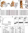
Crystal structure of the C. remanei PGL-1 dimerization domain. (A) C. remanei PGL-1 (Cr-PGL-1) DD crystal structure to 1.6 Å (PDB ID code 5COW). Structure represents Cr-PGL-1 amino acids 202–464 with amino acids 321–335 removed. See Table S1 for data statistics and Fig. S2A for C. elegans PGL-1 DD crystal structure. Helix labeled “α11” encloses the dimer channel in B–E. (B) Cr-PGL-1 DD dimer. See main text for explanation. Subunits in orange and gray. (C) C. remanei PGL-1 DD dimer interface. Salt bridge and hydrogen (H-) bond residues in magenta; other interacting residues that are closely apposed, including hydrophobic residues, in pink. (D) Conservation of dimer interfacing residues by identity (red) and similarity (salmon), as assessed by sequence alignments with PGL-1 homologs, including PGL-3. This analysis does not include interface residues capable of coordinating water molecules between the two subunits. (E) Electrostatic potential of Cr-PGL-1 DD dimer. Color intensity correlates with degree of estimated positive (blue, +) or negative (red, −) charge. Image generated by PDB2PQR (39).
Fig. S2.
Fig. S2.
Further structural analyses of PGL-1 DD. (A) Enlarged image of C. remanei PGL-1 DD. N- and C- termini identified. Helices α9–11 form the dimer channel. (B) C. elegans PGL-1 DD crystal structure to 3.6 Å (PDB ID code 5CV1). See Table S1 for data statistics. Helix-labeled “α11” encloses the dimer channel in B and C. (C) Models for proposed PGL-1 DD dimers of C. elegans (Left) and C. remanei (Right, PDB ID code 5COW). See main text for explanation. (D) Conservation of dimer interfacing residues by identity (red) and similarity (salmon). Conservation assessed by sequence alignments with or without inclusion of C. elegans PGL-2. This analysis does not include interface residues capable of coordinating water molecules between the two subunits.
Fig. 3.
Fig. 3.
PGL DD is a guanosine-specific, single-stranded RNA endonuclease. In A–D and F–I, recombinant MBP::Ce-PGL-1 DD (PGL-1 DD) was incubated with 5′ 32P-labeled RNA oligos and assayed for RNA cleavage products at room temperature (∼20 °C). Substrates were polyX/Y, where X refers to the major base of the oligo and Y refers to a single base at an interior site (Table S2). All nucleic acid oligos were 24 bases long, with the interior site at base number 10. (A and B) Native gels of different RNA oligos (*1 nM) incubated for 1 h with decreasing concentrations of PGL-1 DD (3–0.03 μM). “−” indicates when RNA was incubated for 1 h without PGL-1 DD. The “faster oligo” is observed with (A) polyU/G but not with (B) polyU/U RNA. (C and D) Denaturing gels run after time-course incubation of RNA with 1 μM PGL-1 DD. “0” indicates when sample was immediately taken after addition of PGL-1 DD. “−” indicates when RNA was incubated for 120 min without PGL-1 DD. (C) With polyU/G RNA as a substrate, increasing amounts of cleavage product (“faster oligo”) appear with increasing times of DD incubation. (D) With polyU/U RNA as a substrate, no cleavage product is observed upon DD incubation. (E and F) Comparison of PGL-1 DD enzymatic activity with characterized RNases. (E) RNA cleavage with PGL-1 DD and commercial RNases. Denaturing gel of in vitro-transcribed 32P-guanosine-labeled pos-1 3’UTR (315 bases) (Table S2) incubated with PGL-1 DD (1 and 3 μM), RNase T1 (1.2 nM), or RNase A (0.02 nM) for 1 h; “none” indicates sample incubated without recombinant protein or RNase. PGL-1 DD (3 μM) produces a similar cleavage pattern to RNase T1 (1.2 nM), an ∼2,500-fold concentration difference. RNase A (0.02 nM) completely degrades the radiolabeled RNA. (F) Higher-resolution gel of RNase cleavage products and enzyme sensitivity to inhibitors. Denaturing gel of polyU/G RNA (*10 nM) incubated with PGL-1 DD (1 μM), RNase T1 (1.2 nM), and RNase A (0.02 nM) for 1 h; “none” indicates sample incubated without RNase. Sample incubated without (“−”) or with (“+”) RNase inhibitors. Alkaline hydrolysis fragmentation of polyU/G RNA used to generate a ladder for cleavage product size approximation. (G) One micromolar PGL-1 DD incubated with polyuridine RNA oligos (*1 nM) containing four different interior RNA bases (guanosine, uridine, cytidine, and adenosine) and sampled over time. (H) One micromolar PGL-1 DD incubated with polyU/G RNA, polyU/G RNA with 2′-fluorinated guanosine (“polyU/G-fluoro”) and polyU/G DNA (“DNA”), all at *1 nM. Cleavage percentage in (G and H) calculated as (cleavage product)/[(uncleaved oligo) + (cleavage product)] from measured band density. Average values and SDs of RNase cleavage determined from three separate experiments. (I) Cleavage gel of polyU/G RNA (*1 nM) incubated with complementary polyA/C or polyU/U RNAs in 10:1,1:1, 0.1:1 estimated molar ratios (ramp) before the addition (“+”) of PGL-1 DD. “−” indicates when protein or RNA were excluded from the reaction. *Oligo concentration prior to 5’ radiolabeling.
Fig. S3.
Fig. S3.
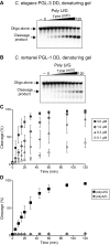
PGL DD cleavage with different protein homologs and RNAs. (A and B) Time-course digestion and denaturing gels of 5′ 32P-labeled polyU/G RNA oligo (Table S2), and (A) 1 μM Ce-PGL-3 DD or (B) 1 μM Cr-PGL-1 DD. “0” indicates when sample was taken immediately after addition of PGL DD. “−” indicates when RNA was incubated for 120 minutes without protein. A single RNA cleavage product appears with either protein. (C) Concentration-dependent cleavage by PGL-1 DD. MBP::Ce-PGL-1 DD (3, 1, 0.3, 0.1 μM) incubated with 5′ 32P-labeled polyU/G RNA oligo (*1 nM) and sampled over time for cleavage efficiency. (D) PGL-1 DD cleavage activity with polyA/C RNA. MBP::Ce-PGL-1 DD (3 μM) incubated with a 5′ 32P-labeled polyA/C RNA oligo (Table S2) (*1 nM) and sampled over time for cleavage efficiency. Cleavage percentage calculated as in Fig. 3 G and H. Average values and standard deviations of RNase cleavage determined from three separate experiments. *Oligo concentration prior to 5′ radiolabeling.
Fig. S4.
Fig. S4.
A histidine mutation diminishes PGL-1 DD cleavage activity. (A) Cleavage efficiency of PGL-1 DD with metals. MBP::PGL-1 DD incubated with polyU/G RNA (Table S2) and either EDTA, MgCl2, or MnCl2. Cleavage ratios were assessed on a native gel. (B and C) Location of the histidine amino acid H478 on the Cr-PGL-1 DD dimer. An identical site is on the opposite end of the dimer. (B) Red box highlights region enlarged in C. (C) Atomic detail of H478 and the surrounding conserved residues (Y439, E442, R481). Numbers correspond to C. elegans PGL-1. (D) Sequence conservation of H478, Y439, E442, and R481 (“*”) among Caenorhabditid. (E) Mutation of PGL-1 DD affects cleavage activity. Cleavage efficiency assessed with 3 μM MBP::Ce-PGL-1 DD wild-type, H478A, and E442Q mutant recombinant protein, and *1 nM 5′ 32P-labeled polyuridine RNA oligo with a single interior guanosine base (polyU/G). Cleavage percentage reported and experiments performed as in Fig. 3 G and H. *Oligo concentration before 5′ radiolabeling.
Fig. 4.
Fig. 4.
A glutamine mutation abrogates PGL-1 endonuclease activity. (A) Location of the relevant glutamine in the Cr-PGL-1 DD dimer. When oriented along the channel, the PGL-1 DD dimer subunits assemble antiparallel to each other. Red line shows the plane of the red box in adjacent orthogonal view. Red box identifies structural region enlarged in B. (B) Atomic detail of Q342 in the Cr-PGL-1 DD dimer. Numbering corresponds to Ce-PGL-1. (C) Sequence conservation of Q342 (“*”) among Caenorhabditid. (D) PGL-1 Q342A abrogates cleavage activity. Cleavage efficiency assessed with 3 μM MBP::Ce-PGL-1 DD wild-type and Q342A mutant protein, and a polyU/G RNA oligo (Table S2). Cleavage percentage calculated as in Fig. 3 G and H. (E) PGL-1 Q342A still binds RNA. Native gels of 5′ 32P-labeled RNA incubated for 30 min without (“−”) or with 3–0.3 μM (ramp) MBP::Ce-PGL-1 DD wild-type (WT) or RNase mutant (Q342A). In all panels, recombinant protein was incubated with *1 nM 5′ 32P-labeled polyU/G or 2′ fluorinated polyU/G (“polyU/G-fluoro”) (see main text and Table S2) RNA oligos before running on a native gel. “−” identifies RNA incubated without PGL-1 DD. *Oligo concentration prior to 5’ radiolabeling. (F) Lack of biological effect in pgl-1 Q342A mutants. Wild-type, pgl-1–null and two independent pgl-1 Q342A RNase mutants were singled and scored for fertility after incubation at either 20 °C or 26.5 °C, as performed in ref. . More details in SI Materials and Methods.
Fig. S5.
Fig. S5.
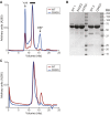
Purification of MBP-tagged PGL-1 DD. (A) Wild-type and mutant PGL-1 DD both dimerize. Sizing column of recombinant MBP::Ce-PGL-1 DD wild-type (WT) and Q342A mutant protein. Column void volume (“void”) and free MBP (“MBP”) labeled with arrows. Black bar indicates fractions pooled and used in RNA cleavage analyses. (B) Coomassie-stained SDS/PAGE gel of purified MBP::Ce-PGL-1 wild-type and mutant proteins E442Q, H478A, and Q342A. Protein pooled from sizing column fractions (A, black bar). The wild-type samples represent two different protein preparations (WT 1, WT 2). (C) MBP::Ce-PGL-1 DD wild-type (WT) and Q342A recombinant protein purified from a sizing column rerun on the same column one day later. No protein is observed in the void, and both WT and Q342A elute at a similar position to that observed in the initial sizing column run. A minor peak is again observed later during the elution and attributed to MBP.

References

    1. Buchan JR, Parker R. Eukaryotic stress granules: The ins and outs of translation. Mol Cell. 2009;36(6):932–941. - PMC - PubMed
    1. Anderson P, Kedersha N. RNA granules: Post-transcriptional and epigenetic modulators of gene expression. Nat Rev Mol Cell Biol. 2009;10(6):430–436. - PubMed
    1. Brangwynne CP, et al. Germline P granules are liquid droplets that localize by controlled dissolution/condensation. Science. 2009;324(5935):1729–1732. - PubMed
    1. Kato M, et al. Cell-free formation of RNA granules: Low complexity sequence domains form dynamic fibers within hydrogels. Cell. 2012;149(4):753–767. - PMC - PubMed
    1. Voronina E, Seydoux G, Sassone-Corsi P, Nagamori I. RNA granules in germ cells. Cold Spring Harb Perspect Biol. 2011;3(12):a002774. - PMC - PubMed

Publication types