. 2016 Feb 2;113(5):1315-20.
doi: 10.1073/pnas.1518752113. Epub 2016 Jan 19.

Integrated data analysis reveals uterine leiomyoma subtypes with distinct driver pathways and biomarkers

Affiliations

Integrated data analysis reveals uterine leiomyoma subtypes with distinct driver pathways and biomarkers

Miika Mehine et al. Proc Natl Acad Sci U S A. .

Abstract

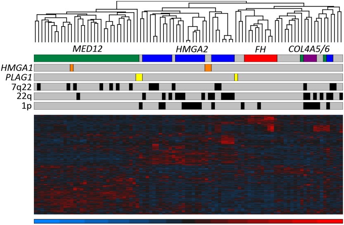
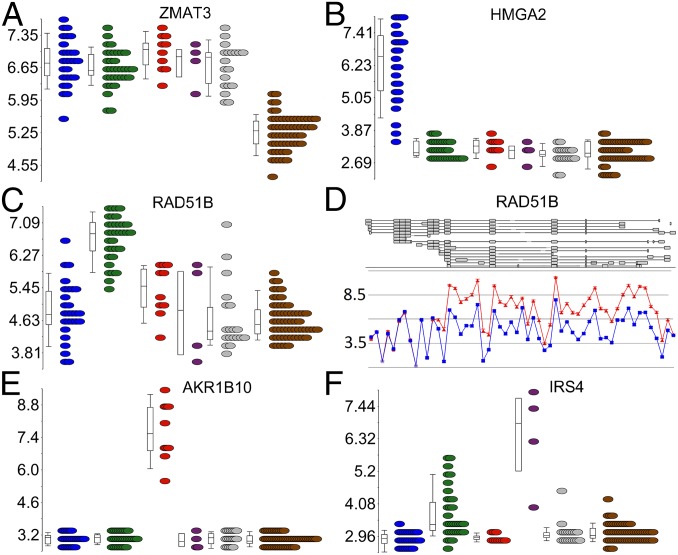
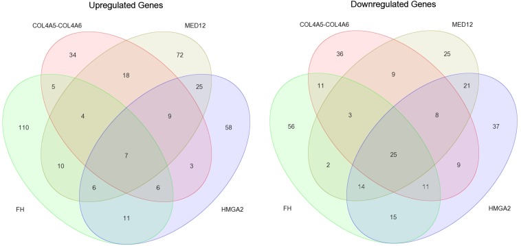
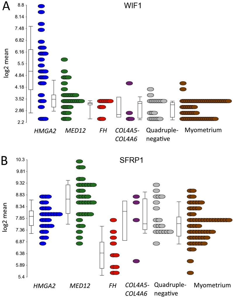
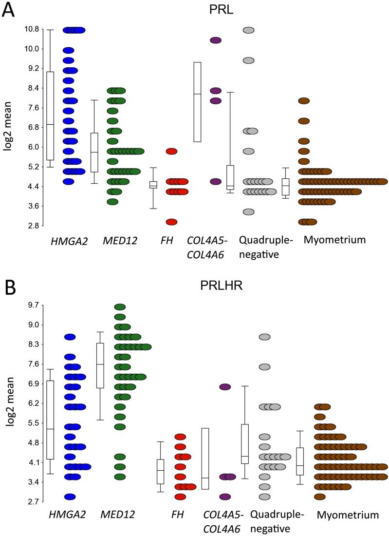
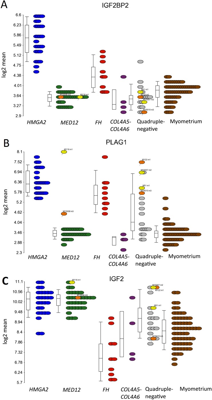
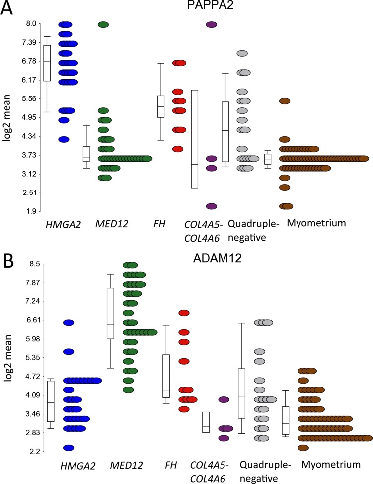
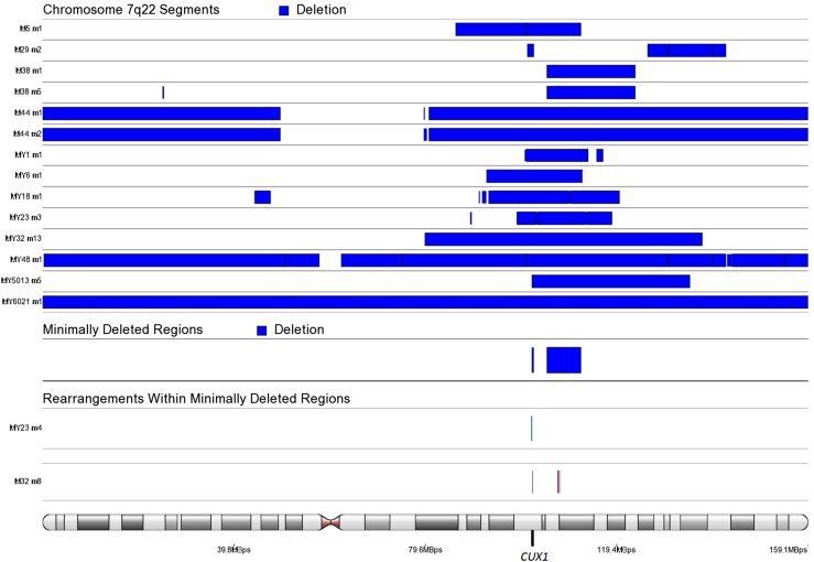
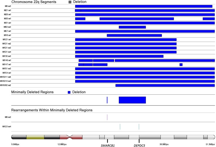
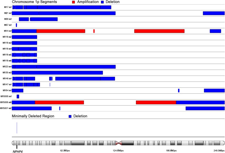
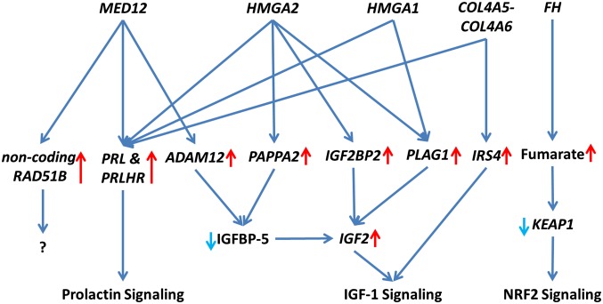
Uterine leiomyomas are common benign smooth muscle tumors that impose a major burden on women's health. Recent sequencing studies have revealed recurrent and mutually exclusive mutations in leiomyomas, suggesting the involvement of molecularly distinct pathways. In this study, we explored transcriptional differences among leiomyomas harboring different genetic drivers, including high mobility group AT-hook 2 (HMGA2) rearrangements, mediator complex subunit 12 (MED12) mutations, biallelic inactivation of fumarate hydratase (FH), and collagen, type IV, alpha 5 and collagen, type IV, alpha 6 (COL4A5-COL4A6) deletions. We also explored the transcriptional consequences of 7q22, 22q, and 1p deletions, aiming to identify possible target genes. We investigated 94 leiomyomas and 60 corresponding myometrial tissues using exon arrays, whole genome sequencing, and SNP arrays. This integrative approach revealed subtype-specific expression changes in key driver pathways, including Wnt/β-catenin, Prolactin, and insulin-like growth factor (IGF)1 signaling. Leiomyomas with HMGA2 aberrations displayed highly significant up-regulation of the proto-oncogene pleomorphic adenoma gene 1 (PLAG1), suggesting that HMGA2 promotes tumorigenesis through PLAG1 activation. This was supported by the identification of genetic PLAG1 alterations resulting in expression signatures as seen in leiomyomas with HMGA2 aberrations. RAD51 paralog B (RAD51B), the preferential translocation partner of HMGA2, was up-regulated in MED12 mutant lesions, suggesting a role for this gene in the genesis of leiomyomas. FH-deficient leiomyomas were uniquely characterized by activation of nuclear factor erythroid 2-related factor 2 (NRF2) target genes, supporting the hypothesis that accumulation of fumarate leads to activation of the oncogenic transcription factor NRF2. This study emphasizes the need for molecular stratification in leiomyoma research and possibly in clinical practice as well. Further research is needed to determine whether the candidate biomarkers presented herein can provide guidance for managing the millions of patients affected by these lesions.

Keywords: HMGA2; MED12; transcriptional profiling; uterine leiomyoma.


Conflict of interest statement

The authors declare no conflict of interest.

Figures

Fig. 1.
Fig. 1.
Unsupervised hierarchical clustering of 94 leiomyomas from 60 patients. Hierarchical clustering using 1% most variable genes revealed that most leiomyomas grouped together according to the mutation status of MED12 (green), HMGA2 (blue), FH (red), and COL4A5-COL4A6 (purple). The remaining quadruple-negative leiomyomas exhibited transcriptional heterogeneity and clustered into several unique branches; however, four of these clustered with leiomyomas of the HMGA2 subtype, and two of them were found to harbor a genetic HMGA1 alteration (orange). One leiomyoma (MY5008 m3) harbored both an HMGA1 alteration and a MED12 mutation, and consequently clustered with leiomyomas of the MED12 subtype. We identified genetic PLAG1 alterations (yellow) in three leiomyomas. Although one of these tumors (MY16 m1) also harbored a MED12 mutation and clustered with the MED12 subtypes, all three tumors also displayed expression signatures similar to those seen in leiomyomas with HMGA2 or HMGA1 alterations. Chromosomal deletions of 7q22, 22q, and 1p had no major influence on the clustering.
Fig. 2.
Fig. 2.
Examples of shared and uniquely expressed genes. (A) ZMAT3 was the most significantly differentially expressed gene in all leiomyomas compared with the normal myometrial tissue (brown). (B) HMGA2 was the most uniquely expressed gene in leiomyomas of the HMGA2 (blue) subtype. (C) RAD51B was the most uniquely expressed gene in leiomyomas of the MED12 (green) subtype. (E) AKR1B10 was the most uniquely expressed gene in leiomyomas of the FH (red) subtype. (F) IRS4 was the most uniquely expressed gene in leiomyomas of the COL4A5-COL4A6 (purple) subtype. (D) Exon-level analysis revealed that the overexpression of RAD51B in MED12 mutant leiomyomas originated predominantly from a noncoding transcript (ENST00000492236).
Fig. S1.
Fig. S1.
Venn diagram illustrating the number of shared and uniquely differentially expressed genes (q <0.05; –2> FC >2) among leiomyomas of the HMGA2, MED12, FH, and COL4A5-COL4A6 subtypes. Leiomyomas of the MED12 and HMGA2 subtypes shared the most differentially expressed genes, and leiomyomas of the FH subtype exhibited the most uniquely differentially expressed genes.
Fig. S2.
Fig. S2.
mRNA expression of the Wnt antagonists WIF1 and SFRP1 in different uterine leiomyoma subtypes. (A) Leiomyomas of the HMGA2 subtype displayed a significant up-regulation of the Wnt inhibitor WIF1 (FC = 4.7). (B) Leiomyomas of the MED12 subtype displayed a significant up-regulation of the Wnt inhibitor SFRP1 (FC = 2.1).
Fig. S3.
Fig. S3.
mRNA expression of PRL and PRLHR in different uterine leiomyoma subtypes. (A) PRL was significantly up-regulated in three subtypes (HMGA2, MED12, and COL4A5-COL4A6), and particularly high expression was seen in leiomyomas of the HMGA2 (FC = 7.6) and COL4A5-COL4A6 (FC = 9.9) subtypes. (B) In contrast, PRLHR was significantly up-regulated in two subtypes (HMGA2 and MED12), and particularly high expression was seen in leiomyomas of the MED12 subtype (FC = 9.0).
Fig. S4.
Fig. S4.
mRNA overexpression of IGF2BP2, PLAG1, and IGF2 in leiomyomas of the HMGA2 subtype. (A) Statistical analysis identified IGF2BP2 as the second-most uniquely expressed gene (FC = 4.4) in leiomyomas of the HMGA2 subtype (blue dots). (B) The proto-oncogene PLAG1 also was among the most uniquely expressed genes (FC = 8.2). Up-regulation of PLAG1 (FC = 5.7) was also seen among the three leiomyomas with genetic HMGA1 alterations (orange dots). Three leiomyomas harbored a genetic PLAG1 alteration (yellow dots) and showed a highly significant up-regulation of PLAG1 (FC = 12.5). (C) PLAG1 is known to regulate the expression of IGF2, and we detected a significant up-regulation (FC = 3.0) of IGF2 in leiomyomas of the HMGA2 subtype. A significant up-regulation of IGF2 was also seen in the leiomyomas with a genetic PLAG1 alteration (FC = 4.3) and in two out of three leiomyomas with a HMGA1 alteration (FC > 2). Of note, leiomyomas of the MED12 subtype also displayed a significant up-regulation of IGF2 (FC = 3.1), and whereas leiomyomas of the FH subtype displayed a significant up-regulation of PLAG1 (FC = 4.8), surprisingly, IGF2 was down-regulated in these lesions (FC = –2.5).
Fig. S5.
Fig. S5.
mRNA expression of the PAPPA2 and ADAM12 in different uterine leiomyoma subtypes. (A) Leiomyomas of the HMGA2 subtype displayed a significant up-regulation of the PAPPA2 (FC = 7.9). (B) Leiomyomas of the MED12 subtype displayed a significant up-regulation of the ADAM12 (FC = 10.5).
Fig. S6.
Fig. S6.
Chromosomal rearrangements in a quadruple-negative leiomyoma (M18 m1) resulting in a fusion gene involving the 5′ end IGFBP5 (A) and the 3′ end of PDGFRB (B). The breakpoints were located in intron 1 of IGFBP5 and in intron 10 of PDGFRB.
Fig. S7.
Fig. S7.
RNA sequencing validation of the up-regulated noncoding transcript of RAD51B in leiomyomas of the MED12 subtype. Illustrated are read alignments at the RAD51B locus in two pooled MED12 mutant samples (MY18 m1 and MY23 m1). Protein-coding transcripts are marked in red; noncoding transcripts, in blue. As observed with the exon arrays, the overexpression of RAD51B originates predominately from a noncoding transcript of RAD51B (ENST00000492236).
Fig. S8.
Fig. S8.
SCNAs affecting chromosome 7q22. We identified 104849448–111900000 as the minimally deleted region. Another, less commonly deleted region was identified on 101732303–102100000. This region contained the putative target gene CUX1. Two additional samples harbored chromosomal rearrangements that disrupted the CUX1 gene.
Fig. S9.
Fig. S9.
SCNAs affecting chromosome 22q. We identified 27111559–33871686 as the minimally deleted region on chromosome 22q. We analyzed this region further, and identified one additional leiomyoma (MY23 m4) harboring chromosomal rearrangements within this region. One of the breakpoints was located ∼14 kbp upstream of the putative target gene DEPDC5. Another, less commonly deleted region was identified on 24087031–24200000. We analyzed this region further, and identified one leiomyoma (M9 m3) as harboring an additional rearrangement within this region, resulting in a second hit disruption of the tumor suppressor SMARCB1.
Fig. S10.
Fig. S10.
SCNAs affecting chromosome 1p. We identified Chr1:5753010–5953574 as the minimally deleted region on chromosome 1p. This region contained only one protein-coding gene: NPHP4.
Fig. 3.
Fig. 3.
Schematic of highlighted driver pathways in leiomyoma development and growth. Leiomyomas display subtype-specific differences in key driver pathways, including Prolactin, IGF1, and NRF2 signaling.

References

    1. Rice KE, Secrist JR, Woodrow EL, Hallock LM, Neal JL. Etiology, diagnosis, and management of uterine leiomyomas. J Midwifery Womens Health. 2012;57(3):241–247. - PubMed
    1. Zhao D, Rogers PAW. Is fibroid heterogeneity a significant issue for clinicians and researchers? Reprod Biomed Online. 2013;27(1):64–74. - PubMed
    1. Mäkinen N, et al. MED12, the mediator complex subunit 12 gene, is mutated at high frequency in uterine leiomyomas. Science. 2011;334(6053):252–255. - PubMed
    1. Mehine M, et al. Characterization of uterine leiomyomas by whole-genome sequencing. N Engl J Med. 2013;369(1):43–53. - PubMed
    1. Mehine M, Mäkinen N, Heinonen HR, Aaltonen LA, Vahteristo P. Genomics of uterine leiomyomas: Insights from high-throughput sequencing. Fertil Steril. 2014;102(3):621–629. - PubMed

Publication types