. 2015 Dec 10;11(6):1164-78.
doi: 10.5114/aoms.2015.56342. Epub 2015 Dec 11.

Brain-derived neurotrophic factor and its clinical implications

Affiliations

Brain-derived neurotrophic factor and its clinical implications

Siresha Bathina et al. Arch Med Sci. .

Abstract

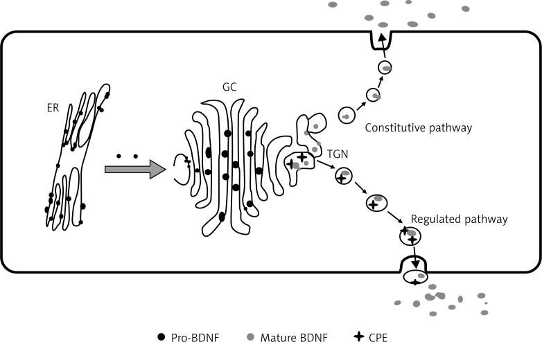
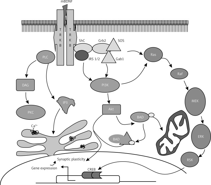
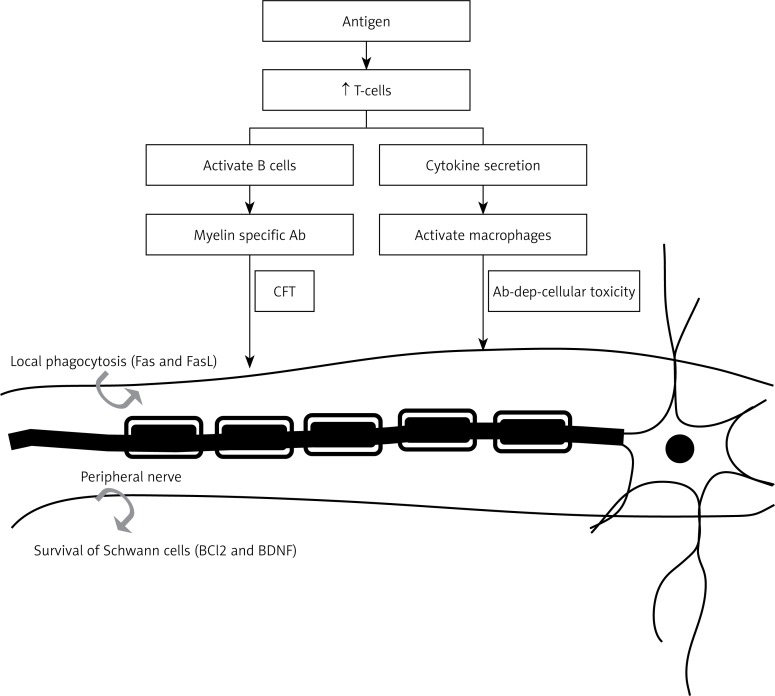
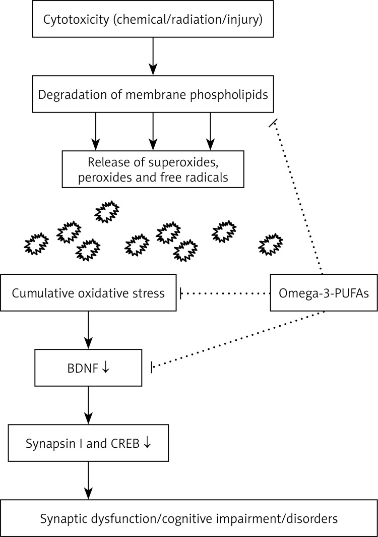
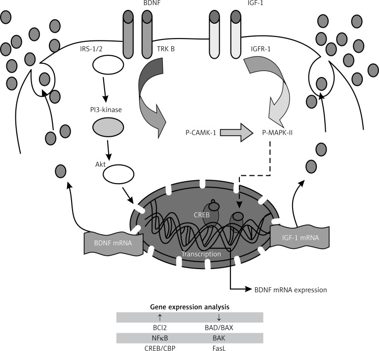
Brain-derived neurotrophic factor (BDNF) plays an important role in neuronal survival and growth, serves as a neurotransmitter modulator, and participates in neuronal plasticity, which is essential for learning and memory. It is widely expressed in the CNS, gut and other tissues. BDNF binds to its high affinity receptor TrkB (tyrosine kinase B) and activates signal transduction cascades (IRS1/2, PI3K, Akt), crucial for CREB and CBP production, that encode proteins involved in β cell survival. BDNF and insulin-like growth factor-1 have similar downstream signaling mechanisms incorporating both p-CAMK and MAPK that increase the expression of pro-survival genes. Brain-derived neurotrophic factor regulates glucose and energy metabolism and prevents exhaustion of β cells. Decreased levels of BDNF are associated with neurodegenerative diseases with neuronal loss, such as Parkinson's disease, Alzheimer's disease, multiple sclerosis and Huntington's disease. Thus, BDNF may be useful in the prevention and management of several diseases including diabetes mellitus.

Keywords: Alzheimer's disease; brain-derived neurotrophic factor; diabetes mellitus; neurotransmission; signal transduction; β cell.


Figures

Figure 1
Figure 1
The above figure illustrates the fact of origin of pro-BDNF in endoplasmic reticulum (ER), which is later transported to the Golgi complex (GC) and then to the trans-Golgi network (TGN). From here in the regulated pathway, by the action of CPE and convertase, 13 KDa mature BDNF (mBDNF) is formed and released outside the plasma membrane. This figure is adapted from ref. [13]
Figure 2
Figure 2
Gene structure of BDNF. Note the presence of four promoters in rat and 9 promoters in mouse. Each of the driving transcripts of BDNF mRNAs containing one of the four 5′ non-coding exons (I, II, III, IV) in promoters is later spliced to the common 3′ protein coding exon. Human BDNF structure and its splicing variant are seen above with arrows indicating alternative polyadenylation sites (PolyA) in the 3′-UTR and internal alternative splice sites in exons 2, 6, 7 and 9a (letters a, b, c and d) [18]. Arrangement of introns and exons on BDNF genes is determined by analyzing genomic and mRNA sequence using bioinformatics, RACE, and RT-PCR [17]
Figure 3
Figure 3
Signaling pathway of BDNF. BDNF binds to its high-affinity receptor tyrosine kinase B (TrkB), resulting in the recruitment of proteins that activate three different signal transduction cascades. One cascade involves sequential activation of insulin receptor substrate-1 (IRS-1/2), phosphatidylinositol-3-kinase (PI-3K) and protein kinase B (Akt). The second is the activation of Shc/Grb2, Ras, Raf, mitogen-activated protein kinase kinases (MEKs) and extracellular signal regulated kinases (ERKs). The third cascade involves phospholipase C (PLC), inositol (1,4,5)-trisphosphate [Ins(1,4,5)P3], diacylglycerol (DAG) and protein kinase C (PKC). BDNF signaling pathways activate one or more transcription factors (cAMP-response-element-binding protein (CREB) and CREB-binding protein (CBP) that regulate expression of genes encoding proteins involved in neural plasticity, stress resistance and cell survival. This figure is adapted and modified from refs. [32] and [33]
Figure 4
Figure 4
Role of BDNF in neural degeneration diseases such as multiple sclerosis. The antigen once it crosses the blood-brain barrier stimulates the production of T cells, which, in turn, activate B cells and macrophages. Damage to nerve fibers may result from either complement fixation or antibody-dependent cell-mediated immunity, resulting in multiple sclerosis. Neuroinflammatory reactions may also trigger neuroprotective events such as secretion of BDNF and anti-apoptotic Bcl2 that may explain the therapeutic role of BDNF in multiple sclerosis. This figure is adapted from refs. [–84]
Figure 5
Figure 5
Interaction between omega 3-fatty acids and BDNF that may underlie their cytoprotective actions. Mechanism of cytoprotection may involve: a) prevention of degradation of membrane phospholipids; b) reduction of oxidative stress that helps maintain synaptic plasticity; and c) normalization of levels of BDNF and its downstream effectors synapsin I and CREB, which are important in learning, memory and LTP. This figure is adapted and modified from refs. [–91]
Figure 6
Figure 6
Possible mechanism(s) involved in cytoprotective action of BDNF. Sequential activation of insulin receptor substrate-1 (IRS-1/2), phosphatidylinositol-3-kinase (PI-3K) and protein kinase B (Akt) result in activation of pro-survival genes. IGF-1 and BDNF were shown to have similar downstream mechanisms, incorporating both CAMK and MAPK, which inactivate cell death machinery (Bad, BAX and FasL) and promote cell survival (Bcl2), neurogenesis and plasticity. Both BDNF and IGF mRNA are re-synthesized to form respective molecules which not only enhance insulin production but also inhibit apoptosis of β cell machinery. This figure is adapted from refs. [–110]

References

    1. Binder DK, Scharfman HE. Brain-derived neurotrophic factor. Growth Factors. 2004;22:123–31. - PMC - PubMed
    1. Acheson A, Conover JC, Fandl JP, et al. A BDNF autocrine loop in adult sensory neurons prevents cell death. Nature. 1995;374:450–3. - PubMed
    1. Huang EJ, Reichardt LF. Neurotrophins: roles in neuronal development and function. Ann Rev Neurosci. 2001;24:677–736. - PMC - PubMed
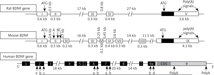
    1. Maisonpierre PC, Le Beau MM, Ip NY, et al. Human and rat brain-derived neurotrophic factor and neurotrophin-3: gene structures, distributions, and chromosomal localizations. Genomics. 1991;10:558–68. - PubMed
    1. Zigova T, Pencea V, Wiegand SJ, Luskin MB. Intraventricular administration of BDNF increases the number of newly generated neurons in the adult olfactory bulb. Mol Cell Neurosci. 1998;11:234–45. - PubMed