. 2016 Jan 26;7(4):3709-25.
doi: 10.18632/oncotarget.6915.

Cooperative TRAIL production mediates IFNα/Smac mimetic-induced cell death in TNFα-resistant solid cancer cells

Affiliations

Cooperative TRAIL production mediates IFNα/Smac mimetic-induced cell death in TNFα-resistant solid cancer cells

Stefanie Roesler et al. Oncotarget. .

Abstract

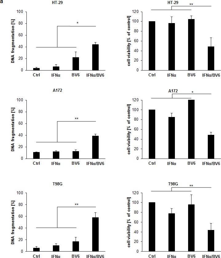
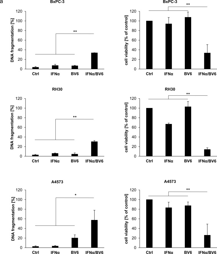
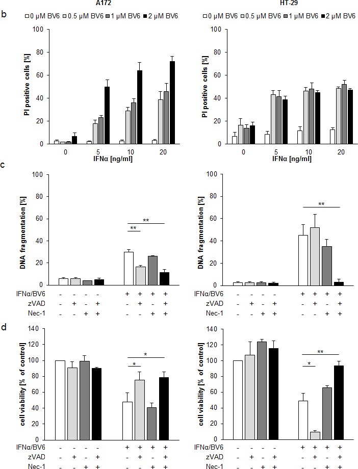
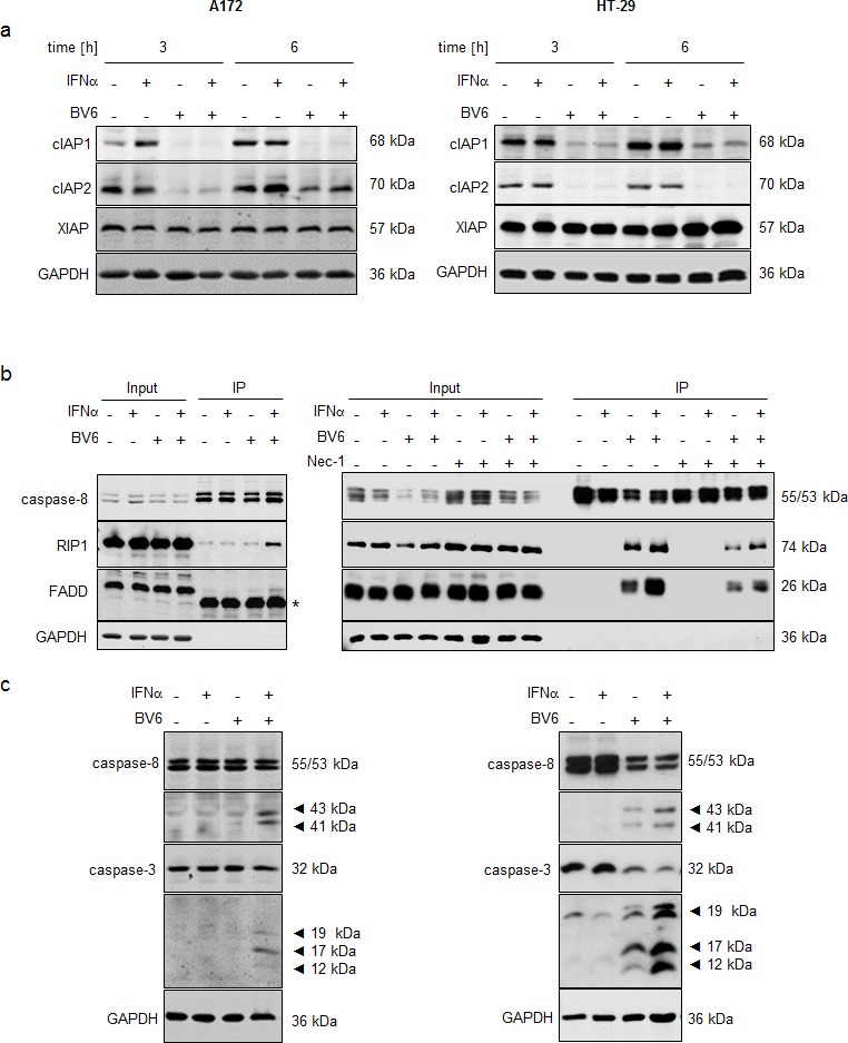
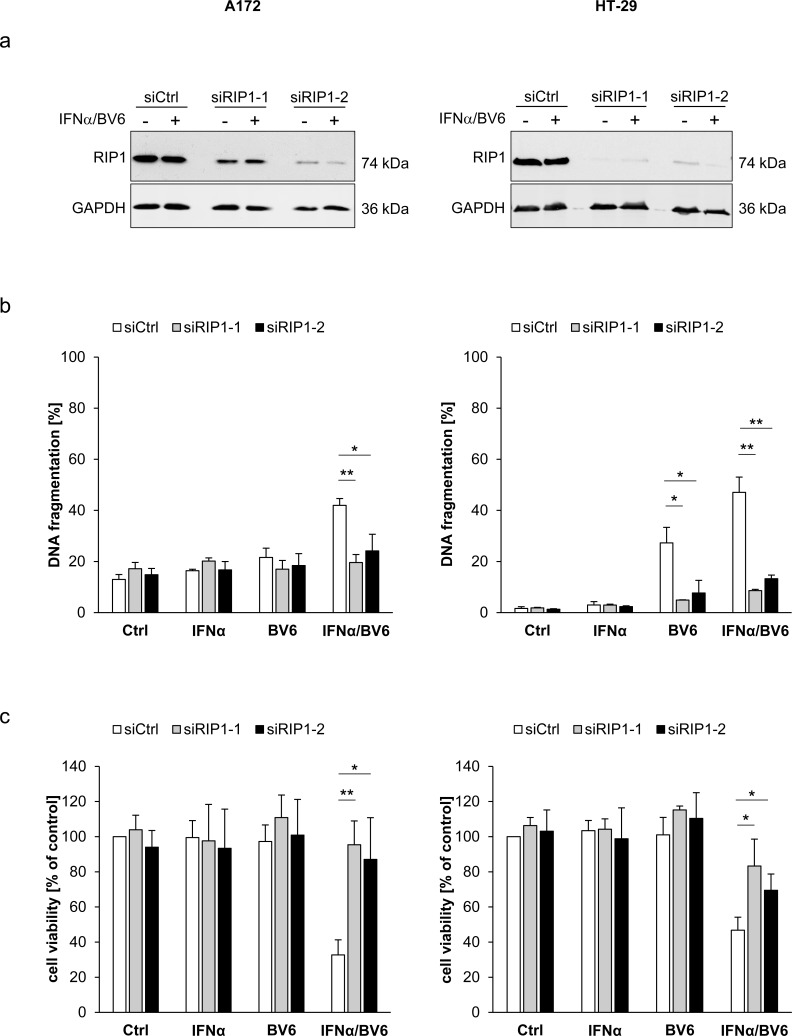
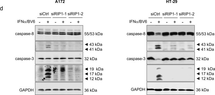
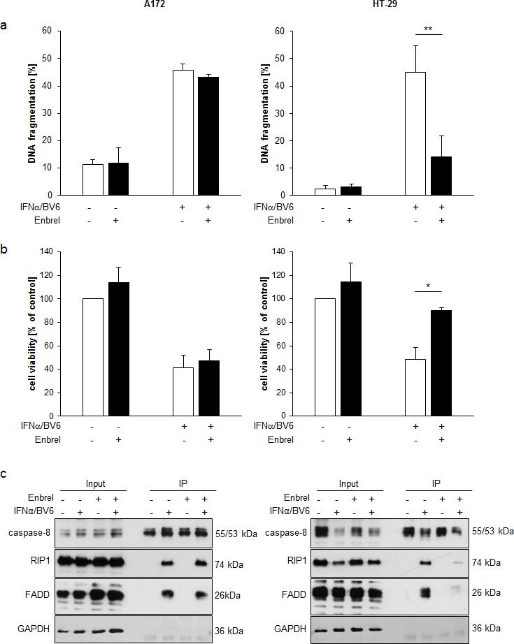
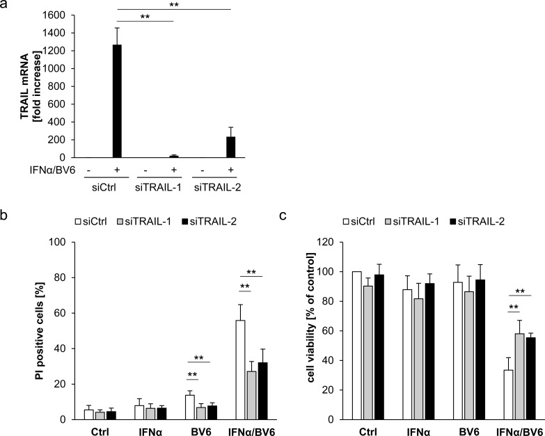
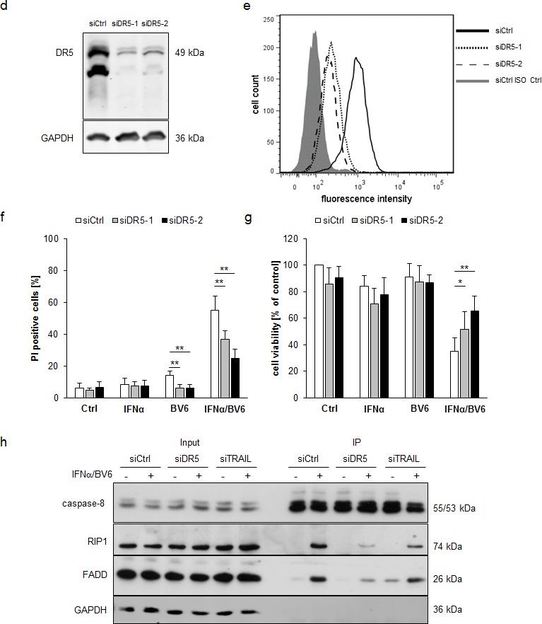
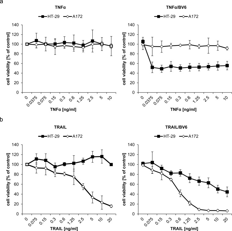
Smac mimetics antagonize IAP proteins, which are highly expressed in several cancers. Recent reports indicate that Smac mimetics trigger a broad cytokine response and synergize with immune modulators to induce cell death. Here, we identify a differential requirement of TRAIL or TNFα as mediators of IFNα/Smac mimetic-induced cell death depending on the cellular context. Subtoxic concentrations of Smac mimetics cooperate with IFNα to induce cell death in various solid tumor cell lines in a highly synergistic manner as determined by combination index. Mechanistic studies show that IFNα/BV6 cotreatment promotes the formation of a caspase-8-activating complex together with the adaptor protein FADD and RIP1. Assembly of this RIP1/FADD/caspase-8 complex represents a critical event, since RIP1 silencing inhibits IFNα/BV6-induced cell death. Strikingly, pharmacological inhibition of paracrine/autocrine TNFα signaling by the TNFα scavenger Enbrel rescues HT-29 colon carcinoma cells, but not A172 glioblastoma cells from IFNα/BV6-induced cell death. By comparison, A172 cells are significantly protected against IFNα/BV6 treatment by blockage of TRAIL signaling through genetic silencing of TRAIL or its cognate receptor TRAIL receptor 2 (DR5). Despite this differential requirement of TNFα and TRAIL signaling, mRNA and protein expression is increased by IFNα/BV6 cotreatment in both cell lines. Interestingly, A172 cells turn out to be resistant to exogenously added recombinant TNFα even in the presence of BV6, whereas they display a high sensitivity towards TRAIL/BV6. In contrast, BV6 efficiently sensitizes HT-29 cells to TNFα while TRAIL only had limited efficacy. This demonstrates that a differential sensitivity towards TRAIL or TNFα determines the dependency on either death receptor ligand for IFNα/Smac mimetic-induced cell death. Thus, by concomitant stimulation of both death receptor systems IFNα/Smac mimetic combination treatment is an effective strategy to induce cell death in TNFα- or TRAIL-responsive cancers.

Keywords: Smac; TRAIL; apoptosis; cell death; interferon.


Conflict of interest statement

CONFLICTS OF INTEREST

None to declare.

Figures

Figure 1
Figure 1. IFNα and BV6 synergistically induce cell death in various cancer cell lines
A. Cells were treated with IFNα (HT-29, BxPC-3, A4573: 10 ng/ml; A172, T98G, RH30: 5 ng/ml) and/or BV6 (HT-29, A172: 1 μM; T-98G: 7 μM; BxPC-3: 2 μM; RH30: 500 nM; A4573: 4 μM) for 72 hours (except BxPc-3 were treated for 24 hours). Cell death was determined by analysis of DNA fragmentation of PI-stained nuclei using flow cytometry and mean + SD of three independent experiments performed in duplicate are shown (left panels). Cell viability was determined by MTT assay and data are shown as percentage of untreated control cells with mean + SD of three independent experiments performed in triplicate (right panel). B. A172 and HT-29 cells were treated for 72 hours with indicated concentrations of IFNα and/or BV6. Cell death was determined by PI staining using flow cytometry and mean + SD of three independent experiments performed in duplicate are shown. C., D. Cells were treated for 72 hours with IFNα (A172: 5 ng/ml, HT-29: 10 ng/ml) and 1 μM BV6 in the presence or absence of 20 μM zVAD.fmk and/or 30 μM Nec-1 without renewal of inhibitors. Cell death was determined by analysis of DNA fragmentation of PI-stained nuclei using flow cytometry and mean + SD of three independent experiments performed in duplicate are shown C.. Cell viability was determined by MTT assay and data are shown as percentage of untreated control cells with mean + SD of three independent experiments performed in triplicate D.. *P < 0.05; **P < 0.01.
Figure 2
Figure 2. IFNα/BV6 cotreatment induces IAP depletion, complex II formation and caspase activation
A. Cells were treated for 3 and 6 hours with IFNα (A172: 5 ng/ml, HT-29: 10 ng/ml) and/or 1 μM BV6. Protein levels of cIAP1/2 and XIAP were assessed by Western blotting, GAPDH was used as loading control. B. Cells were treated for 12 hours with IFNα (A172: 5 ng/ml, HT-29: 10 ng/ml) and/or 1 μM BV6 in the presence of 20 μM zVAD.fmk and/or 30 μM Nec-1 (HT-29 only). Caspase-8 was immunoprecipitated using an anti-caspase-8 antibody and detection of RIP1, FADD and caspase-8 was done by Western blotting, GAPDH served as loading control. Asterisk indicates IgG light chain. C. Cells were treated for 24 hours (A172) and 18 hours (HT-29) with IFNα (A172: 5 ng/ml, HT-29: 10 ng/ml) and/or 1 μM BV6. Cleavage of caspase-8 and 3 was assessed by Western blotting, active cleavage products are indicated by arrows. GAPDH was used as loading control.
Figure 3
Figure 3. IFNα/BV6-induced cell death depends on RIP1
Cells were transiently transfected with 5 nM siRNA targeting RIP1 (siRIP1-1, siRIP1-2) or control siRNA. A. Cells were treated with IFNα (A172: 5 ng/ml, HT-29: 10 ng/ml) and 1 μM BV6. Protein expression of RIP1 was analyzed by Western blotting, GAPDH was used as loading control. B. Cells were treated for 72 hours with IFNα (A172: 5 ng/ml, HT-29: 10 ng/ml) and/or 1 μM BV6. Cell death was determined by analysis of DNA fragmentation of PI-stained nuclei using flow cytometry. Mean + SD of three independent experiments performed in duplicate are shown; *P < 0.05; **P < 0.01. C. Cell viability was determined by MTT assay. Data are shown as percentage of untreated control cells with mean + SD of three independent experiments performed in triplicate; *P < 0.05; **P < 0.01. D. Cells were treated for 24 hours with IFNα (A172: 5 ng/ml, HT-29: 10 ng/ml) and 1 μM BV6. Cleavage of caspase-8 and 3 was assessed by Western blotting, GAPDH was used as loading control.
Figure 4
Figure 4. Differential requirement of TNFα for IFNα/BV6-induced cell death
A., B. Cells were treated for 72 hours with IFNα (A172: 5 ng/ml, HT-29: 10 ng/ml) and 1 μM BV6 in the presence or absence of 50 μg/ml Enbrel. Cell death was determined by analysis of DNA fragmentation of PI-stained nuclei using flow cytometry and mean + SD of three independent experiments performed in duplicate are shown A.. Cell viability was determined by MTT assay and data are shown as percentage of untreated control cells with mean + SD of three independent experiments performed in triplicate B.; *P < 0.05; **P < 0.01. C. Cells were treated for 12 hours with IFNα (A172: 5 ng/ml, HT-29: 10 ng/ml) and 1 μM BV6 and/or 50 μg/ml Enbrel in the presence of 20 μM zVAD.fmk. Caspase-8 was immunoprecipitated using an anti-caspase-8 antibody and detection of RIP1, FADD and caspase-8 was done by Western blotting, GAPDH was used as loading control.
Figure 5
Figure 5. IFNα and BV6 cooperate to increase TNFα and TRAIL production
A. Cells were treated for 12 hours with IFNα (A172: 5 ng/ml, HT-29: 10 ng/ml) and/or 1 μM BV6. TNFα mRNA levels were determined by qRT-PCR and are shown as fold increase with mean + SD of four independent experiments performed in duplicate; 28S rRNA was used as loading control, *P < 0.05; **P < 0.01. B. Cells were treated for 14 hours with IFNα (A172: 5 ng/ml, HT-29: 10 ng/ml) and/or 1 μM BV6. TNFα protein levels in cell culture supernatants were determined by ELISA and are shown as protein concentration with mean + SD of four independent experiments performed in duplicate; *P < 0.05; **P < 0.01. C. Cells were treated for 12 hours with IFNα (A172: 5 ng/ml, HT-29: 10 ng/ml) and/or 1 μM BV6. TRAIL mRNA levels were determined by qRT-PCR and are shown as fold increase with mean + SD of three (A172) or four (HT-29) independent experiments performed in duplicate; 28S rRNA was used as loading control, *P < 0.05; **P < 0.01. D. Cells were treated for 16 hours with IFNα (A172: 5 ng/ml, HT-29: 10 ng/ml) and/or 1 μM BV6. TRAIL protein levels from cell lysates were determined by ELISA and are shown as protein concentration with mean + SD of four independent experiments performed in duplicate; *P < 0.05; **P < 0.01.
Figure 6
Figure 6. IFNα/BV6-induced cell death depends on TRAIL signaling in A172 cells
A.-C. A172 cells were transiently transfected with 5 nM siRNA targeting TRAIL (siTRAIL1, siTRAIL2) or control siRNA. TRAIL mRNA levels were analyzed by qRT-PCR and are shown as fold increase with mean + SD of three (A172) or five (HT-29) independent experiments performed in duplicate, 28S rRNA was used as loading control A.. Cell death was determined after treatment for 72 hours with 5 ng/ml IFNα and/or 1 μM BV6 by PI staining using flow cytometry, mean + SD of five independent experiments performed in duplicate are shown; *, P < 0.05; **P < 0.01 B.. Cell viability was determined by MTT assay and data are shown as percentage of untreated control cells with mean + SD of five independent experiments performed in triplicate; *P < 0.05; **P < 0.01 C..D.-G. A172 cells were transiently transfected with 5 nM siRNA targeting DR5 (siDR5-1, siDR5-2) or control siRNA. Protein expression of DR5 was analyzed by Western blot analysis, GAPDH was used as loading control D., and by immunostaining of DR5 surface expression using flow cytometry showing the isotype control (ISO Ctrl) in grey E.. Cell death was determined after treatment for 72 hours with 5 ng/ml IFNα and/or 1 μM BV6 by PI staining using flow cytometry and mean + SD of three independent experiments performed in duplicate are shown (F). Cell viability was determined by MTT assay and data are shown as percentage of untreated control cells with mean + SD of three independent experiments performed in triplicate; *P < 0.05; **P < 0.01 (G). H. A172 cells were transiently transfected with 5 nM siRNA targeting DR5 (siDR5-2), TRAIL (siTRAIL-1) or control siRNA and treated with 5 ng/ml IFNα and 1 μM BV6 for 18 hours. Caspase-8 was immunoprecipitated using an anti-caspase-8 antibody and detection of RIP1, FADD and caspase-8 was done by Western blotting, GAPDH served as loading control.
Figure 7
Figure 7. Differential sensitivity to TNFα and TRAIL is responsible for the differential requirement of TNFα and TRAIL for IFNα/BV6-induced cell death
A172 and HT-29 cells were treated for 72 hours with indicated concentrations of TNFα A. or TRAIL B. with (right panel) or without (left panel) 1 μM BV6. Cell viability was determined by MTT assay. Data are shown as percentage of untreated control cells with mean + SD of three independent experiments performed in triplicate; *P < 0.05; **P < 0.01.

Similar articles

Cited by

References

    1. Fulda S, Vucic D. Targeting IAP proteins for therapeutic intervention in cancer. Nat Rev Drug Discov. 2012;11:109–124. - PubMed
    1. Varfolomeev E, Blankenship JW, Wayson SM, Fedorova AV, Kayagaki N, Garg P, Zobel K, Dynek JN, Elliott LO, Wallweber HJ, Flygare JA, Fairbrother WJ, Deshayes K, et al. IAP antagonists induce autoubiquitination of c-IAPs, NF-kappaB activation, and TNFalpha-dependent apoptosis. Cell. 2007;131:669–681. - PubMed
    1. Vince JE, Wong WW, Khan N, Feltham R, Chau D, Ahmed AU, Benetatos CA, Chunduru SK, Condon SM, McKinlay M, Brink R, Leverkus M, Tergaonkar V, et al. IAP antagonists target cIAP1 to induce TNFalpha-dependent apoptosis. Cell. 2007;131:682–693. - PubMed
    1. Petersen SL, Wang L, Yalcin-Chin A, Li L, Peyton M, Minna J, Harran P, Wang X. Autocrine TNFalpha signaling renders human cancer cells susceptible to Smac-mimetic-induced apoptosis. Cancer Cell. 2007;12:445–456. - PMC - PubMed
    1. Christofferson DE, Li Y, Yuan J. Control of life-or-death decisions by RIP1 kinase. Annu Rev Physiol. 2014;76:129–150. - PubMed

Publication types

MeSH terms