. 2016 Mar 15:547:234-243.
doi: 10.1016/j.scitotenv.2015.12.095. Epub 2016 Jan 11.

Release and characteristics of fungal fragments in various conditions

Affiliations

Release and characteristics of fungal fragments in various conditions

Jacob Mensah-Attipoe et al. Sci Total Environ. .

Abstract

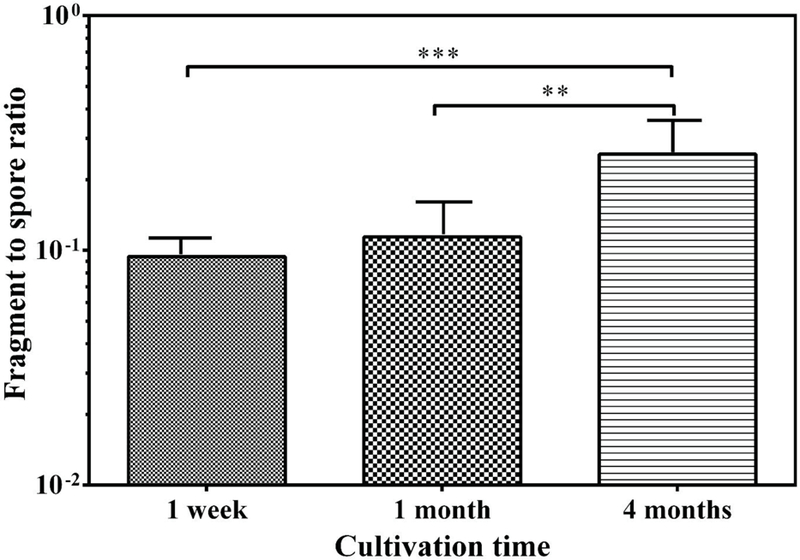
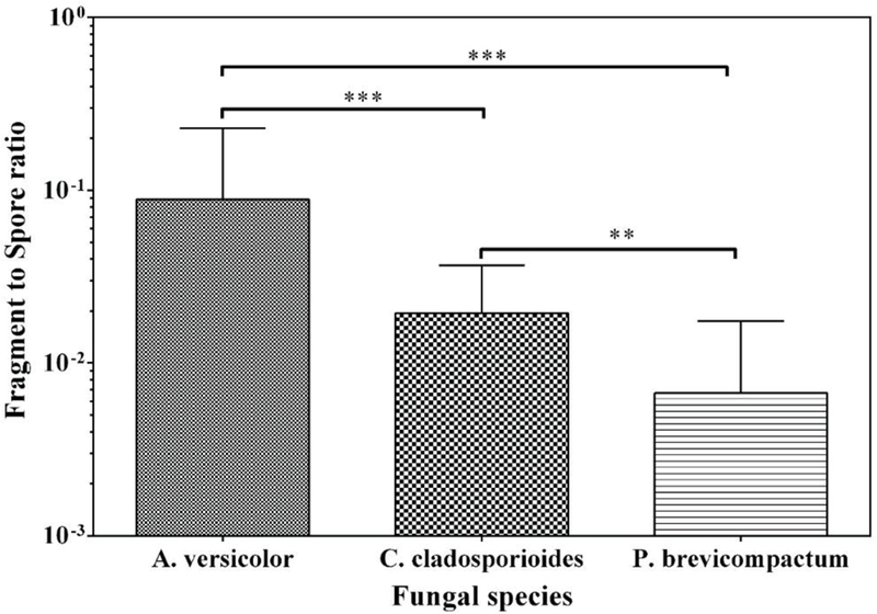
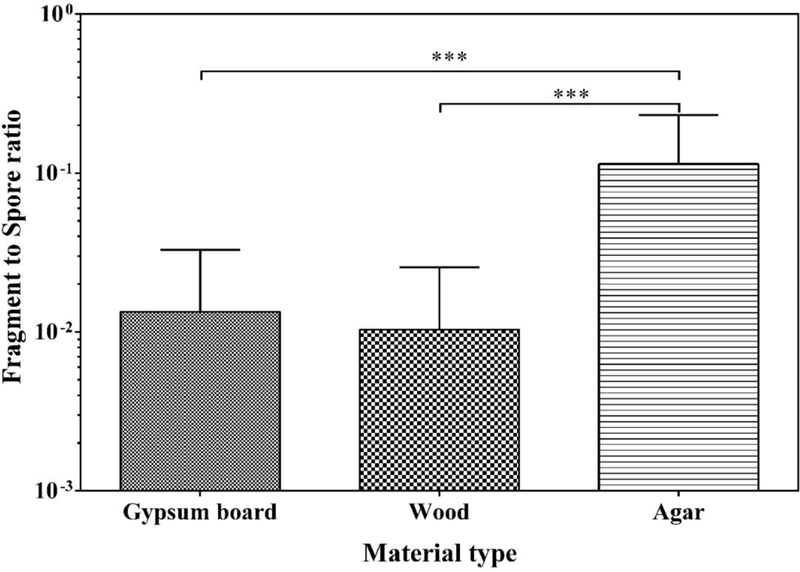
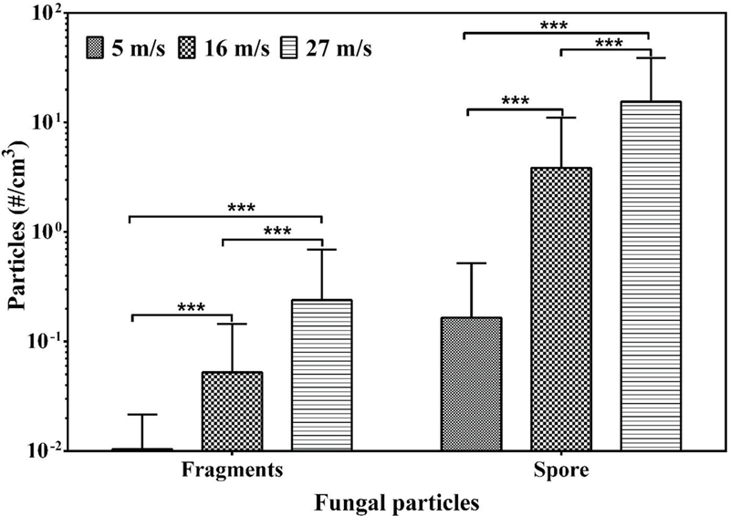
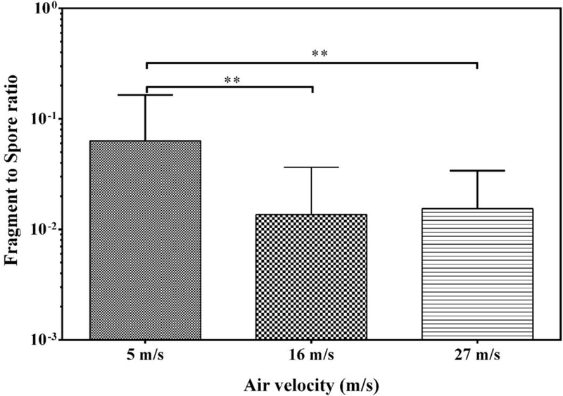
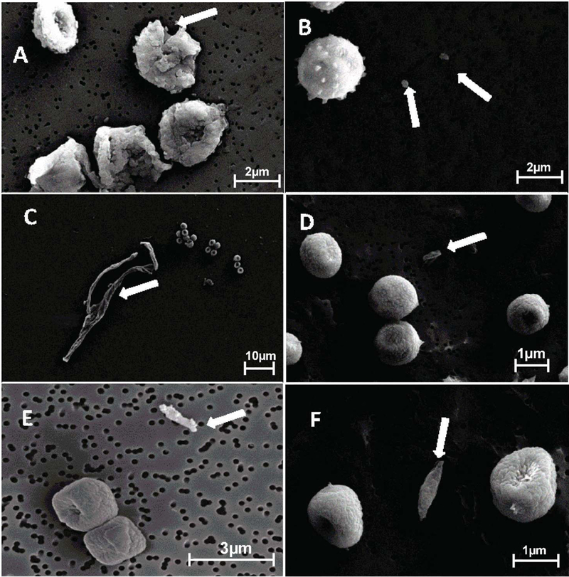
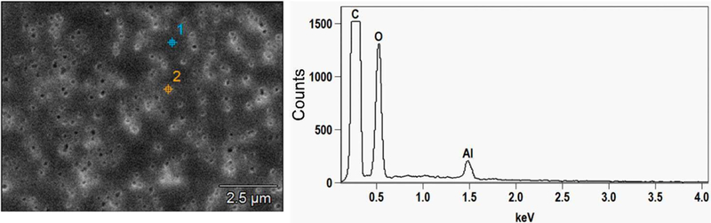
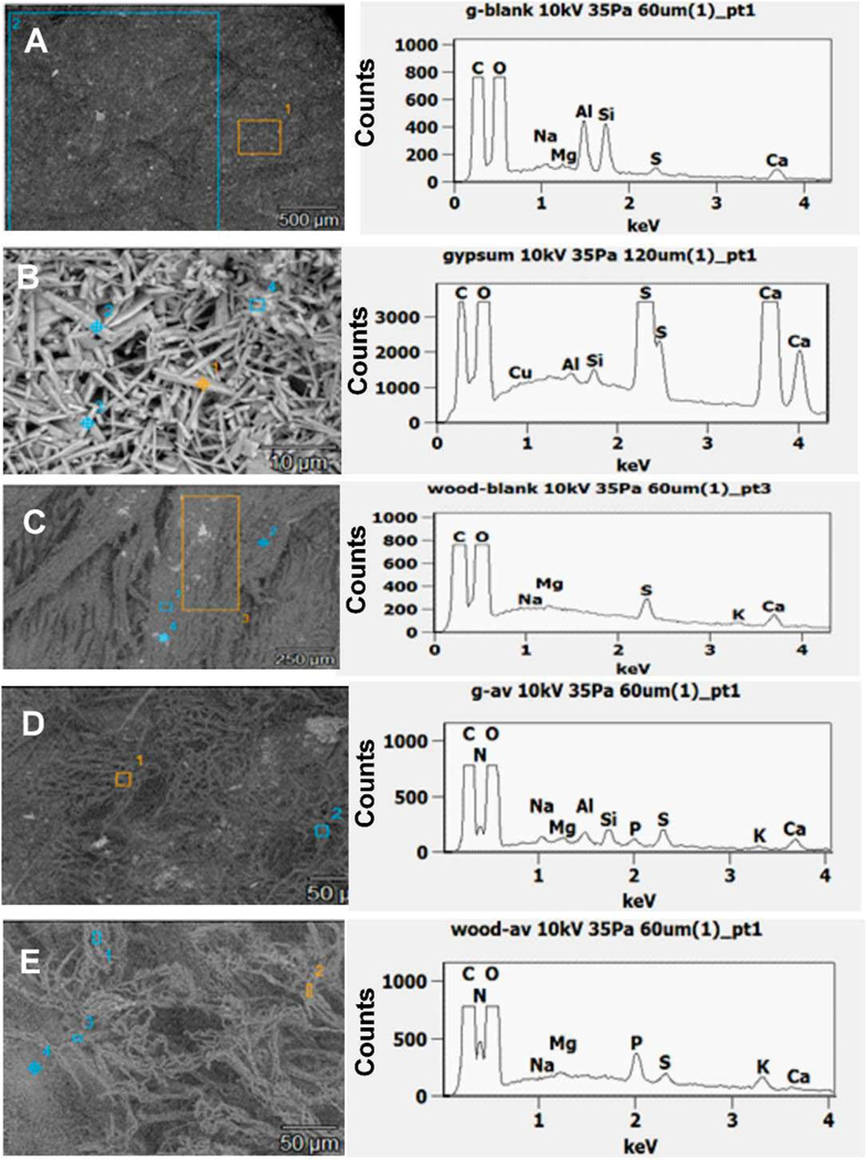
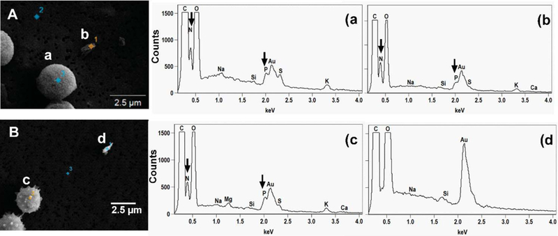
Intact spores and submicrometer size fragments are released from moldy building materials during growth and sporulation. It is unclear whether all fragments originate from fungal growth or if small pieces of building materials are also aerosolized as a result of microbial decomposition. In addition, particles may be formed through nucleation from secondary metabolites of fungi, such as microbial volatile organic compounds (MVOCs). In this study, we used the elemental composition of particles to characterize the origin of submicrometer fragments released from materials contaminated by fungi. Particles from three fungal species (Aspergillus versicolor, Cladosporium cladosporioides and Penicillium brevicompactum), grown on agar, wood and gypsum board were aerosolized using the Fungal Spore Source Strength Tester (FSSST) at three air velocities (5, 16 and 27 m/s). Released spores (optical size, dp ≥ 0.8 μm) and fragments (dp ≤ 0.8 μm) were counted using direct-reading optical aerosol instruments. Particles were also collected on filters, and their morphology and elemental composition analyzed using scanning electron microscopes (SEMs) coupled with an Energy-Dispersive X-ray spectroscopy (EDX). Among the studied factors, air velocity resulted in the most consistent trends in the release of fungal particles. Total concentrations of both fragments and spores increased with an increase in air velocity for all species whereas fragment-spore (F/S) ratios decreased. EDX analysis showed common elements, such as C, O, Mg and Ca, for blank material samples and fungal growth. However, N and P were exclusive to the fungal growth, and therefore were used to differentiate biological fragments from non-biological ones. Our results indicated that majority of fragments contained N and P. Because we observed increased release of fragments with increased air velocities, nucleation of MVOCs was likely not a relevant process in the formation of fungal fragments. Based on elemental composition, most fragments originated from fungi, but also fragments from growth material were detected.

Keywords: Air velocity; Elemental analysis; Energy Dispersive X-ray spectroscopy; Fragments; Scanning electron microscope.


Figures

Fig. 1.
Fig. 1.
Fragment/spore-ratios obtained from agar at three cultivation time points. Bars represent the geometric means of data from all species inoculated on agar and released at all air velocities combined, n= 27. The error bars represent the geometric standard deviations. Asterisks indicate the level of statistical significance (**p < 0.01; *** p < 0.001).
Fig. 2.
Fig. 2.
Fragment/spore-ratio obtained from three fungal species at 1 month of cultivation. Bars represent the geometric means of data from all materials, and all air velocities combined, n=9. The error bars represent the geometric standard deviations. Asterisks indicate the level of statistical significance (**p < 0.01; *** p < 0.001).
Fig. 3.
Fig. 3.
Fragment/spore-ratio obtained from gypsum board, wood and agar at 1 month of cultivation. The bars represent the geometric means of data from all species and air velocities combined, n = 9. Error bars represent the geometric standard deviations. Asterisks indicate the statistical significance (***, p < 0.001).
Fig. 4.
Fig. 4.
Total concentration of fragments and spores released at three air velocities at 1 month of cultivation. Bars are the geometric means of all data obtained from all fungal species and materials, n = 9. Error bars represent geometric standard deviation. Asterisks indicate the level of statistical significance (*, p < 0.05; ***, p < 0.001).
Fig 5.
Fig 5.
Fragment/spore-ratio obtained at three air velocities at 1 month of cultivation. The bars represent the geometric means of data from all species and materials combined, n= 9. The error bars represent the geometric standard deviations. Asterisks indicate the level of statistical significance (**, p < 0.01).
Fig 6.
Fig 6.
SEM micrographs of gold coated spores and fragments originating from A. versicolor spore breakdown (A), spiny structures on A. versicolor spore surface (B), P. brevicompactum hyphal breakdown (C), C. cladosporioides and P. brevicompactum spores and fragments (D, E respectively) and P. brevicompactum spores with possible fragment from gypsum board (F) (Arrowed).
Fig. 7.
Fig. 7.
SEM image and elemental composition (EDX spectra) of non gold coated blank polycarbonate membrane filter.
Fig. 8.
Fig. 8.
SEM images and elemental composition (SEM-EDX spectra) of non gold coated top of blank gypsum board (A), blank inner core of gypsum (B), top of blank wood (C), A. versicolor growth on gypsum board (D) and A. versicolor growth on wood (E). Figures D and E have the same scale.
Fig 9.
Fig 9.
SEM images and EDX spectra of elemental composition of gold coated samples of (A) P. brevicompactum spore (a) and possible biological fragment (b) and (B) A. versicolor spore (c) and non-biological fragment (d) aerosolized from gypsum board. N and P detected in the samples are arrowed.

References

    1. Adan OCG. On the fungal defacement of interior furnishes 1994.
    1. Adhikari A, Reponen T, Rylander R. Airborne fungal cell fragments in homes in relation to total fungal biomass. Indoor Air 2013;23:142–7. - PubMed
    1. Afanou KA, Straumfors A, Skogstad A, Skaar I, Hjeljord L, Skare Ø et al. Profile and Morphology of Fungal Aerosols Characterized by Field Emission Scanning Electron Microscopy (FESEM). Aerosol Sci Tech 2015: 49:6, 423–435. - PMC - PubMed
    1. Afanou KA, Straumfors A, Skogstad A, Nilsen T, Synnes O, Skaar I et al. Submicronic fungal bioaerosols: high-resolution microscopic characterization and quantification. Appl Environ Microbiol 2014;80:7122–30. - PMC - PubMed
    1. Brandl H, von Däniken A, Hitz C, Krebs W. Short-term dynamic patterns of bioaerosol generation and displacement in an indoor environment. Aerobiologia 2008;24:203–9.

Publication types