. 2016 Jan 20;12(1):e1005798.
doi: 10.1371/journal.pgen.1005798. eCollection 2016 Jan.

Short Telomeres in Key Tissues Initiate Local and Systemic Aging in Zebrafish

Affiliations

Short Telomeres in Key Tissues Initiate Local and Systemic Aging in Zebrafish

Madalena C Carneiro et al. PLoS Genet. .

Abstract

Telomeres shorten with each cell division and telomere dysfunction is a recognized hallmark of aging. Tissue proliferation is expected to dictate the rate at which telomeres shorten. We set out to test whether proliferative tissues age faster than non-proliferative due to telomere shortening during zebrafish aging. We performed a prospective study linking telomere length to tissue pathology and disease. Contrary to expectations, we show that telomeres shorten to critical lengths only in specific tissues and independently of their proliferation rate. Short telomeres accumulate in the gut but not in other highly proliferative tissues such as the blood and gonads. Notably, the muscle, a low proliferative tissue, accumulates short telomeres and DNA damage at the same rate as the gut. Together, our work shows that telomere shortening and DNA damage in key tissues triggers not only local dysfunction but also anticipates the onset of age-associated diseases in other tissues, including cancer.


Conflict of interest statement

The authors have declared that no competing interests exist.

Figures

Fig 1
Fig 1. Different zebrafish tissues have different telomere lengths and follow specific dynamics of shortening with aging.
A) Representative images of telomere restriction fragment (TRF) analysis of genomic DNA by Southern Blot (random primer-labelled telomeric probe (TTAGGG)n 32P-dCTP). Aa) WT telomeres are longer in whole larvae (“L”, ca. 12 kb) and shorter in 3 month-old zebrafish gut (G), testis (T) and muscle (M), but show significant variation in length between tissues (shown for two independent WT zebrafish “1” and “2” in Fig 1Aa, except for testis where only zebrafish “1” is shown; densitometry shown for zebrafish “1”, Fig 1Ab). WT have longer telomeres in testis (ca.9.9 Kb), followed by muscle (ca. 9.4 Kb) and, finally, gut (8.4 Kb). These differences are globally maintained in tert-/- tissues (ca. 8.3 Kb in the testis, ca. 8.5 in the muscle and ca. 7.8 Kb in the gut; shown for two 3-month old zebrafish “1” and “2” in Fig 1Aa; densitometry shown for individual “1”, Fig 1Ab). Yellow line indicates median telomere length, mTL, for each tissue sample/lane. B) WT telomeres significantly shorten in the fin and kidney marrow with age, but follow different shortening kinetics with time (black star represents WT larvae telomeres; two zebrafish shown for each age after sexual maturation– 3, 12, 17, 24, 28, 36 months for the fin and 3, 18, 24, 36 months for the kidney marrow). Ba) The telomere shortening rate within the same individual (measured by cutting different fins in different time points) is of 45 bp/month and 90 bp/month for two independent zebrafish (in Fig Ba, lanes 1 and 2 of each time point are the same individual over time between the ages of 3 and 24 months). WT telomeres in the fin match the shorter length of 12-month old tert-/- mutants by 18–24 months (N = 4 for WT and N = 3 for tert-/- fin; red star represents tert-/- larvae telomeres; Fig 1Ba), but WT kidney marrow telomeres never reach tert-/- levels (N = 3–6 per time point for adult WT and N = 3 for adult tert-/- mutants, Fig 1Bb). TRF mean sizes were calculated as previously described [54]. mTL data is represented as mean +/- SEM.
Fig 2
Fig 2. Telomeres shorten in WT gut and muscle, but not in testis, with aging reaching the length of tert-/- tissues.
A-C) Representative images of telomere restriction fragment (TRF) analysis of genomic DNA by Southern Blot (random primer-labelled telomeric probe (TTAGGG)n 32P-dCTP) and quantifications of median telomere length. Black and red stars represent WT and tert-/- larvae telomeres, respectively, in quantifications of median TRF over time. WT telomeres shorten linearly with aging from 3 to 24 months, in A) gut (N = 3–4 per time point) and B) muscle (N = 3–4 per time point), stabilizing in later ages. C) No significant telomere shortening is detected in the testis (N = 5–6 per time point). Around 20 months of age WT telomeres reach the shorter length of 12 month-old tert-/- in the gut (graph in fig A–ca. 6.8 Kb) and muscle (graph in fig B–ca. 8.5 Kb) but not in testis (graph in fig C), indicated by blue arrow. N = 3–4 per time point for tert-/- gut and muscle, N = 4–6 per time point for tert-/- testis. TRF mean sizes were calculated as previously described [54]. Data are represented as mean +/- SEM.
Fig 3
Fig 3. Shortening of mTL anticipates accumulation of DDR markers, decrease in cell proliferation and increase in apoptotis with age.
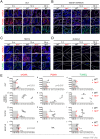
A-D) Representative immunofluorescence images of DNA damage (γH2AX), proliferation (PCNA) and apoptosis (TUNEL) for gut (A—dashed outline identifies the villi), kidney marrow (B), testis (C) and muscle (D—dashed outline identifies the fibers) of WT (at 3, 12 and 36 months) and tert-/- mutant siblings (at 3 and 12 months). All tissues show a significant increase in cells bearing five or more γH2AX foci by 12 months of age in tert-/- zebrafish (panels b, d) and by 36 months in WT zebrafish (panels a, c and e). Increased DNA Damage Response (DDR) correlates with shorter mTL in Ea) Gut (p = 0.021) and Ej) Muscle (p = 0.044) but not Ed) Kidney Marrow or Eg) Testis. Grey shaded area identifies the median telomeric length at which significant DDR activation is observed in tert-/- mutants (Ea, Ed, Eg, Ej). Proliferative tissues, A) gut B) kidney marrow and C) testis, show a sustained decrease in cell proliferation by 12 months of age in tert-/- (panels g, i) and by 36 months in WT tissues (panels f, h and j). Eb) Decrease in proliferation correlates with shorter mTL in the gut (p = 0.015). Grey shaded area identifies the median telomeric length at which significant proliferation defects are observed in tert-/- mutants (Eb, Ee, Eh). tert-/- mutants’ A) gut B) kidney marrow and C) testis show increased apoptotic responses at 3 months when compared with WT controls (panel l). WT zebrafish show a continuous accumulation of apoptotic signals with age (panels k, m, o). Apoptotic responses are not anticipated by shorter mTL (Ec, Ef, Ei, El). Grey shaded area identifies the median telomeric length at which significant apoptotic responses are observed in tert-/- mutants (Ec, Ef, Ei, El). Most DDRs and apoptotic signals locate to the proliferative zone of maturing spermatocytes (C—dashed outline). WT and tert-/- age groups are indicated in each graph by black and red colored numbers, respectively. Immunofluorescence (IF) quantifications were performed in at least 3 different fields of view for 3–5 different individuals per time point per genotype. Gut IF quantifications were calculated as number of positive cells per “crypt” zone. Other tissues' IF was quantified as overall % positive cells. Scale bar = 50 μm. N = 3–6 for tissue mTL quantifications per genotype per time point (x-axis in graphs of Fig 3E). IF and mTL quantifications are represented as mean +/- SEM.
Fig 4
Fig 4. Telomere shortening culminates in tissue defects associated with aging.
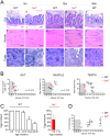
A) Representative haematoxylin and eosin-stained sections of gut, muscle and testis from WT and tert-/- siblings. By 12 months of age, tert-/- mutants (N = 3) show inflammatory cell infiltration of the lamina propria of the gut (panel b, d), myocyte atrophy and degeneration (characteristic of sarcopenia—panels i), and reduced numbers of mature spermatozoa (panels l, n). Similarly, by 36 months, WT show similar lesions in the gut (N = 5, panels e) and muscle (N = 5, panels j); and with aging, WT testis show a gradual decrease in the number of spermatozoa (panels k, m, o, N = 5). B) Among these age-related histological lesions, intestinal inflammation and sarcopenia correlate with shortening of mTL (p = 0.002 and p = 0.028, respectively), while no correlation is found for reduction in mature spermatozoa numbers. Grey shaded area identifies the median telomeric length at which significant intestinal inflammation, sarcopenia and defects in production of mature spermatozoa are observed in tert-/- mutants. WT and tert-/- age groups are indicated in each graph by black and red colored numbers, respectively. C) In accordance with lower levels of spermatozoa production at later ages, WT show a decreased ability to successfully fertilize female eggs from 24 months of age onwards (N = 4). tert-/- have impairment of reproductive ability by 6 months and complete lack of function by 12 months (N = 3). D) Erythrocyte levels (N = 5) decrease with aging in WT and tert-/- mutants’ kidney marrow, indicative of anemia, but this is not predicted by mTL decline in this tissue. Grey shaded area identifies the median telomeric length at which a significant decrease in erythrocyte levels is observed in tert-/- mutants. Scale bar = 50 μm. N = 3–6 for tissue mTL quantifications per genotype per time point (x-axis in graphs of Fig 4B and 4D). Data are represented as mean +/- SEM.
Fig 5
Fig 5. tert-/- mutants accelerate the onset of age-associated cachexia and neoplasia.
A) Representative images of WT and tert-/- mutants show that, at time-of-death (TOD), zebrafish exhibit signs of cachexia and deformation of the spine, with very low body mass indexes. B) In WT population the incidence of these alterations increases with age, while in tert-/- mutants, at TOD, 100% of the population is affected. C) Sustained cachexia in older zebrafish is accompanied by an increase in mortality as shown by Kaplan Meier curves of WT and tert-/- zebrafish. WT zebrafish have a half-life of 30.8m, 3x greater than the half-life of 10.6m of tert-/- (p<0.0001). N = 426 WT; N = 98 tert-/-, for Fig 5A–5C. TOD corresponds to the interval comprising the second and third quartiles of survival (25 to 75%). Scale bar = 1 cm. D) Tumorigenesis exponentially accelerates with age in both WT and tert-/- zebrafish, reaching cumulative incidences of ca. 10% and ca. 8%, respectively. N = 23/238 WT; N = 5/66 tert-/-. tert-/- mutants have an earlier onset of neoplasia starting at 4 months of age (p = 0.003). E) Tumors in WT male zebrafish affect mainly the reproductive system, following the appearance of Periodic Acid Shift (PAS+) stained cells in the tissue stroma (F), (quantified in G). WT females show an even distribution of tumors between reproductive and hematopoietic systems, followed by liver, intestine and pancreas. N = 118 WT males; N = 120 WT females; N = 58 tert-/- males; N = 8 tert-/- females. Quantifications of %PAS+ cells was performed in 3 fields of view for each individual in the graph. Scale bar = 50 μm. Data are represented as mean +/- SEM.
Fig 6
Fig 6. Working model—short mTL and DDR in key tissues establish the rate of local and systemic aging.
Expression of telomerase is restricted in most somatic tissues resulting in telomere shortening with aging (1). In gut and muscle, shortening of telomeres to critical levels results in increased DDRs with aging (2) which disrupt local homeostasis, culminating in organ and tissue-specific lesions such as inflammation and sarcopenia (3). Gut and muscle DDRs act in a cell non-autonomous manner by inducing defects in organs where telomeres do not shorten appreciably, such as testis and kidney marrow (4). DDR signals trigger systemic damage resulting in cachexia and fueling tumorigenesis. Overall, cellular damage creates an amplifying positive feedback loop accelerating tissue dysfunction with aging (5).

References

    1. de Lange T. Shelterin: the protein complex that shapes and safeguards human telomeres. Genes & development. 2005;19(18):2100–10. 10.1101/gad.1346005 . - DOI - PubMed
    1. Greider CW, Blackburn EH. A telomeric sequence in the RNA of Tetrahymena telomerase required for telomere repeat synthesis. Nature. 1989;337(6205):331–7. 10.1038/337331a0 . - DOI - PubMed
    1. Olovnikov AM. A theory of marginotomy. The incomplete copying of template margin in enzymic synthesis of polynucleotides and biological significance of the phenomenon. Journal of theoretical biology. 1973;41(1):181–90. . - PubMed
    1. Watson JD. Origin of concatemeric T7 DNA. Nature: New biology. 1972;239(94):197–201. . - PubMed
    1. Harley CB, Futcher AB, Greider CW. Telomeres shorten during ageing of human fibroblasts. Nature. 1990;345(6274):458–60. 10.1038/345458a0 . - DOI - PubMed

Publication types