. 2016 Jan 21;374(3):223-32.
doi: 10.1056/NEJMoa1510791.

Mutations in TUBB8 and Human Oocyte Meiotic Arrest

Affiliations

Mutations in TUBB8 and Human Oocyte Meiotic Arrest

Ruizhi Feng et al. N Engl J Med. .

Abstract

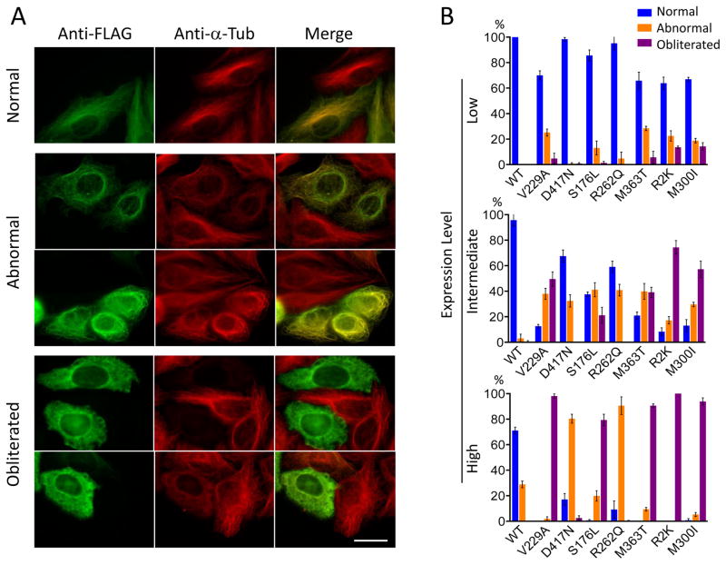
Background Human reproduction depends on the fusion of a mature oocyte with a sperm cell to form a fertilized egg. The genetic events that lead to the arrest of human oocyte maturation are unknown. Methods We sequenced the exomes of five members of a four-generation family, three of whom had infertility due to oocyte meiosis I arrest. We performed Sanger sequencing of a candidate gene, TUBB8, in DNA samples from these members, additional family members, and members of 23 other affected families. The expression of TUBB8 and all other β-tubulin isotypes was assessed in human oocytes, early embryos, sperm cells, and several somatic tissues by means of a quantitative reverse-transcriptase-polymerase-chain-reaction assay. We evaluated the effect of the TUBB8 mutations on the assembly of the heterodimer consisting of one α-tubulin polypeptide and one β-tubulin polypeptide (α/β-tubulin heterodimer) in vitro, on microtubule architecture in HeLa cells, on microtubule dynamics in yeast cells, and on spindle assembly in mouse and human oocytes. Results We identified seven mutations in the primate-specific gene TUBB8 that were responsible for oocyte meiosis I arrest in 7 of the 24 families. TUBB8 expression is unique to oocytes and the early embryo, in which this gene accounts for almost all the expressed β-tubulin. The mutations affect chaperone-dependent folding and assembly of the α/β-tubulin heterodimer, disrupt microtubule behavior on expression in cultured cells, alter microtubule dynamics in vivo, and cause catastrophic spindle-assembly defects and maturation arrest on expression in mouse and human oocytes. Conclusions TUBB8 mutations have dominant-negative effects that disrupt microtubule behavior and oocyte meiotic spindle assembly and maturation, causing female infertility. (Funded by the National Basic Research Program of China and others.).


Figures

Figure 1
Figure 1. Pedigree and mutations in TUBB8 in oocytes maturation arrest patients
Pedigrees of seven families with inherited or de novo TUBB8 mutations. Family 1 is a large four-generation family that includes five affected women with long-term primary infertility. The V229A mutation in TUBB8 was inherited from the patients’ father. Family member IV-1 is a 7-year-old girl carrying the mutation who cannot be evaluated for fertility. The D417N, M363T, R2K and M300I mutations were identified in families 2, 5, 6 and 7, respectively. These four mutations were inherited from the respective fathers. The S176L and R262Q mutations identified in families 3 and 4 are de novo. Sanger sequencing chromatograms are shown defining the nature and location of the mutations in each family. Squares denote male family members and circles female family members. Black circles represent affected individuals. Slashes indicate deceased individuals. The arrow indicates the index patient. WT, wild type.
Figure 2
Figure 2. Phenotypes of oocytes from patients with maturation arrest
(A–E) Normal (A) and patient (B–E) oocytes were separated from granulosa cells and examined by light and polarization microscopy. Note that a normal MII oocyte has a first polar body (black arrow in A) and that normal MI and MII oocytes have visible spindles (white arrows in A). All patient oocytes are at MI and none have a first polar body. Oocytes in patients from families 1 (B), 2 (C), and 6 (E) have no visible spindle (polarization microscopy data was not obtained for oocytes from the patient in family 3). (F) Oocytes from control, V229A, D417N, S176L, R262Q and R2K patients were fixed, permeabilized, immunolabeled and examined by confocal microscopy using antibodies against β-tubulin to visualize the spindle (shown in green) and counterstained with Hoechst 33342 (shown in blue) to visualize DNA. Note that the oocyte from the D417N patient has an abnormal (disorganized) spindle, while oocytes from all other patients have no visible spindle.
Figure 3
Figure 3. Microtubule phenotypes resulting from expression of wild type and mutant forms of TUBB8 in cultured cells
(A) Expression of wild type and mutant forms of TUBB8 in cultured cells. Constructs engineered for the expression of C-terminally FLAG-tagged TUBB8 (wild type and mutant) were expressed by transfection into HeLa cells. After 42 h, the cells were fixed, permeabilized and labeled with antisera to the FLAG epitope (to detect expression of the transgene, shown in green) and α-tubulin (to detect the endogenous microtubule network, shown in red). Examples are shown of cells expressing the various transgenes in which there was incorporation into a normal interphase microtubule network, an abnormal microtubule network with either a conspicuously reduced filament density or with a wavy, tangled or disorganized appearance, or cells in which the microtubule network had been completely obliterated. Note that in the latter cases, the FLAG and α-tubulin labels appear as a diffuse mottled pattern throughout the cytoplasm. Bar = 10 μm. (B) Quantification of microtubule phenotypes exemplified in (A) resulting from expression of wild type and each TUBB8 mutation at low, intermediate or high levels of expression. About 200 transfected cells expressing either wild type or mutant TUBB8 were examined in each of three separate experiments; the result shown is the mean percentage of cells assigned to each category (normal, abnormal or obliterated) ± s. d. The propensity of the TUBB8 mutations to confer microtubule obliteration follows the order V229A = R2K = M300I = M363T >S176L > R262Q = D417N.
Figure 4
Figure 4. Mutant TUBB8 RNAs affect spindle assembly in mouse and human oocytes
(A) Compared with the wild type control, polar body extrusion rates in mouse oocytes injected with mutant RNA (Figure S11) was significantly decreased; the experiment was performed 3 times; error bars represent mean ± s. d. ** P<0.01, **P<0.001. Unpaired T-test. (B) Immunostaining of mouse oocytes 12h after GVBD. GV oocytes were injected with a higher (1000 ng/μl) concentration of wild type RNA (n=25), S176L mutant RNA (n=36) or D417N mutant RNA (n=28). (C) Immunostaining of human oocytes 16h after GVBD. GV oocytes were injected with a higher concentration (1000 ng/μl) of TUBB8 wild type (n=28), S176L mutant RNA (n=8) or D417N mutant RNA (n=7). The MI oocytes were stained to visualize chromosomes (Hoechst 33342; blue), spindles (β-tubulin; green) and TUBB8 (FLAG; red).

Comment in

References

    1. Eppig JJ. Regulation of mammalian oocyte maturation. In: Adash PCKL, editor. The Ovary. San Diego: Elsevier/Academic Press; 1993. pp. 113–29.
    1. Mehlmann LM. Stops and starts in mammalian oocytes: recent advances in understanding the regulation of meiotic arrest and oocyte maturation. REPRODUCTION. 2005;130:791–9. - PubMed
    1. Park JY, Su YQ, Ariga M, Law E, Jin SL, Conti M. EGF-like growth factors as mediators of LH action in the ovulatory follicle. SCIENCE. 2004;303:682–4. - PubMed
    1. Eppig JJ. Coordination of nuclear and cytoplasmic oocyte maturation in eutherian mammals. Reprod Fertil Dev. 1996;8:485–9. - PubMed
    1. Santos MA, Kuijk EW, Macklon NS. The impact of ovarian stimulation for IVF on the developing embryo. REPRODUCTION. 2010;139:23–34. - PubMed

Publication types