. 2016 May 15;110(2):178-87.
doi: 10.1093/cvr/cvw017. Epub 2016 Jan 19.

Lack of MG53 in human heart precludes utility as a biomarker of myocardial injury or endogenous cardioprotective factor

Affiliations

Lack of MG53 in human heart precludes utility as a biomarker of myocardial injury or endogenous cardioprotective factor

Frances A Lemckert et al. Cardiovasc Res. .

Abstract

Aims: Mitsugumin-53 (MG53/TRIM72) is an E3-ubiquitin ligase that rapidly accumulates at sites of membrane injury and plays an important role in membrane repair of skeletal and cardiac muscle. MG53 has been implicated in cardiac ischaemia-reperfusion injury, and serum MG53 provides a biomarker of skeletal muscle injury in the mdx mouse model of Duchenne muscular dystrophy. We evaluated the clinical utility of MG53 as a biomarker of myocardial injury.

Methods and results: We performed Langendorff ischaemia-reperfusion injury on wild-type and dysferlin-null murine hearts, using dysferlin deficiency to effectively model more severe outcomes from cardiac ischaemia-reperfusion injury. MG53 released into the coronary effluent correlated strongly and significantly (r = 0.79-0.85, P < 0.0001) with functional impairment after ischaemic injury. We initiated a clinical trial in paediatric patients undergoing corrective heart surgery, the first study of MG53 release with myocardial injury in humans. Unexpectedly, we reveal although MG53 is robustly expressed in rat and mouse hearts, MG53 is scant to absent in human, ovine, or porcine hearts. Absence of MG53 in 11 human heart specimens was confirmed using three separate antibodies to MG53, each subject to epitope mapping and confirmed immunospecificity using MG53-deficient muscle cells.

Conclusion: MG53 is an effective biomarker of myocardial injury and dysfunction in murine hearts. However, MG53 is not expressed in human heart and therefore does not hold utility as a clinical biomarker of myocardial injury. Although cardioprotective roles for endogenous myocardial MG53 cannot be extrapolated from rodents to humans, potential therapeutic application of recombinant MG53 for myocardial membrane injury prevails.

Keywords: Biomarker of myocardial injury; Ischaemia–reperfusion injury; Ischaemic preconditioning/postconditioning; MG53; TRIM72.


Figures

Figure 1
Figure 1
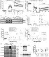
MG53 accurately reflects the degree of ischaemia–reperfusion injury to murine A/J hearts. A/J WT and dysferlin-null hearts were subjected to a Langendorff I/R protocol comprising 10 min baseline perfusion, 20 min global no-flow ischaemia, and 30 min reperfusion. (A) Following equivalent baseline function, on reperfusion A/J dysferlin-null hearts demonstrated higher diastolic left ventricular pressure than WT hearts (Diastolic-LVP), and (B) lower left ventricular developed pressure (LVDP), indicating poorer functional recovery from ischaemic injury. (C) Levels of MG53 detected in the coronary effluent increases following ischaemia–reperfusion injury, with higher levels released from dysferlin-null hearts, consistent with increased membrane permeability in this model and (D) supported by a corresponding increase in LDH content of coronary effluent (*P < 0.05, **P < 0.01, ***P < 0.005). (E) Comparison of levels of released MG53 and LDH gave a strong positive correlation (Pearson r = 0.85, P < 0.0001), while (F) shows a strong inverse correlation of MG53 with LVDP (Pearson r = −0.77, P < 0.0001), demonstrating hearts with a poorer LVDP released more MG53 (n = 3). These data highlight the potential of MG53 as a serum biomarker of cardiac ischaemic burden.
Figure 2
Figure 2
Murine MG53 is an accurate biomarker of cardiac ischaemic burden in the C57BL/6 heart. The C57BL/6 mouse line shows greater intrinsic resistance to cardiac I/R challenge, displaying little functional impairment following 20 min of global ischaemia. We therefore utilized two consecutive 20 min ischaemic challenges to obtain a consistent and measureable functional deficit in both WT and dysferlin-null hearts following ischaemic challenge. C57BL/6 dysferlin-null hearts subjected to an extended I/R challenge [10 min baseline perfusion (b), 20 min global no-flow ischaemia (I1), 10 min reperfusion (R1), 20 min global no-flow ischaemia (I2), 30 min final reperfusion (R2)] showed a deficit in functional recovery in agreement with hearts on the A/J background. (A) Dysferlin-null hearts showed significantly higher diastolic left ventricular pressure throughout the second reperfusion period when the ischaemic insult primarily manifests (R2, 60–90 min) and showed much poorer recovery of baseline LVDP compared with WT hearts (B). Accordingly, dysferlin-null Bla/J hearts released more MG53 to the coronary effluent than WT hearts as detected by western blot (C) in parallel with greater LDH release (D) (*P < 0.05). Levels of released MG53 showed a strong and highly significant positive correlation with LDH release (E) and a strong and highly significant inverse correlation with LVDP (F). Dysferlin-null hearts on either the A/J (G) or C57BL/6 genetic background (H) express equivalent MG53 to WT hearts, and LDH content of WT and dysferlin-null hearts is not different in the A/J background (I).
Figure 3
Figure 3
Wild-type and dysferlin-null cardiomyocytes show identical contractile and calcium handling properties under conditions of baseline superfusion. (A) Sarcomeric contractile properties of A/J WT (black lines) and A/J dysferlin-null (grey lines) cardiac myocytes were measured at baseline (solid) and after 20 min (dashed) of pacing at 1 Hz. All parameters of sarcomeric contractility were equivalent in wild-type and dysferlin-null myocytes; time to peak shortening (or 50% peak shortening, not shown), resting and peak sarcomere length, and maximum velocities of contraction and relaxation. (B) Calcium transients initiating contraction in A/J WT (black lines) and A/J dysferlin-null (grey lines) cardiac myocytes were measured at baseline (solid) and after 20 min (dashed) of pacing at 1 Hz. All parameters of calcium transients were equivalent in wild-type and dysferlin-null myocytes; time to peak calcium (or 50% peak calcium, not shown), resting and peak intracellular calcium, maximum velocity of calcium release, and clearance (A/J null n = 16, A/J WT n = 16, mean ± 95% CI).
Figure 4
Figure 4
Wild-type murine hearts subjected to a Langendorff ischaemia/reperfusion protocol release ∼10–15% of their total MG53 to the coronary effluent. (A) Comparative western analyses of coronary effluent vs. naive murine heart lysate as a loading standard. Forty microlitres of coronary effluent collected during reperfusion from C57BL/6 WT hearts contained as much MG53 as up to 2 µg of total protein lysate from naive hearts and was readily detectable by immunoblot methodology. The temporal MG53 release profile (A, B) for each heart varied, but the total amount of MG53 released over 30 min of reperfusion was remarkably consistent (C), in the order of 10–15% of the total MG53 contained in a naive WT heart (calculated by extrapolation of the MG53 content in 40 µL effluent to the total volume of effluent flowing through the heart during the 30 min reperfusion) (n = 4). (D) Wild-type hearts subjected to no perfusion (U), an I/R protocol (I/R, dysferlin-null and WT hearts both shown), baseline perfusion (B) for an equivalent period as the I/R protocol, or hearts harvested early in the reperfusion phase of an I/R protocol (I/R arrest, time into reperfusion shown) displayed similar levels of MG53 protein, suggesting MG53 expression did not vary in response to an ischaemic insult; MG53 was neither induced nor substantially degraded in this period of time.
Figure 5
Figure 5
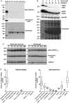
MG53 is not expressed in human, pig, or sheep myocardium using three validated antibodies. (A) Western Blotting of 10 μg total protein from heart (H) and skeletal muscle (SM) samples from mouse, rat, human, pig, and sheep. MG53 (arrow) is present in mouse and rat skeletal muscle and heart, though pAbs 108 and 288 show lower affinity for mouse than rat. In contrast, although MG53 is readily detected in skeletal muscle of human, pig, and sheep, MG53 is absent in cardiac muscle of these species (Lanes 5, 7, and 9). (B) Schematic representation of MG53 protein domain structure. (C) The antigenic regions of antibodies pAb-mMG53144, pAb-hMG53108, and pAb-hMG53288 were confirmed using deletion constructs expressed in 3T3 cells. Untf = untransfected control 3T3 cells (D) The specificity of pAb-mMG53144, pAb-hMG53108, and pAb-hMG53288 was confirmed via western blotting of wild-type C2C12 and three separate CRISPR/Cas9 gene-edited MG53-null lines. (E) MG53 was found at very low levels or not detected in 10 human heart samples from patients with congenital heart defects [1LV—ventricular septal defect (VSD), patent ductus arteriosus (PDA), male 5 years; 2LV—Tetralogy of Fallot (TOF), male 2 years; 3LV—levo-transoposition of the great arteries, pulmonary atresia (PA), VSD, PDA, male 3 years; 4LV—double outlet right ventricle, VSD, pulmonary stenosis, PDA, patent foramen ovale, female 4 years; 5LV—PA, VSD, male 4 years; 6LV—truncus arteriosus type 1, VSD, atrial septal defect, Dandy–Walker Syndrome, female 5 years; 7LV—VSD, male 11 years; 8LV—TOF, male 9 months; 9LV—PDA, Shone's complex, Noonan Syndrome, female 8 years; 10RV—PA, VSD, PDA, male 2 years), and a control donor heart (11donor, female 23 years) but was detected in three human skeletal muscle samples (Q = quadricep, female, 5 years; HF = hip flexor, female, 5 years; VM = vastus medialis, male, 18 years) as well as mouse cardiac (LV) and skeletal muscle (Quad, 10 weeks, 10 μg total protein). In contrast, dysferlin is strongly expressed in both human skeletal muscle and heart (left bottom panel, Dysferlin). (F) MG53 was absent in left and right atria and ventricles of porcine (38 days) and ovine (12 weeks) hearts (10 μg total protein). LV, left ventricle, RV, right ventricle, LA, left atrium, RA, right atrium.
Figure 6
Figure 6
MG53 is not detected in serum from paediatric patients undergoing heart surgery, demonstrates no overt lability over 48 h, and protein expression in human heart correlates with mRNA expression levels according to the GTEx Portal. (A) Western blot analysis of 0.25 µL serum (containing ∼15–20 µg total protein) from two controls (C1, C2) and two patients (P1pre, P1post, P2pre, P2post) undergoing corrective surgery for structural hearts defects, and with significantly elevated troponin T levels post-surgery failed to detect serum MG53 in both pre- and post-surgical samples. (B) Western blot of equivalent amounts of human skeletal muscle lysate with increasing volumes of human serum demonstrate the difficulty of detecting MG53 in serum against the huge background levels of serum proteins, in particular albumin (MW∼65 kDa) which physically deforms the gel matrix as seen in the Coomassie panel at bottom. (C) Murine MG53 from both heart and skeletal muscle tissue left at ∼4°C for up to 48 h displays no overt lability, consistent with MG53 being absent from our human heart samples rather than lost due to degradation of the protein. (D) Genotype Tissue Expression (GTEx) Project RNA-Seq data from a range of human tissues shows MG53 (TRIM72) expression is greatest in skeletal muscle, and >100-fold greater than in heart [median value for skeletal muscle 29.21 vs. left ventricle 0.157 (>180-fold) vs. right atrial appendage 0.032 (>900-fold)], while for dysferlin, mRNA is highly expressed in both skeletal muscle and heart [median value for skeletal muscle 22.44 vs. left ventricle 12.89 (<2-fold) vs. right atrial appendage 10.06 (<2.5-fold)], consistent with our western blot analyses of MG53 and dysferlin in these two tissues.

References

    1. Cai C, Masumiya H, Weisleder N, Matsuda N, Nishi M, Hwang M, Ko J-K, Lin P, Thornton A, Zhao X, Pan Z, Komazaki S, Brotto M, Takeshima H, Ma J. MG53 nucleates assembly of cell membrane repair machinery. Nat Cell Biol 2009;11:56–64. - PMC - PubMed
    1. Lek A, Evesson FJ, Lemckert FA, Redpath GMI, Lueders A-K, Turnbull L, Whitchurch CB, North KN, Cooper ST. Calpains, cleaved mini-dysferlinC72, and L-type channels underpin calcium-dependent muscle membrane repair. J Neurosci 2013;33:5085–5094. - PMC - PubMed
    1. Waddell LB, Lemckert FA, Zheng XF, Tran J, Evesson FJ, Hawkes JM, Lek A, Street NE, Lin P, Clarke NF, Landstrom AP, Ackerman MJ, Weisleder N, Ma J, North KN, Cooper ST. Dysferlin, annexin A1, and mitsugumin 53 are upregulated in muscular dystrophy and localize to longitudinal tubules of the T-system with stretch. J Neuropath Exp Neur 2011;70:302–313. - PMC - PubMed
    1. Wang X, Xie W, Zhang Y, Lin P, Han L, Han P, Wang Y, Chen Z, Ji G, Zheng M, Weisleder N, Xiao R-P, Takeshima H, Ma J, Cheng H. Cardioprotection of ischemia/reperfusion injury by cholesterol-dependent MG53-mediated membrane repair. Circ Res 2010;107:76–83. - PubMed
    1. Cao C-M, Zhang Y, Weisleder N, Ferrante C, Wang X, Lv F, Zhang Y, Song R, Hwang M, Jin L, Guo J, Peng W, Li G, Nishi M, Takeshima H, Ma J, Xiao R-P. MG53 constitutes a primary determinant of cardiac ischemic preconditioning. Circulation 2010;121:2565–2574. - PubMed

Publication types

MeSH terms