. 2016 Jan 20:13:12.
doi: 10.1186/s12985-016-0467-9.

Evidence for a biphasic mode of respiratory syncytial virus transmission in permissive HEp2 cell monolayers

Affiliations

Evidence for a biphasic mode of respiratory syncytial virus transmission in permissive HEp2 cell monolayers

Tra Nguyen Huong et al. Virol J. .

Abstract

Background: During respiratory syncytial virus (RSV) infection filamentous virus particles are formed on the cell surface. Although the virus infectivity remains cell-associated, low levels of cell-free virus is detected during advanced infection. It is currently unclear if this cell-free virus infectivity is due to a low-efficiency specific cell-release mechanism, or if it arises due to mechanical breakage following virus-induced cell damage at the advanced stage of infection. Understanding the origin of this cell-free virus is a prerequisite for understanding the mechanism of RSV transmission in permissive cells. In this study we describe a detailed examination of RSV transmission in permissive HEp2 cell monolayers.

Methods: HEp2 cell monolayers were infected with RSV using a multiplicity of infection of 0.0002, and the course of infection monitored over 5 days. The progression of the virus infection within the cell monolayers was performed using bright-field microscopy to visualise the cell monolayer and immunofluorescence microscopy to detect virus-infected cells. The cell-associated and cell-free virus infectivity were determined by virus plaque assay, and the virus-induced cell cytotoxicity determined by measuring cell membrane permeability and cellular DNA fragmentation.

Results: At 2 days-post infection (dpi), large clusters of virus-infected cells could be detected indicating localised transmission in the cell monolayer, and during this stage we failed to detect either cell-free virus or cell cytotoxicity. At 3 dpi the presence of much larger infected cell clusters correlated with the begining of virus-induced changes in cell permeability. The presence of cell-free virus correlated with continued increase in cell permeability and cytotoxicity at 4 and 5 dpi. At 5 dpi extensive cell damage, syncytial formation, and increased cellular DNA fragmentation was noted. However, even at 5 dpi the cell-free virus constituted less than 1 % of the total virus infectivity.

Conclusions: Our data supports a model of RSV transmission that initially involves the localised cell-to-cell spread of virus particles within the HEp2 cell monolayer. However, low levels of cell free-virus infectivity was observed at the advanced stages of infection, which correlated with a general loss in cell monolayer integrity due to virus-induced cytotoxicity.


Figures
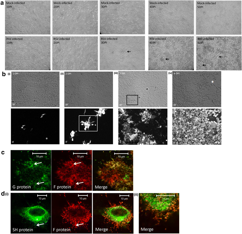
Fig. 1
Fig. 1
Localized RSV transmission occurs within the HEp2 cell monolayers. HEp2 cell monolayers were either mock-infected or RSV-infected using a multiplicity of infection (moi) of 0.0002. a At between 1 and 5 days post-infection (dpi) the monolayers in the tissue culture dish were fixed using 3 % glutaraldehyde and viewed using an inverted light microscope (objective x4 magnification). The virus-induced morphological changes in the monolayer are highlighted (black arrows). b In a parallel analysis the virus-infected cells were stained using anti-RSV and anti-mouse IgG conjugated to Alexa 488 at (i) 1 dpi, (ii) 2 dpi, (iii) 3 dpi and (iv) 4 dpi. The stained cells were then viewed using fluorescence microscopy (IF) and by bright-field (BF) microscopy (objective x20 magnification). The infected cell cluster (open white box), virus-induced morphological changes in the monolayer (open black box) and region of the monolayer cleared of cells (*) are highlighted. c and d Distribution of the F, G and SH virus glycoproteins in the infected cell clusters. HEp2 cell monolayers were infected with RSV using infection moi of 0.01 and 30 h post-infection the cells were fixed and stained using either (c) anti-G and anti-F or (d) anti-SH and anti-F. d(ii) is an enlarged image from (d) (i)). The stained cells were viewed using confocal microscopy at an optical plane that allowed imaging of the virus filaments. The virus filaments (white arrow) and the typical Golgi staining in the SH stained cells (*) are indicated
Fig. 2
Fig. 2
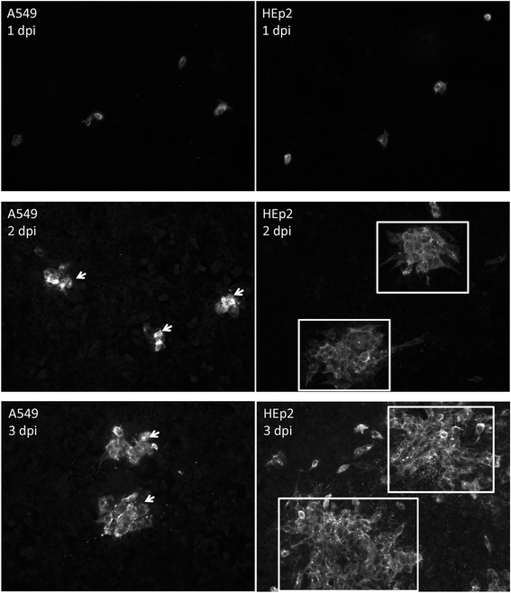
Localized RSV transmission occurs with the A549 cell monolayers. A549 and HEp2 cell monolayers were infected with RSV using a multiplicity of infection (moi) of 0.0002 and at between 1 and 3 days post-infection (dpi) the virus-infected cells were stained using anti-RSV and anti-mouse IgG conjugated to Alexa 488 and viewed using fluorescence microscopy (objective x20 magnification). The infected cell clusters in the A549 (white arrows) and HEp2 (open white box) cell monolayers are highlighted
Fig. 3
Fig. 3
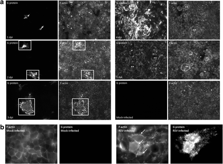
RSV-infection induces localized changes in the F-actin distribution in HEp2 cell monolayers. a HEp2 cell monolayers were infected with RSV using a multiplicity of infection of 0.0002 and at between 1 and 5 days post-infection (dpi) the cells were co-stained using anti-G and phalloidin-FITC (to detect F-actin). The mock-infected sample was stained at 5 dpi. The stained cell monolayers were viewed by immunofluorescence microscopy (objective x20 magnification). Individual infected cells at 1 dpi (white arrows), infected cell clusters (open white box), cell detachment (*) and syncytia (**) are indicated. b Virus-induced increased phalloidin-FITC staining at the intercellular junctions. Mock and virus-infected cells at 2 dpi were stained with anti-G and phalloidin-FITC. The increased phalloidin-FITC staining in the virus infected cells is highlighted (white arrows). The stained cell monolayers were viewed by immunofluorescence microscopy (objective 100x magnification)
Fig. 4
Fig. 4
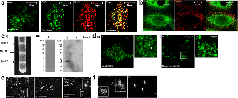
The rac-1 and rho A kinases are required for localised cell-to-cell transmission in the HEp2 cell monolayer. a Mock and RSV-infected cells (multiplicity of infection (moi) of 0.1) were stained using anti-F and anti-rac-1 at 24 h post-infection (hpi) and imaged by confocal microscopy (objective x100 magnification). Only the merged image is shown in the mock-infected cells. b Co-stained cells showing virus filaments bridging two cells. In all cases the virus filaments are highlighted (white arrows). c Virus particles were dissociated from infected cells and infectious virus particles isolated using discontinuous sucrose gradient centrifugation as described previously [21]. c (i) A representative gradient showing the locations of the fractions at the 20-35 % sucrose (band-1), the 35-45 % sucrose (band-2) and 45-55%sucrose (band-3) interfaces. (ii) Each fraction was examined by immunoblotting using anti-rac-1. A protein band corresponding in size to the rac-1 protein is highlighted (black arrow). d and eThe rac-1 inhibitor NSC23766 prevents virus filament formation and virus transmission. d RSV-infected HEp2 cells (moi = 0.1) were either non-treated or NSC23766-treated and the cells stained using anti-G. The virus filaments in non-treated (white arrows) and punctuate staining pattern in NSC23766-treated cells (*) are highlighted. In each case (ii) is an enlarged image of the region highlighted by the white box in (i). e rac-1 activity is required for cell-to-cell transmission. HEp2 cell monolayer were infected using a moi of 0.0002 and at 5 hpi the cell were either non-treated or NSC23766 treated. At 18 hpi the inhibitor was either removed (washout) or maintained. At 36 hpi the cells were fixed and stained using anti-RSV. The cells were imaged using immunofluorescence microscopy (objective x20 magnification). The infected cell clusters in the NT cells (open white box) and smaller clusters in the NSC23766-treated cells (white arrows) are indicated. f HEp2 cell monolayers were infected with RSV using a moi of 0.0002 and at 18 hpi the cells were either non-treated (NT) or treated with Y-27632 (YT). At 48 hpi the cells were fixed and stained using anti-RSV and imaged using immunofluorescence microscopy (objective x20 magnification). The infected cell clusters in the NT cells (open white box) and smaller clusters in the YT cells (white arrows) are indicated
Fig. 5
Fig. 5
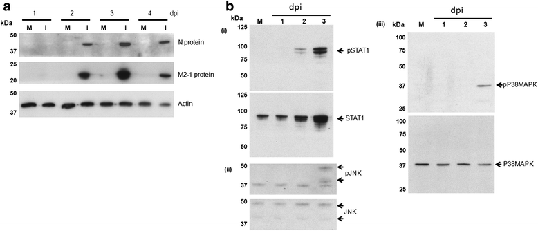
Virus infection induces cell signaling pathways associated with cell death at 3 dpi. a HEp2 cell monolayers were either mock-infected (M) or infected with RSV using a multiplicity of infection of 0.0002. At between 1 and 4 days post-infection (dpi) cell lysates were prepared and the presence of the N and M2-1 proteins detected by immunoblotting. Actin is the loading control. b The presence of (i) STAT1 and pSTAT1, (ii) JNK and pJNK and (iii) MAPKp38 and pMAPKp38 were detected in the RSV-infected cell lysates by immunoblotting with the relevant antibody at 1, 2 and 3 dpi. In this case the infected cells were compared with the mock-infected cell lysate harvested at 3 dpi
Fig. 6
Fig. 6
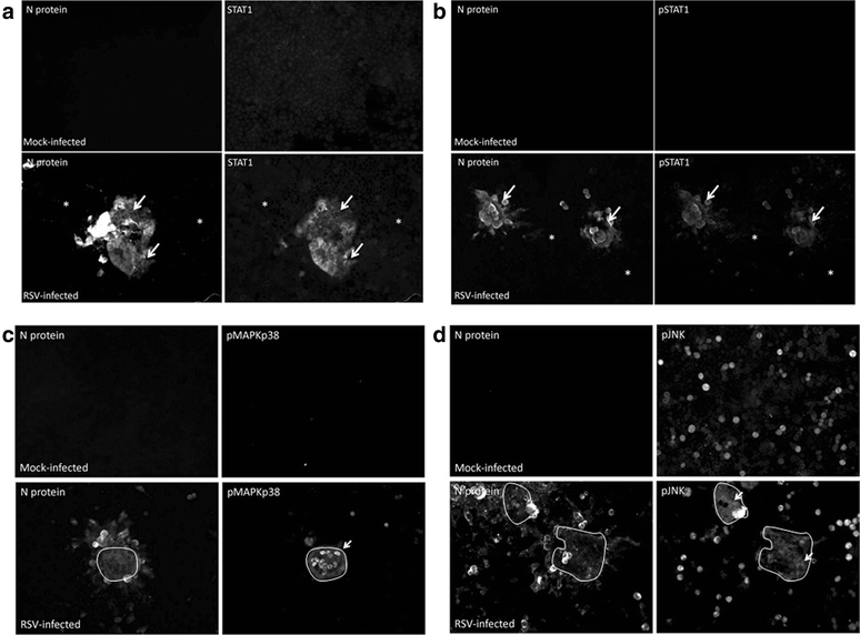
Distribution of STAT1, pSTAT1, pMAPKp38 and pJNK in the RSV-infected monolayers. HEp2 cell monolayers were mock-infected or infected with RSV using a multiplicity of infection of 0.0002. a and b At 2 days post-infection (dpi) the cells were stained using anti-N and either (a) anti-STAT1 or (b) anti-pSTAT1. The cells were viewed by IF microscopy (objective x20 magnification). The STAT1 and pSTAT1 regions in the cell monolayer are highlighted (white arrow), and the region of the monolayer containing non-infected cells is indicated (*) (c and d) HEp2 cell monolayers were mock-infected or infected with RSV using a multiplicity of infection of 0.0002. At 3 dpi the cells were stained using anti-N and either (c) anti-pMAPKp38 or (d) anti-pJNK. The cells were viewed by fluorescence microscopy (objective x20 magnification). The pMAPKp38 and pJNK regions in the cell monolayer are indicated (white arrow). The region of pMAPKp38 and pJNK staining in each image is demarcated (white outline)
Fig. 7
Fig. 7
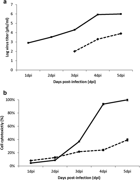
The presence of cell-free virus particles occurs late in infection and correlates with increased cell permeability and cytotoxicity. HEp2 cell monolayers were infected with RSV using an multiplicity of infection of 0.0002 and (a) between 1 and 5 days post-infection (dpi) the cell-associated virus infectivity (solid line) and the cell-free virus infectivity (broken line) in the tissue culture supernatant was measured by plaque assay. Average values (duplicates) for the infectivity measurements are shown and for all times the standard error for each of the infectivity measurements was ≤ 10 %. Representative data obtained from one experiment is shown. b Lactate dehydrogenase (LDH) release from RSV-infected HEp2 cells. HEp2 cell monolayers were either mock-infected (broken line) or RSV-infected (solid line) using a multiplicity of infection of 0.0002 and at between 1 and 5 dpi the levels of LDH was measured in the tissue culture supernatant. The values are average values of triplicate measurements at each time point. Representative data from one experiment is shown
Fig. 8
Fig. 8
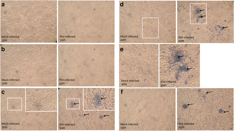
Trypan blue staining is detected in the HEp2 cell monolayers. HEp2 cell monolayers were either mock-infected or RSV-infected using an multiplicity of infection of 0.0002 and (a-f) at between 1 and 5 days post-infection (dpi) the monolayers were stained using trypan blue and imaged using an inverted light microscope (objective x10 magnification). The regions of the cell monolayer showing trypan blue staining is highlighted (black arrows). In plate (c) selected regions in the monolayer (indicated by open white box) are enlarged in the inset. Plate (e) shows enlarged images from the region indicated in plate (d) (indicated by open white box)
Fig. 9
Fig. 9
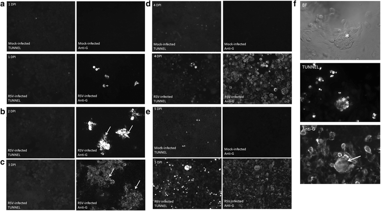
TUNEL assay of virus-infected HEp2 cell monolayers. HEp2 cell monolayers were either mock-infected or infected with RSV using a multiplicity of infection of 0.0002. At (a) 1 day post infection (dpi), (b) 2 dpi, (c) 3 dpi, (d) 4 dpi and (e) 5 dpi the monolayers were co-stained using anti-G and TUNEL. The stained cells were then viewed using fluorescence microscopy. The anti-G infected cell clusters (white arrows) and the increased TUNEL staining (*) are indicated (objective x20 magnification). (f) Enlarged image showing virus-induced syncytia viewed using bight field (BF), TUNEL staining, and anti-G staining at 5 dpi. The syncytia (*), TUNEL staining (**) and anti-G staining (white arrow) are highlighted. Mock infected monolayers are shown at 1, 4 and 5 dpi

Similar articles

Cited by

References

    1. Nair H, Nokes DJ, Gessner BD, Dherani M, Madhi SA, Singleton RJ, et al. Global burden of acute lower respiratory infections due to respiratory syncytial virus in young children: a systematic review and meta-analysis. The Lancet. 2010;375:1545–55. doi: 10.1016/S0140-6736(10)60206-1. - DOI - PMC - PubMed
    1. Grosfeld H, Hill MG, Collins PL. RNA replication by respiratory syncytial virus (RSV) is directed by the N, P, and L proteins; transcription also occurs under these conditions but requires RSV superinfection for efficient synthesis of full-length mRNA. J Virol. 1995;69:5677–86. - PMC - PubMed
    1. Yu Q, Hardy RW, Wertz GW. Functional cDNA clones of the human respiratory syncytial (RS) virus N, P, and L proteins support replication of RS virus genomic RNA analogs and define minimal trans-acting requirements for RNA replication. J Virol. 1995;69:2412–9. - PMC - PubMed
    1. Carromeu C, Simabuco FM, Tamura RE, Farinha Arcieri LE, Ventura AM. Intracellular localization of human respiratory syncytial virus L protein. Arch Virol. 2007;152:2259–63. doi: 10.1007/s00705-007-1048-4. - DOI - PubMed
    1. Garcia J, Garcia-Barreno B, Vivo A, Melero JA. Cytoplasmic inclusions of respiratory syncytial virus-infected cells: formation of inclusion bodies in transfected cells that coexpress the nucleoprotein, the phosphoprotein, and the 22 K protein. Virology. 1993;195:243–7. doi: 10.1006/viro.1993.1366. - DOI - PubMed

Publication types