. 2016 Jan 15;340(2):259-73.
doi: 10.1016/j.yexcr.2016.01.003. Epub 2016 Jan 12.

Rab11-FIP1A regulates early trafficking into the recycling endosomes

Affiliations

Rab11-FIP1A regulates early trafficking into the recycling endosomes

Jenny C Schafer et al. Exp Cell Res. .

Abstract

The Rab11 family of small GTPases, along with the Rab11-family interacting proteins (Rab11-FIPs), are critical regulators of intracellular vesicle trafficking and recycling. We have identified a point mutation of Threonine-197 site to an Alanine in Rab11-FIP1A, which causes a dramatic dominant negative phenotype when expressed in HeLa cells. The normally perinuclear distribution of GFP-Rab11-FIP1A was condensed into a membranous cisternum with almost no GFP-Rab11-FIP1A(T197A) remaining outside of this central locus. Also, this condensed GFP-FIP1A(T197A) altered the distribution of proteins in the Rab11a recycling pathway including endogenous Rab11a, Rab11-FIP1C, and transferrin receptor (CD71). Furthermore, this condensed GFP-FIP1A(T197A)-containing structure exhibited little movement in live HeLa cells. Expression of GFP-FIP1A(T197A) caused a strong blockade of transferrin recycling. Treatment of cells expressing GFP-FIP1A(T197A) with nocodazole did not disperse the Rab11a-containing recycling system. We also found that Rab5 and EEA1 were accumulated in membranes by GFP-Rab11-FIP1A but Rab4 was unaffected, suggesting that a direct pathway may exist from early endosomes into the Rab11a-containing recycling system. Our study of a potent inhibitory trafficking mutation in Rab11-FIP1A shows that Rab11-FIP1A associates with and regulates trafficking at an early step in the process of membrane recycling.

Keywords: EEA1; Endocytosis; Membrane recycling; Rab11; Rab11-FIP1; Rab11-FIP2; Rab11-FIP5; Rab11a; Rab14; Rab4; Rab5; Rab8a.


Figures

Figure 1
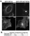
Figure 1. GFP-Rab11-FIP1A(T197A) localizes to a collapsed perinuclear membrane cisternum
A. HeLa cells were transfected with either GFP-Rab11-FIP1A or GFP-Rab11-FIP1A(T197A) and imaged live for deconvolution. Separately, cells were transfected, fixed, and imaged by Structured Illumination Microscopy (SIM). Wild-type GFP-Rab11-FIP1A shows vesicles concentrated in the perinuclear region as well as extended tubules throughout the cell, typical of vesicular proteins involved in endosomal trafficking. Mutant GFP-Rab11-FIP1A(T197A), however, was redistributed almost entirely to the perinuclear area and shows no tubule localization. See Supplementary Videos 1 and 2. B. The distribution of GFP-Rab11-FIP1A and GFP-Rab11-FIP1A(T197A) was evaluated in 50 cells and the distribution of transfected protein was scored as located in diffuse vesicles, in one to two condensed puncta, or in multiple large puncta.
Figure 2
Figure 2. T197A mutation in Rab11-FIP1A causes increased β-sheet structure
Diagram of Rab11-FIP1A protein with putative 14-3-3 binding site (A). CD spectra (B) of recombinant human Rab11-FIP1A (GREEN) and Rab11-FIP1A(T197A) (BLUE).The difference between the wild type protein and mutant protein is quite dramatic. The mutant protein shows a characteristic peak at 196 nm and a low at 218 nm, which is characteristic of a beta-sheet rich protein. Overall, the mutant protein displays much more secondary structure than the wild type protein.
Figure 3
Figure 3. GFP-Rab11-FIP1A(T197A) inhibits transferrin recycling
HeLa cells transfected with either GFP-Rab11-FIP1A or GFP-Rab11-FIP1A(T197A) were serum starved for one hour and then loaded with Transferrin-Alexa568 for 15 minutes on ice. Cells were chased with unlabeled Transferrin for 40 minutes and imaged in real time. Seven time points over 40 minutes are shown in the Transferrin-Alexa568 channel. Most of the Transferrin-Alexa568 was trafficked out of cells expressing wild type Rab11-FIP1A by 40 minutes, while GFP-FIP1A(T197A) cells still showed a concentration of Transferrin at 40 minutes. See Supplementary Videos 3 and 4.
Figure 4
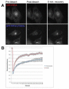
Figure 4. GFP-Rab11-FIP1A(T197A) expressing membranes show slow recovery after photobleaching
Fluorescence Recovery After Photobleaching (FRAP) was performed on HeLa cells transfected with GFP-Rab11-FIP1A or GFP-Rab11-FIP1A(T197A). Three images were taken before bleach, cells were bleached for 5 seconds, and then cells were imaged every 5 seconds for a total of 5 minutes. A. Representative images pre-bleach, post bleach and after 5 minutes of recovery. B. Fluorescence recovery along with SEM is plotted for cells expressing GFP-Rab11-FIP1A or GFP-Rab11-FIP1A(T197A). Fluorescence in GFP-FIP1A cells recovered more rapidly than in GFP-FIP1A(T197A) cells. See Supplementary Videos 5 and 6.
Figure 5
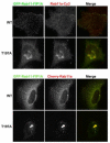
Figure 5. Rab11a accumulates with GFP-Rab11-FIP1A(T197A) in a collapsed membranous cisternum
HeLa cells were transfected with either GFP-Rab11-FIP1A or GFP-Rab11-FIP1A(T197A), stained for endogenous Rab11a, and imaged by SIM. Cells imaged by Structured Illumination Microscopy showed a redistribution of Rab11a to the GFP-Rab11-FIP1A(T197A) spot compared to wild type (A). HeLa cells were transfected with Cherry-Rab11a along with either GFP-Rab11-FIP1A or GFP-Rab11-FIP1A(T197A) and were imaged in time lapse to observe the movement of vesicles (B). Cherry-Rab11a was condensed in the perinuclear area in the presence of the mutant protein, while it remains vesicular in the presence of the wild type protein. Cherry-Rab11a and GFP-Rab11-FIP1A vesicles were seen throughout the cell for wild type, but few vesicles were only found in the perinuclear region with the mutant. See Supplementary Videos 7 and 8.
Figure 6
Figure 6. Alteration of endosomal protein localization with GFP-Rab11-FIP1A(T197A) expression
HeLa cells were transfected with either GFP-Rab11-FIP1A or GFP-Rab11-FIP1A(T197A), stained for Rab5, Rab8a, Rab14, or Rab11-FIP5, and imaged by SIM. Rab5 was seen in vesicles in the presence of GFP-Rab11-FIP1A, but Rab5 localization was altered to the condensed regions in the presence of GFP-Rab11-FIP1A(T197A). Rab8a was seen in vesicles in the presence of GFP-Rab11-FIP1A, but Rab8a localization was altered to the condensed regions in the presence of GFP-Rab11-FIP1A(T197A) . Rab14 was seen in vesicles in the presence of GFP-Rab11-FIP1A, but Rab14 localization was altered to the condensed regions in the presence of GFP-Rab11-FIP1A(T197A). Rab11-FIP5 was seen in vesicles in the presence of GFP-Rab11-FIP1A, but Rab11-FIP5 localization was altered to the condensed regions in the presence of GFP-Rab11-FIP1A(T197A). Quantitation is shown in Figure 7B.
Figure 7
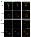
Figure 7. Expression of GFP-Rab11-FIP1A(T197A) does not alter the distribution of Rab4
HeLa cells were transfected with either GFP-Rab11-FIP1A or GFP-Rab11-FIP1A(T197A), immunostained for Rab4a and Rab11a, and imaged by SIM (A). In the presence of wild type GFP-Rab11-FIP1A, Rab4a was found on vesicles and rarely overlapped with GFP-Rab11-FIP1A and Rab11a. In the presence of GFP-Rab11-FIP1A(T197A), Rab4a distribution was unchanged and remained in dispersed vesicles despite the redistribution of GFP-Rab11-FIP1A(T197A) and Rab11a. The Rab4a immunostaining showed a non-specific variable nuclear background. Pearson’s Correlation Coefficients (PCC) were calculated for GFP-Rab11-FIP1A(T197A) and endogenous Rabs and Rab11-FIPs shown in Figures 6- (B). PCC were plotted along with SEM. Rab11a showed the highest PCC and Rab4 showed the lowest. Rab4 was decreased compared to other Rabs and Rab11-FIP5. We found a statistically significant difference between groups (Rab4, Rab5, Rab8a, Rab14, and Rab11-FIP5) as determined by one-way ANOVA (F(5,42) = 2.412, p = .05).
Figure 8
Figure 8. Rab11-FIP2 does not associate with membranes containing GFP-Rab11-FIP1A(T197A)
HeLa cells were transfected with GFP-Rab11-FIP1A, stained for Rab11-FIP2, and imaged by widefield deconvolution (A). GFP-Rab11-FIP1A and Rab11-FIP2 were not found on the same vesicles. HeLa cells were transfected with GFP-Rab11-FIP1A, stained for Rab11-FIP2, and imaged by widefield deconvolution (B). GFP-Rab11-FIP1A and Rab11-FIP2 were not found on the same vesicles. HeLa cells were transfected with GFP-Rab11-FIP1A(T197A) along with Rab11-FIP2-Cherry and imaged by widefield deconvolution (C). GFP-Rab11-FIP1A(T197A) and Rab11-FIP2-Cherry were not found on the same vesicles.
Figure 9
Figure 9. Rab5 accumulated on GFP-Rab11-FIP1A(T197A)-containing membranes
HeLa cells were transfected with either GFP-Rab11-FIP1A or GFP-Rab11-FIP1A(T197A), stained for Rab5 and EEA1, and imaged by widefield deconvolution (A). HeLa cells were transfected with either GFP-Rab11-FIP1A or GFP-Rab11-FIP1A(T197A) along with Cherry-Rab5a, and imaged live by widefield deconvolution. Still images from movies are shown (B). Both endogenous Rab5 and Cherry-Rab5a as well as EEA1 localization were altered in the presence of GFP-Rab11-FIP1A(T197A). See Supplementary Videos 9 and 10.
Figure 10
Figure 10. Disruption of microtubules only partially affects GFP-Rab11-FIP1A(T197A)
HeLa cells were transfected with either GFP-Rab11-FIP1A or GFP-Rab11-FIP1A(T197A) and treated with nocodazole or DMSO (control). Cells were then stained for Rab11a and imaged on a widefield microscope. Disruption of microtubules caused a dispersion of GFP-Rab11-FIP1A and Rab11a vesicles (A). In the presence of GFP-Rab11-FIP1A(T197A), this dispersion was incomplete and the GFP-Rab11-FIP1A(T197A) cisternum was partially retained (B).
Figure 11
Figure 11. Expression of GFP-Rab11-FIP1A(T197A) alters the distribution of microtubules, but not F-actin
HeLa cells were transfected with either GFP-Rab11-FIP1A or GFP-Rab11-FIP1A(T197A). One set of cells was stained for a-tubulin (A) to view microtubules and a second set was stained for phalloidin to view F-actin (B). Note the peripheral distribution of microtubules in cells expressing GFP-Rab11-FIP1A(T197A) (A).

References

    1. Schwartz SL, Cao C, Pylypenko O, Rak A, Wandinger-Ness A. Rab GTPases at a glance. J Cell Sci. 2007;120:3905–3910. - PubMed
    1. Wandinger-Ness A, Zerial M. Rab proteins and the compartmentalization of the endosomal system. Cold Spring Harbor perspectives in biology. 2014;6:a022616. - PMC - PubMed
    1. Sonnichsen B, De Renzis S, Nielsen E, Reitdorf J, Zerial M. Distinct membrane domains on endosomes in the recycling pahtway visualized by multicolor imaging of Rab4, Rab5 and Rab11. J Cell Biol. 2000;149:901–914. - PMC - PubMed
    1. Ullrich O, Reinsch S, Urbe S, Zerial M, Parton RG. Rab11 regulates recycling through the pericentriolar recycling endosome. J.Cell Biol. 1996;135:913–924. - PMC - PubMed
    1. Green EG, Ramm E, Riley NM, Spiro DJ, Goldenring JR, Wessling-Resnick M. Rab11 is associated with transferrin-containing recycling compartments in K562 cells. Biochem.Biophys.Res.Commun. 1997;239:612–616. - PubMed

Publication types