

A technique for continuous bedside monitoring of global cerebral energy state
- PMID: 26791144
- PMCID: PMC4720625
- DOI: 10.1186/s40635-016-0077-2
A technique for continuous bedside monitoring of global cerebral energy state
Abstract
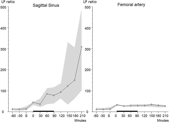
Background: Cerebral cytoplasmatic redox state is a sensitive indicator of cerebral oxidative metabolism and is conventionally evaluated from the extracellular lactate/pyruvate (LP) ratio. In the present experimental study of global cerebral ischemia induced by hemorrhagic shock, we investigate whether the LP ratio obtained from microdialysis of cerebral venous blood may be used as a surrogate marker of global cerebral energy state.
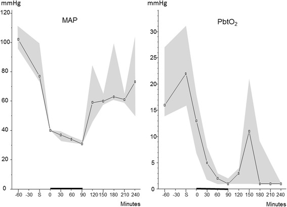
Methods: Six female pigs were anesthetized and vital parameters were recorded. Microdialysis catheters were placed in the left parietal lobe, the superior sagittal sinus, and the femoral artery. Hemorrhagic shock was achieved by bleeding the animals to a mean arterial pressure (MAP) of approximately 40 mmHg and kept at a MAP of about 30-40 mmHg for 90 min. The animals were resuscitated with autologous whole blood followed by 3 h of observation.
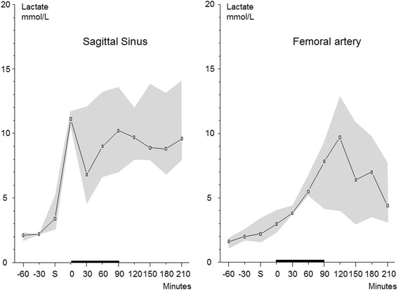
Results: The LP ratio obtained from the intracerebral and intravenous catheters immediately increased during the period of hemorrhagic shock while the LP ratio in the arterial blood remained close to normal levels. At the end of the experiment, median LP ratio (interquartile range) obtained from the intracerebral, intravenous, and intra-arterial microdialysis catheters were 846 (243-1990), 309 (103-488), and 27 (21-31), respectively. There was a significant difference in the LP ratio obtained from the intravenous location and the intra-arterial location (P < 0.001).
Conclusions: During cerebral ischemia induced by severe hemorrhagic shock, intravascular microdialysis of the draining venous blood will exhibit changes of the LP ratio revealing the deterioration of global cerebral oxidative energy metabolism. In neurocritical care, this technique might be used to give information regarding global cerebral energy metabolism in addition to the regional information obtained from intracerebral microdialysis catheters. The technique might also be used to evaluate cerebral energy state in various critical care conditions when insertion of an intracerebral microdialysis catheter may be contraindicated, e.g., resuscitation after cardiac standstill, open-heart surgery, and multi-trauma.
Keywords: Cerebral energy state; Hemorrhagic shock; Ischemia; Microdialysis.
Figures
Similar articles
-
Moderately prolonged permissive hypotension results in reversible metabolic perturbation evaluated by intracerebral microdialysis - an experimental animal study.Intensive Care Med Exp. 2019 Dec 4;7(1):67. doi: 10.1186/s40635-019-0282-x. Intensive Care Med Exp. 2019. PMID: 31802303 Free PMC article.
-
Bedside interpretation of cerebral energy metabolism utilizing microdialysis in neurosurgical and general intensive care.Front Neurol. 2022 Aug 10;13:968288. doi: 10.3389/fneur.2022.968288. eCollection 2022. Front Neurol. 2022. PMID: 36034291 Free PMC article. Review.
-
Aspects on the Physiological and Biochemical Foundations of Neurocritical Care.Front Neurol. 2017 Jun 19;8:274. doi: 10.3389/fneur.2017.00274. eCollection 2017. Front Neurol. 2017. PMID: 28674514 Free PMC article. Review.
-
Design paper of the "Blood pressure targets in post-resuscitation care and bedside monitoring of cerebral energy state: a randomized clinical trial".Trials. 2019 Jun 10;20(1):344. doi: 10.1186/s13063-019-3397-1. Trials. 2019. PMID: 31182135 Free PMC article. Clinical Trial.
-
Bedside Monitoring of Cerebral Energy State During Cardiac Surgery-A Novel Approach Utilizing Intravenous Microdialysis.J Cardiothorac Vasc Anesth. 2017 Aug;31(4):1166-1173. doi: 10.1053/j.jvca.2016.11.001. Epub 2016 Nov 2. J Cardiothorac Vasc Anesth. 2017. PMID: 28089142 Clinical Trial.
Cited by
-
Moderately prolonged permissive hypotension results in reversible metabolic perturbation evaluated by intracerebral microdialysis - an experimental animal study.Intensive Care Med Exp. 2019 Dec 4;7(1):67. doi: 10.1186/s40635-019-0282-x. Intensive Care Med Exp. 2019. PMID: 31802303 Free PMC article.
-
A Prospective Observational Feasibility Study of Jugular Bulb Microdialysis in Subarachnoid Hemorrhage.Neurocrit Care. 2020 Aug;33(1):241-255. doi: 10.1007/s12028-019-00888-0. Neurocrit Care. 2020. PMID: 31845174
-
Effects of norepinephrine infusion on cerebral energy metabolism during experimental haemorrhagic shock.Intensive Care Med Exp. 2022 Feb 4;10(1):4. doi: 10.1186/s40635-022-00432-z. Intensive Care Med Exp. 2022. PMID: 35118520 Free PMC article.
-
Bedside interpretation of cerebral energy metabolism utilizing microdialysis in neurosurgical and general intensive care.Front Neurol. 2022 Aug 10;13:968288. doi: 10.3389/fneur.2022.968288. eCollection 2022. Front Neurol. 2022. PMID: 36034291 Free PMC article. Review.
-
Aspects on the Physiological and Biochemical Foundations of Neurocritical Care.Front Neurol. 2017 Jun 19;8:274. doi: 10.3389/fneur.2017.00274. eCollection 2017. Front Neurol. 2017. PMID: 28674514 Free PMC article. Review.
References
-
- Siesjö BK. Brain energy metabolism. Chichester,[Eng.] ; New York: Wiley; 1978.
LinkOut - more resources
Full Text Sources
Other Literature Sources
