. 2016 Dec;4(1):4.
doi: 10.1186/s40635-016-0075-4. Epub 2016 Jan 21.

Cost of surviving sepsis: a novel model of recovery from sepsis in Drosophila melanogaster

Affiliations

Cost of surviving sepsis: a novel model of recovery from sepsis in Drosophila melanogaster

Ata Murat Kaynar et al. Intensive Care Med Exp. 2016 Dec.

Abstract

Background: Multiple organ failure, wasting, increased morbidity, and mortality following acute illness complicates the health span of patients surviving sepsis. Persistent inflammation has been implicated, and it is proposed that insulin signaling contributes to persistent inflammatory signaling during the recovery phase after sepsis. However, mechanisms are unknown and suitable pre-clinical models are lacking. We therefore developed a novel Drosophila melanogaster model of sepsis to recapitulate the clinical course of sepsis, explored inflammation over time, and its relation to impaired mobility, metabolic disturbance, and changes in lifespan.

Methods: We used wild-type (WT), Drosomycin-green fluorescent protein (GFP), and NF-κB-luc reporter male Drosophila melanogaster 4-5 days of age (unmanipulated). We infected Drosophila with Staphylococcus aureus (infected without treatment) or pricked with aseptic needles (sham). Subsets of insects were treated with oral linezolid after the infection (infected with antibiotics). We assessed rapid iterative negative geotaxis (RING) in all the groups as a surrogate for neuromuscular functional outcome up to 96 h following infection. We harvested the flies over the 7-day course to evaluate bacterial burden, inflammatory and metabolic pathway gene expression patterns, NF-κB translation, and metabolic reserve. We also followed the lifespan of the flies.

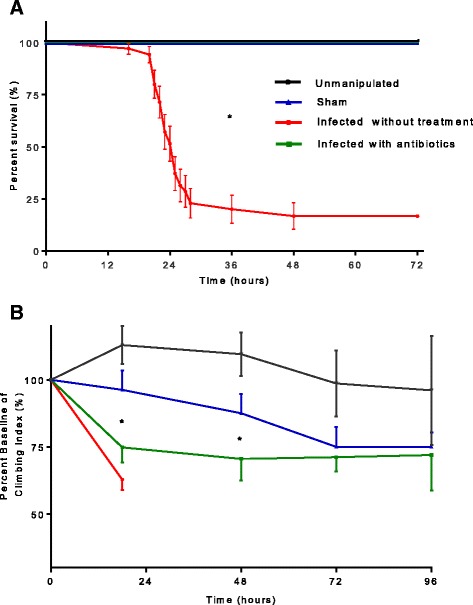
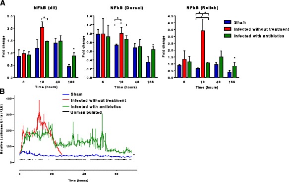
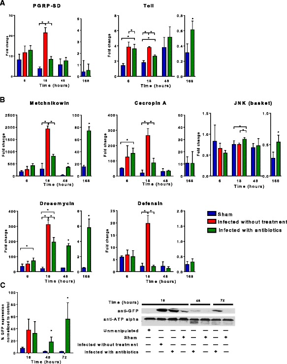
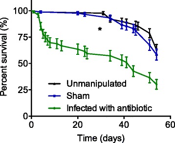
Results: Our results showed that when treated with antibiotics, flies had improved survival compared to infected without treatment flies in the early phase of sepsis up to 1 week (81 %, p = 0.001). However, the lifespan of infected with antibiotics flies was significantly shorter than that of sham controls (p = 0.001). Among infected with antibiotic sepsis survivors, we observed persistent elevation of NF-κB in the absence of any obvious infection as shown by culturing flies surviving sepsis. In the same group, geotaxis had an early (18 h) and sustained decline compared to its baseline. Geotaxis in infected with antibiotics sepsis survivors was significantly lower than that in sham and age-matched unmanipulated flies at 18 and 48 h. Expression of antimicrobial peptides (AMP) remained significantly elevated over the course of 7 days after sepsis, especially drosomycin (5.7-fold, p = 0.0145) on day 7 compared to that of sham flies. Infected with antibiotics flies had a trend towards decreased Akt activation, yet their glucose stores were significantly lower than those of sham flies (p = 0.001). Sepsis survivors had increased lactate levels and LDH activity by 1 week, whereas ATP and pyruvate content was similar to that of the sham group.

Conclusions: In summary, our model mimics human survivors of sepsis with persistent inflammation, impaired motility, dysregulated glucose metabolism, and shortened lifespan.

Keywords: Akt; Drosomycin; Drosophila; Insulin; Recovery; Sepsis.


Figures

Figure 1
Figure 1
a Survival in Drosophila melanogaster after septic injury with Staphylococcus aureus. Flies were infected using needle pricking assay with S. aureus; survival was assessed by observing flies every hour for the first 72 h from 8 a.m. until 11 p.m. Of the infected without treatment flies, 75 % died by the 28th hour after infection; infected with antibiotics group received oral linezolid for 18 h and showed improved survival without any death due to infection in the first 72 h after infection. Sham group pricked with sterile needle showed no death due to pricking-related trauma. We used 25 flies in each group; survival experiment was repeated three times for the Kaplan-Meyer survival analysis (*p < 0.05). b Geotaxis in Drosophila melanogaster after septic injury with Staphylococcus aureus. Each geotaxis experiment was performed 1 h before the needle pricking (0-h baseline) and at 18, 48, 72, and 96 h after needle pricking. Geotaxis in sham flies was significantly lower at 72 h compared to its baseline. Infected with antibiotics group showed significantly lower geotaxis compared to baseline at 18, 48, and 72 h. While there was significant difference in geotaxis between sham and infected with antibiotics groups at 18 and 48 h, this difference was lost by 72 h. Geotaxis of unmanipulated healthy flies remained at the baseline through experiment. We repeated each geotaxis experiment six times (*p < 0.05)
Fig. 2
Fig. 2
NF-κB activity in Drosophila melanogaster after septic injury with Staphylococcus aureus. a We determined NF-κB expression at 6, 18, 48, and 168 h (1 week) after needle pricking with qRT-PCR. Expression of dif and Dorsal 18 h after infection was upregulated in infected with antibiotics group and was significantly higher compared to that of sham; expression of Relish was upregulated in all three groups with significant difference; all three NF-κB components (dorsal, Dif, Relish) remained elevated by 1 week following survival from sepsis (*p < 0.05). b Luciferase activity in live flies expressing NF-κB luciferase reporter was measured for 72 h after infection. Flies demonstrated elevated levels of NF-κB activation in both infected without treatment and infected with antibiotics groups in the first 24 h and sustained elevation of NF-κB activity up to 72 h in infected with antibiotics flies, while activity in sham group was not upregulated after pricking (*p < 0.05)
Fig. 3
Fig. 3
Inflammatory genes expression in Drosophila melanogaster after septic injury with Staphylococcus aureus. We determined inflammatory gene expression, pattern recognition receptors, antimicrobial peptides at 6, 18, 48, and 168 h (1 week) after needle pricking with qRT-PCR (*p < 0.05). a PGRP-SD and Toll were upregulated as early as 6 h after infection. By 18 h, PGRP-SD and Toll expression in infected without treatment flies were significantly elevated compared to infected with antibiotics flies. Toll remained elevated 48 h after infection with treatment; however, there was no difference between infected with antibiotics and sham; 1 week after infection expression of Toll was downregulated. b Antimicrobial peptides (AMP) were upregulated by 6 h in a similar way between infected without treatment vs. infected with antibiotics groups. By 18 h, the infected without treatment flies had significantly elevation of drosomycin, metchnikowin, defensin, JNK, and cecropin A. Drosomycin and metchnikowin remained significantly elevated in the survivors 48 h and 7 days after surviving sepsis. c Expression of Drs-GFP construct was detected at baseline and significantly increased as early as 18 h following infection using Western immunoblot. While its expression came back to baseline in sham flies by 48 h, the infected with antibiotics group had sustained elevation of Drs-GFP expression by 72 h
Fig. 4
Fig. 4
Insulin signaling pathway genes expression in Drosophila melanogaster after septic injury with Staphylococcus aureus. a We determined insulin pathway gene (InR, IRS, PTEN, Akt1, Foxo, and mTOR) expression at 6, 18, 48, and 168 h (1 week) after needle pricking. By 18 h, InR was upregulated and significantly higher in infected without treatment group compared to sham and Akt was upregulated with significant difference between all three groups. By 48 h in infected with antibiotics and sham groups, all insulin pathway genes showed sustained elevation of expression without significant difference between groups. In infected with antibiotics group 1 week after infection, levels of InR, IRS, Akt, and mTORC1 were upregulated and significantly higher than those in the sham group. (*p < 0.05). b We determined phosphorylated-Akt/total-Akt ratio with Western immunoblot at 6, 18, 48, and 168 h after needle pricking. Akt phosphorylation significantly decreased by 18 h in infected without treatment and infected with antibiotics compared to sham (asterisk). Akt phosphorylation significantly increased in sham between 18 and 48 h (dollar sign), yet then it was significantly lower at 168 h compared to 48 h (percent). Akt phosphorylation at 168 h after infection was not significantly different compared to 18 h after infection. Akt phosphorylation significantly increased in infected with antibiotics group 48 h after needle pricking compared to 18 h (number sign), yet then it was significantly lower at 168 h compared to 48 h (ampersand). Akt phosphorylation significantly increased in infected with antibiotics group 168 h after needle pricking compared to 18 h after infection (commercial at sign)
Fig. 5
Fig. 5
Carbohydrate metabolites in Drosophila melanogaster after septic injury with Staphylococcus aureus. a We measured glucose and glycogen concentrations at 18, 48, and 168 h in triplicate. We determined glycogen by measuring glucose level after digesting glycogen with amyloglucosidase into free glucose. Level of glucose significantly increased by 168 h in sham flies compared to 18 h. By 168 h, glucose content was significantly lower in infected with antibiotics flies compared to the sham group. Glycogen, triglyceride, protein, and pyruvate levels at 168 h were similar between the two groups. Lactate and LDH activity were significantly elevated by 168 h after surviving sepsis (infected with antibiotics group) (*p < 0.05). Interestingly, the ATP content was significantly lower in infected with antibiotics flies by 48 h compared to sham; however, this difference was lost by 168 h. b There was no difference in the expression of Glut-1 and Glut-3 by 168 h between sham and infected with antibiotics flies
Fig. 6
Fig. 6
Lifespan of Drosophila melanogaster after septic injury with Staphylococcus aureus. Drosophila surviving sepsis induced by Staphylococcus aureus and treated with linezolid had significantly shorter lifespan compared to sham and unmanipulated flies over the course of 60 days (*p < 0.0001). The food intake has not been measured in different groups; however, the weight of flies by 1 week was unchanged in between groups (Additional file 2: Figure S2). Sham and unmanipulated flies had a similar lifespan. The antibiotic-containing food had no impact on the survival of flies as compared to the regular food
Fig. 7
Fig. 7
Summary illustration: interplay between impaired carbohydrate metabolism and sustained inflammation in Drosophila melanogaster surviving sepsis. Drosophila surviving sepsis induced by Staphylococcus aureus and treated with linezolid had impaired glucose metabolism with increased lactate production, increased LDH activity, sustained inflammation, and significantly shorter lifespan. The switch between oxidative phosphorylation and aerobic glycolysis could be the metabolic regulator of inflammation

References

    1. Angus DC, Linde-Zwirble WT, Lidicker J, et al. Epidemiology of severe sepsis in the United States: analysis of incidence, outcome, and associated costs of care. Crit Care Med. 2001;29(7):1303–1310. doi: 10.1097/00003246-200107000-00002. - DOI - PubMed
    1. Iwashyna TJ, Ely EW, Smith DM, et al. Long-term cognitive impairment and functional disability among survivors of severe sepsis. JAMA. 2010;304(16):1787–1794. doi: 10.1001/jama.2010.1553. - DOI - PMC - PubMed
    1. Kaukonen KM, Bailey M, Suzuki S, et al. Mortality related to severe sepsis and septic shock among critically ill patients in Australia and New Zealand, 2000-2012. JAMA. 2014;311(13):1308–1316. doi: 10.1001/jama.2014.2637. - DOI - PubMed
    1. Yende S, Angus DC. Long-term outcomes from sepsis. Curr Infect Dis Rep. 2007;9(5):382–386. doi: 10.1007/s11908-007-0059-3. - DOI - PubMed
    1. Yende S, D’Angelo G, Mayr F, et al. Elevated hemostasis markers after pneumonia increases one-year risk of all-cause and cardiovascular deaths. PLoS One. 2011;6(8) doi: 10.1371/journal.pone.0022847. - DOI - PMC - PubMed

LinkOut - more resources