. 2016 Jan 20;36(3):837-50.
doi: 10.1523/JNEUROSCI.4047-15.2016.

Rescue of Impaired mGluR5-Driven Endocannabinoid Signaling Restores Prefrontal Cortical Output to Inhibit Pain in Arthritic Rats

Affiliations

Rescue of Impaired mGluR5-Driven Endocannabinoid Signaling Restores Prefrontal Cortical Output to Inhibit Pain in Arthritic Rats

Takaki Kiritoshi et al. J Neurosci. .

Abstract

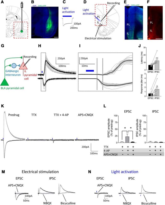
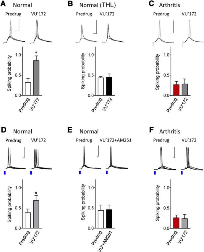
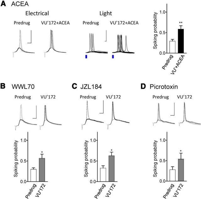
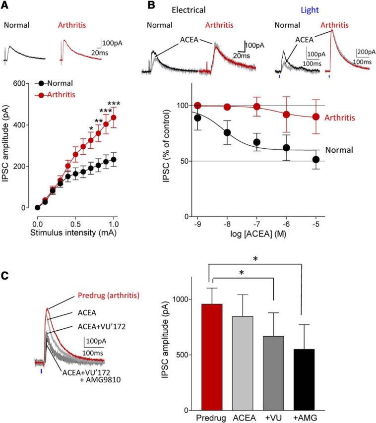
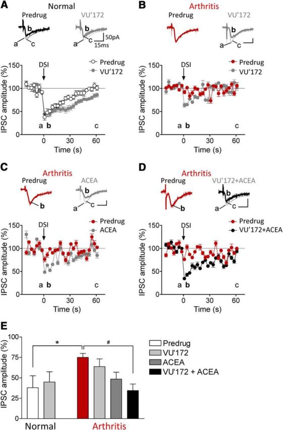
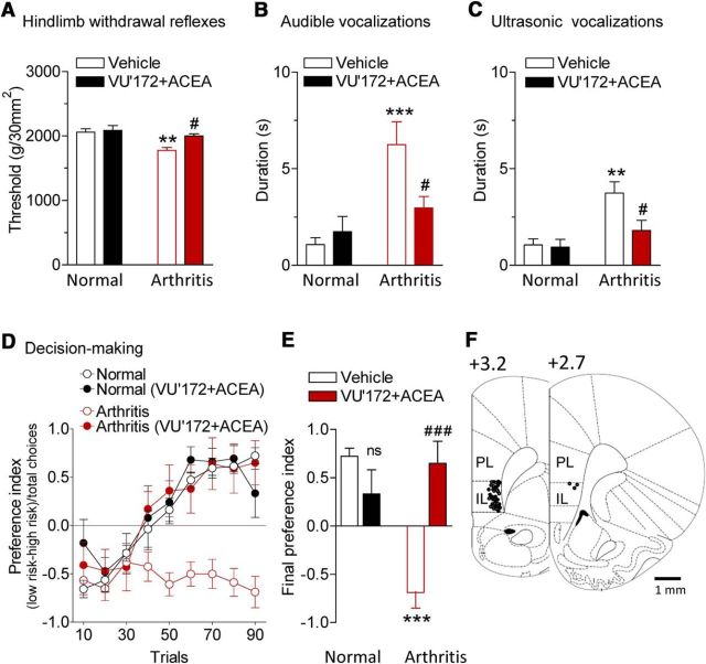
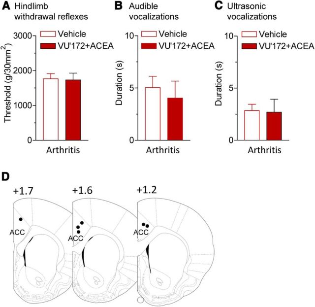
The medial prefrontal cortex (mPFC) serves executive functions that are impaired in neuropsychiatric disorders and pain. Underlying mechanisms remain to be determined. Here we advance the novel concept that metabotropic glutamate receptor 5 (mGluR5) fails to engage endocannabinoid (2-AG) signaling to overcome abnormal synaptic inhibition in pain, but restoring endocannabinoid signaling allows mGluR5 to increase mPFC output hence inhibit pain behaviors and mitigate cognitive deficits. Whole-cell patch-clamp recordings were made from layer V pyramidal cells in the infralimbic mPFC in rat brain slices. Electrical and optogenetic stimulations were used to analyze amygdala-driven mPFC activity. A selective mGluR5 activator (VU0360172) increased pyramidal output through an endocannabinoid-dependent mechanism because intracellular inhibition of the major 2-AG synthesizing enzyme diacylglycerol lipase or blockade of CB1 receptors abolished the facilitatory effect of VU0360172. In an arthritis pain model mGluR5 activation failed to overcome abnormal synaptic inhibition and increase pyramidal output. mGluR5 function was rescued by restoring 2-AG-CB1 signaling with a CB1 agonist (ACEA) or inhibitors of postsynaptic 2-AG hydrolyzing enzyme ABHD6 (intracellular WWL70) and monoacylglycerol lipase MGL (JZL184) or by blocking GABAergic inhibition with intracellular picrotoxin. CB1-mediated depolarization-induced suppression of synaptic inhibition (DSI) was also impaired in the pain model but could be restored by coapplication of VU0360172 and ACEA. Stereotaxic coadministration of VU0360172 and ACEA into the infralimbic, but not anterior cingulate, cortex mitigated decision-making deficits and pain behaviors of arthritic animals. The results suggest that rescue of impaired endocannabinoid-dependent mGluR5 function in the mPFC can restore mPFC output and cognitive functions and inhibit pain. Significance statement: Dysfunctions in prefrontal cortical interactions with subcortical brain regions, such as the amygdala, are emerging as important players in neuropsychiatric disorders and pain. This study identifies a novel mechanism and rescue strategy for impaired medial prefrontal cortical function in an animal model of arthritis pain. Specifically, an integrative approach of optogenetics, pharmacology, electrophysiology, and behavior is used to advance the novel concept that a breakdown of metabotropic glutamate receptor subtype mGluR5 and endocannabinoid signaling in infralimbic pyramidal cells fails to control abnormal amygdala-driven synaptic inhibition in the arthritis pain model. Restoring endocannabinoid signaling allows mGluR5 activation to increase infralimbic output hence inhibit pain behaviors and mitigate pain-related cognitive deficits.

Keywords: amygdala; cannabinoids; mGluR; pain; plasticity; prefrontal cortex.


Figures

Figure 1.
Figure 1.
Optogenetic activation of BLA inputs to infralimbic mPFC pyramidal cells in brain slices from normal rats. AC, Amygdala. A, Schematic representation of viral injection site into the BLA. B, Viral vector-mediated ChR2-eYFP expression in the BLA (green, ChR2-eYFP; blue, DAPI). Scale bar, 500 μm. C, ChR2-mediated current in a BLA pyramidal neuron evoked by a 1 s blue laser light pulse in an amygdala brain slice. DF, mPFC. D, Schematic representation of recording and stimulation sites in an mPFC slice. E, ChR2-eYFP-expressing BLA fibers in an mPFC slice in experiments using optogenetic synaptic stimulation (green, ChR2-eYFP; blue, DAPI). Scale bar, 500 μm. F, Anterogradely labeled amygdala projections in the mPFC by DiI injection into the BLA for experiments using electrical synaptic stimulation. Scale bar, 1 mm. G, Hypothesized synaptic circuitry of BLA-driven monosynaptic excitatory and feedforward inhibitory inputs to mPFC neurons tested with optogenetic strategies. HL, Synaptic responses of mPFC neurons generated by optogenetic activation of BLA axon terminals. H, Traces show individual examples of monosynaptic EPSCs (recorded at −70 mV) and polysynaptic IPSCs (at 0 mV) generated in an infralimbic layer V pyramidal cell by light activation (blue symbols) of ChR2-expressing BLA pyramidal cell axons. I, Enlarged view of the boxed area in H shows differences in latencies. J, Summary of onset latencies and jitter of light-evoked EPSC and IPSC. Each symbol shows one neuron. Bar histograms show mean ± SE (n = 11 neurons); *p < 0.05, **p < 0.01, compared with EPSCs, paired t test. K, L, Individual examples and summary of data showing synaptic nature of the optical-evoked responses. TTX (1 μm) abolished light-evoked EPSC and IPSC. 4-AP (1 mm) partially rescued EPSC but not IPSC. EPSC was blocked by AP5 (50 μm) and CNQX (20 μm). Summary of the effect of TTX, TTX + 4-AP, and TTX + 4-AP + AP5 + CNQX on light-evoked EPSC and IPSC in the same neurons (n = 5). *p < 0.05, repeated-measures ANOVA with Bonferroni posttests. M, N, Comparison of synaptic responses evoked by focal electrical (M) and optical (N) activation. Both techniques generated monosynaptic EPSCs that were blocked by AP5 (50 μm) and CNQX (20 μm) and glutamate-driven IPSCs that were blocked by NBQX (10 μm) or bicuculline (10 μm). Light intensity in the optogenetic experiments was set to evoke synaptic responses of submaximal amplitude (0.11–0.65 mW at brain slice; power density of 0.46–2.7 mW/mm2).
Figure 2.
Figure 2.
Impaired CB1 receptor-mediated facilitatory effects of mGluR5 in the arthritis pain model. Synaptically evoked spiking (E–S coupling) in infralimbic pyramidal cells using electrical (AC) and optical (DF) stimulations of BLA afferents (see Materials and Methods). Stimulation intensity was set to evoke three to four spikes in a series of 10 consecutive trials. Voltage traces show individual examples recorded in current-clamp mode at −60 mV. Scale bars, 50 mV, 5 ms. Bar histograms show number of spikes (mean ± SEM) evoked in a series of 10 trials before and during application of VU'172 (1 μm). A, In brain slices from normal rats, VU'172 (1 μm) alone increased spiking probability significantly (n = 5 neurons). B, In brain slices from normal rats VU'172 (1 μm) alone had no effect when THL (10 μm) was included in the patch-pipette (n = 6 neurons). C, In brain slices from arthritic rats (5 h postinduction) VU'172 (1 μm) alone had no effect (n = 6 neurons). D, In brain slices from normal rats, VU'172 (1 μm) alone increased spiking evoked by optical stimulation (blue laser light) of BLA afferents (n = 6 neurons). E, Coapplication of a CB1 receptor antagonist (AM251, 10 μm) blocked the effect of VU'172 (n = 5 neurons) in brain slices from normal rats. F, In brain slices from arthritic rats (5 h postinduction) VU'172 (1 μm) alone had no effect (n = 5 neurons). *p < 0.05 compared with predrug, paired t test.
Figure 3.
Figure 3.
Activation of endocannabinoid-CB1 receptor signaling restores impaired facilitatory effects of mGluR5 in the arthritis pain model. Synaptically evoked spiking (E–S coupling) was measured in infralimbic pyramidal cells in brain slices from arthritic rats (same display as in Fig. 2). A, Coapplication of a CB1 receptor agonist (ACEA, 10 nm) restored the facilitatory effect of VU'172 (1 μm). Results obtained with electrical or optical stimulation (see individual examples) were not different, and so the data were pooled (bar histograms; n = 14 neurons). B, Including an inhibitor of postsynaptic 2-AG hydrolyzing enzyme ABHD6 (WWL70, 10 μm) in the patch pipette restored the facilitatory effect of VU'172 (n = 6 neurons). C, An inhibitor of monoacylglycerol lipase MGL (JZL184, 1 μm) restored the effect of VU'172 (n = 7 neurons). Slices were preincubated with JZL184 (1 μm) for 30 min, and recordings were performed in the continued presence of the inhibitor. D, Including a GABAA receptor antagonist (picrotoxin, 50 μm) in the patch-pipette also restored the effect of VU'172 (n = 5 neurons). *p < 0.05, **p < 0.01 compared with predrug; paired t tests.
Figure 4.
Figure 4.
Coactivation of CB1 and mGluR5 inhibits enhanced inhibitory synaptic transmission in the arthritis pain model. A, Input-output functions of IPSCs recorded in slices from arthritic rats (n = 34 neurons) were significantly different (F(1,605) = 40.84, p < 0.0001, main effect of arthritis, two-way ANOVA) from normal controls (n = 23 neurons). IPCSs could be blocked with bicuculline or NBQX (data not shown). *,**,***p < 0.05–0.001, compared with normal (Bonferroni posttests). B, ACEA (10 nm) decreased IPSCs evoked by electrical or optical stimulation under normal condition but had no significant effect in the arthritis pain model. Concentration-response curves for ACEA under normal conditions (n = 4–8 neurons) and in the arthritis pain model (n = 3–8 neurons) were significantly different (p < 0.001; F(1,39) = 16.33, two-way ANOVA). C, Application of ACEA (10 nm) alone had no effect but addition of VU'172 (1 μm) decreased IPSCs in the pain model. The inhibitory effect of the combination persisted in the presence of a TRPV1 receptor antagonist AMG9810 (10 μm, n = 5 neurons; same neurons were tested with ACEA alone, ACEA and VU'172, and addition of AMG9810). *p < 0.05, F(1,12) = 56.23, repeated-measures ANOVA with Bonferroni posttests compared with predrug. AC, Current traces show IPSCs (average of 8–10) evoked with electrical stimulation of 0.6 and 0.8 mA (A, B) and with optical activation of BLA terminals with 40 mW (at laser source; power density, 2.17 mW/mm2).
Figure 5.
Figure 5.
Coactivation of CB1 and mGluR5 restores DSI in the arthritis pain model. IPSCs were recorded at −70 mV with a high chloride internal solution. Brief (4 s) depolarization of pyramidal cells decreased IPSCs (=DSI). AD, Current traces show IPSCs (average of 8–10) before (a) and 1–3 s (b), and 60 s after depolarization (c). Graphs show IPSC amplitudes normalized to pre-DSI control values averaged for the sample of neurons (mean ± SE). A, VU'172 (1 μm) prolonged the duration of DSI in slices from normal rats (n = 5 neurons). BD, DSI could not be induced in arthritis. Application of VU'172 (B; n = 5 neurons) or ACEA (C, 10 nm, n = 6 neurons) alone partially rescued DSI in the arthritis pain model. D, Coapplication of VU'172 with ACEA fully restored DSI in arthritis (n = 6 neurons). E, Bar histogram summarizes the data shown in AD. Magnitude of DSI at 1 s after depolarization is shown as IPSC amplitude normalized to pre-DSI values. *p < 0.05, one-way ANOVA with Bonferroni post-tests.
Figure 6.
Figure 6.
Coactivation of CB1 and mGluR5 in infralimbic mPFC inhibits pain-related behaviors. VU'172 (100 μm) and ACEA (10 μm) or ACSF (vehicle control) were applied stereotaxically into the infralimbic mPFC by microdialysis for 20 min. Note that numbers refer to concentration in the microdialysis probe. Tissue concentrations are estimated to be 100 times lower (see Materials and Methods). A, Mechanical thresholds for hindlimb withdrawal reflexes were measured by compressing the knee joint with a calibrated forceps (n = 6 rats). ***p < 0.001, compared with normal; #p < 0.05, compared with predrug in arthritis (Bonferroni posttests). B, C, Duration of audible and ultrasonic vocalizations evoked by compression of the knee with a calibrated forceps for 15 s (n = 6 rats). **p < 0.01, ***p < 0.001, compared with normal; #p < 0.05, compared with predrug in arthritis (Bonferroni posttests). D, Results of rodent gambling task (see Materials and Methods). Preference index for “high-risk” (3 food pellets in 3/10 trials) or “low-risk” (1 food pellet in 9/10 trials) choices of levers providing food rewards was calculated and averaged for every 10 consecutive trials (session of 90 trials; see Materials and Methods). Negative preference indicates high-risk decision making. Normal rats (n = 6 rats) changed their strategy to prefer low-risk lever, but arthritic rats (n = 8 rats) did not. Intra-mPFC administration of VU'172 and ACEA had no effect in normal rats (n = 5 rats) but restored decision making (ability to switch preference) in arthritic rats (n = 5 rats). E, Final preference index was calculated for the last 10 trials of each session for statistical analysis. ns, p > 0.05 compared with normal vehicle, ***p < 0.001, compared with normal vehicle, ###p < 0.001 compared with arthritis vehicle (Bonferroni posttests). F, Histologic verification of drug application sites. Diagrams show coronal brain slices. Numbers indicate distance from the bregma. Symbols show the positions of the microdialysis probes in the mPFC of normal (○; n = 17) and arthritic animals (•; n = 19).
Figure 7.
Figure 7.
Coactivation of CB1 and mGluR5 in anterior cingulate cortex has no effect on pain-related behaviors. VU'172 (100 μm) and ACEA (10 μm) or ACSF (vehicle control) were applied stereotaxically into the ACC (area 24b) by microdialysis for 20 min (Fig. 6; see Materials and Methods). A, Mechanical thresholds for hindlimb withdrawal reflexes evoked by compressing the knee joint with a calibrated forceps (n = 6 rats). B, C, Duration of audible and ultrasonic vocalizations evoked by compression of the knee with a calibrated forceps for 15 s (n = 6 rats). D, Histologic verification of drug application sites. Diagrams show coronal brain slices. Number indicates distance from the bregma.

References

    1. Akirav I, Maroun M. The role of the medial prefrontal cortex-amygdala circuit in stress effects on the extinction of fear. Neural Plast. 2007;2007:30873. doi: 10.1155/2007/30873. - DOI - PMC - PubMed
    1. Amir A, Amano T, Pare D. Physiological identification and infralimbic responsiveness of rat intercalated amygdala neurons. J Neurophysiol. 2011;105:3054–3066. doi: 10.1152/jn.00136.2011. - DOI - PMC - PubMed
    1. Apkarian AV, Sosa Y, Krauss BR, Thomas PS, Fredrickson BE, Levy RE, Harden RN, Chialvo DR. Chronic pain patients are impaired on an emotional decision-making task. Pain. 2004;108:129–136. doi: 10.1016/j.pain.2003.12.015. - DOI - PubMed
    1. Apkarian AV, Baliki MN, Geha PY. Towards a theory of chronic pain. Prog Neurobiol. 2009;87:81–97. doi: 10.1016/j.pneurobio.2008.09.018. - DOI - PMC - PubMed
    1. Bisogno T, Cascio MG, Saha B, Mahadevan A, Urbani P, Minassi A, Appendino G, Saturnino C, Martin B, Razdan R, Di Marzo V. Development of the first potent and specific inhibitors of endocannabinoid biosynthesis. Biochim Biophys Acta. 2006;1761:205–212. doi: 10.1016/j.bbalip.2005.12.009. - DOI - PubMed

Publication types

MeSH terms

Substances

LinkOut - more resources