Review
. 2016 Jan 21;529(7586):307-15.
doi: 10.1038/nature17039.

Reparative inflammation takes charge of tissue regeneration

Affiliations
Review

Reparative inflammation takes charge of tissue regeneration

Michael Karin et al. Nature. .

Abstract

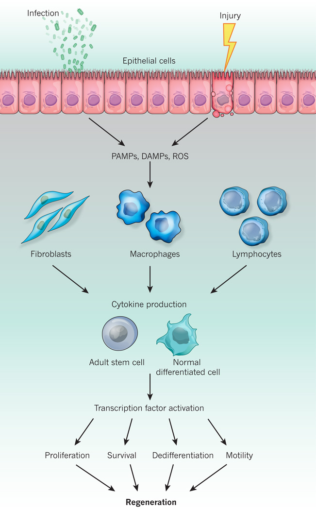
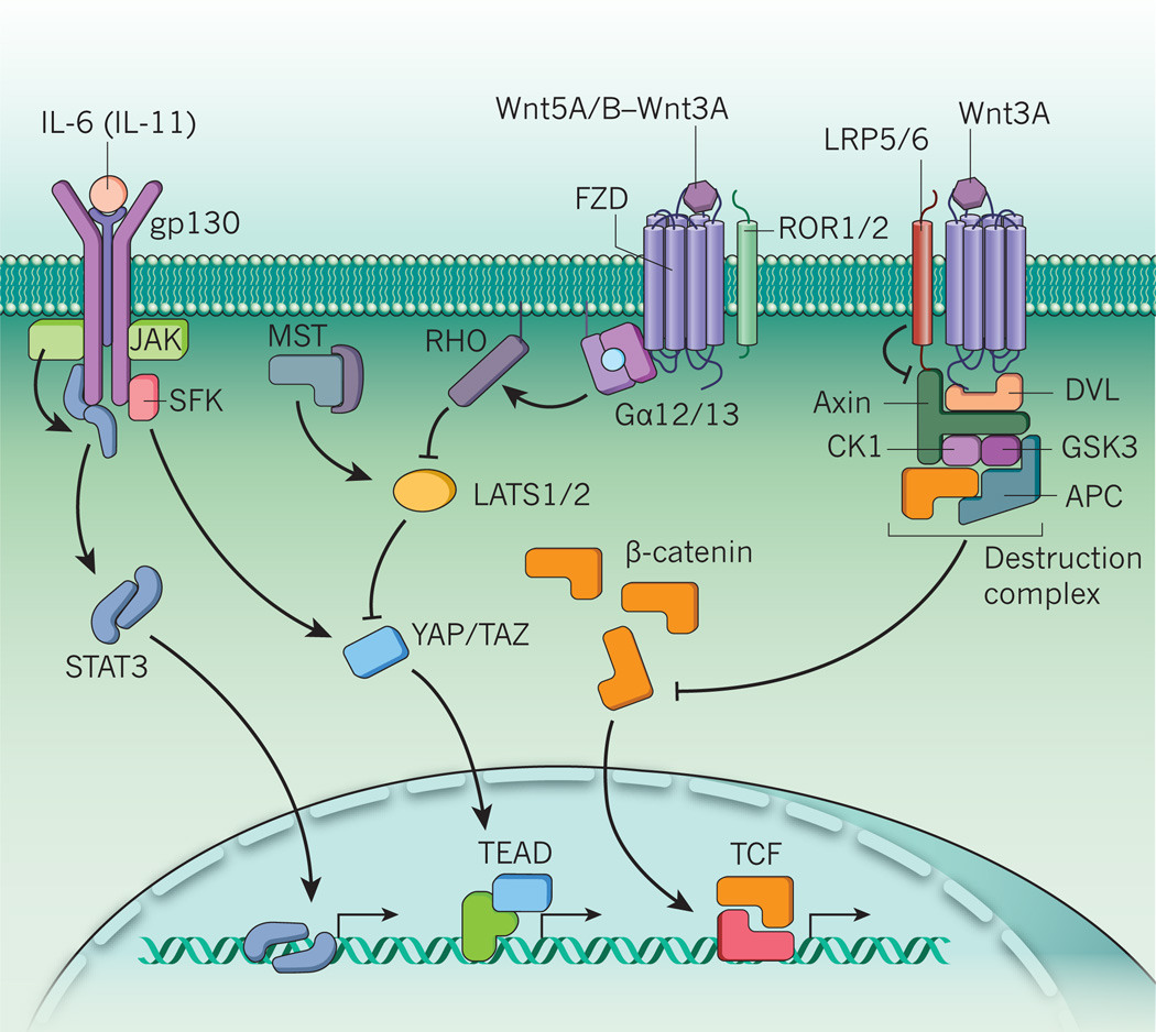
Inflammation underlies many chronic and degenerative diseases, but it also mitigates infections, clears damaged cells and initiates tissue repair. Many of the mechanisms that link inflammation to damage repair and regeneration in mammals are conserved in lower organisms, indicating that it is an evolutionarily important process. Recent insights have shed light on the cellular and molecular processes through which conventional inflammatory cytokines and Wnt factors control mammalian tissue repair and regeneration. This is particularly important for regeneration in the gastrointestinal system, especially for intestine and liver tissues in which aberrant and deregulated repair results in severe pathologies.


Figures

Figure 1
Figure 1. The microanatomy of the cellular compartments responsible for intestinal and liver regeneration
a, Intestinal stem cells (ISCs) are located at the bottom of crypts in the stem-cell zone. They sit wedged between Paneth cells, which provide niche factors. The stem-cell positions are denoted by +1 to +3 from the bottom of the crypt. The label-retaining cells at the +4 position can act as reserve stem cells when crypt damage occurs. The transit-amplifying zone above the stem-cell zone contains rapidly proliferating lineage-committed progenitors that mature as they move up the crypt–villus axis. On injury, ISCs expand and repair the mucosa to restore the gut permeability barrier. b, The liver is made up of roughly hexagonal functional units called lobules. Within these, cords of hepatocytes stretch from the central vein to the portal triad. Hepatocytes differ in function, depending on their position (pericentral to periportal). On the apical side, hepatocytes enclose canaliculi, bile channels that lead outwards to intrahepatic bile ducts. Along the way, bile passes through the canal of Hering, which is lined by hepatocytes and cholangiocytes. Basolaterally, hepatocytes face the perisinusoidal space. This transitional compartment between hepatocyte and sinusoid is delimited by discontinuous, fenestrated endothelial cells and contains hepatic stellate cells. Kupffer cells, a specialized population of liver macrophages, reside at the interface between the perisinusoidal space and sinusoid. Blood from the portal vein and the hepatic artery mixes in sinusoids and flows towards the central vein, transporting both oxygen from the lungs and nutrients from the digestive tract to the liver. Although liver stem cells have not been identified, pericentral diploid hepatocytes were suggested to be responsible for homeostatic hepatocyte renewal. Chronic liver injury, however, leads to expansion of periportal hybrid hepatocytes, which repair the injured liver parenchyma.
Figure 2
Figure 2. Mechanisms through which infection and injury induce a regenerative inflammatory response
Injury or infection of epithelial tissues leads to the generation of pathogen-associated molecular patterns (PAMPs), damage-associated molecular patterns (DAMPs) and reactive oxygen species (ROS). These induce the production of cytokines (tumour necrosis factor, interleukin (IL)-6, IL-11, IL-17 or IL-22) through tissue constituents such as immune cells. Some of the most common inflammatory cytokines trigger signalling pathways in adult stem cells or normal differentiated cells that culminate in the activation of transcription factors (AP-1, NF-κB, STAT3, YAP or Notch intracellular domain). These then mount a regenerative response by inducing genes that encode growth factors, stimulate cell-cycle progression, prevent cell death, promote dedifferentiation and the acquisition of ‘stemness’, and enhance cell motility and migration.
Figure 3
Figure 3. Signalling pathways in inflammation-driven regeneration
Cytokine signalling, the Wnt pathway (canonical and non-canonical) and Hippo-independent Src family kinase (SFK)-YAP signalling connects inflammatory signals to the regenerative response. Interleukin (IL)-6, and other members of the cytokine family, signal through receptors that use the gp130 subunit. STAT3 is activated by the tyrosine kinases JAK1 and JAK2 in response to gp130 or IL-22R signalling. In addition, cytokine signalling recruits SFK to gp130, which leads to phosphorylation, stabilization and nuclear translocation of YAP and its orthologue TAZ. Nuclear YAP and TAZ act as transcriptional co-activators of TEAD transcription factors. In non-stimulated epithelial cells, YAP and TAZ remain in the cytoplasm and undergo proteasomal degradation as a result of phosphorylation of inhibitory serine residues by LATS1/2 kinases. LATS1/2 is activated by Hippo/MST and inhibited by non-canonical Wnt signalling (FZD-ROR1/2 complex) owing to Gα12/13-dependent Rho activation. Canonical Wnt signalling (FZD-LPR5/6 complex), however, stabilizes β-catenin by inhibiting its continuous degradation by the destruction complex. Stabilized β-catenin can then translocate to the nucleus and stimulate TCF-mediated gene transcription.

Similar articles

Cited by

References

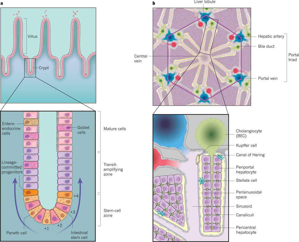
    1. Clevers H. The intestinal crypt, a prototype stem cell compartment. Cell. 2013;154:274–284. - PubMed
    1. Sato T, et al. Paneth cells constitute the niche for Lgr5 stem cells in intestinal crypts. Nature. 2011;469:415–418. This paper outlines how Paneth cells provide support for ISCs.

    1. Durand A, et al. Functional intestinal stem cells after Paneth cell ablation induced by the loss of transcription factor Math1 (Atoh1) Proc. Natl Acad. Sci. USA. 2012;109:8965–8970. - PMC - PubMed
    1. Yang J, et al. β-catenin signaling in murine liver zonation and regeneration: a Wnt-Wnt situation! Hepatology. 2014;60:964–976. - PMC - PubMed
    1. Wang B, Zhao L, Fish M, Logan CY, Nusse R. Self-renewing diploid Axin2+ cells fuel homeostatic renewal of the liver. Nature. 2015;524:180–185. This paper describes a population of diploid pericentral hepatocytes that may act as adult liver stem cells.

Publication types