Review
. 2016 Jan 21;529(7586):316-25.
doi: 10.1038/nature17040.

Angiocrine functions of organ-specific endothelial cells

Affiliations
Review

Angiocrine functions of organ-specific endothelial cells

Shahin Rafii et al. Nature. .

Abstract

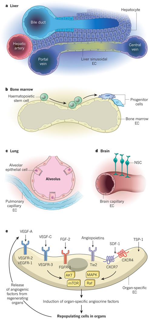
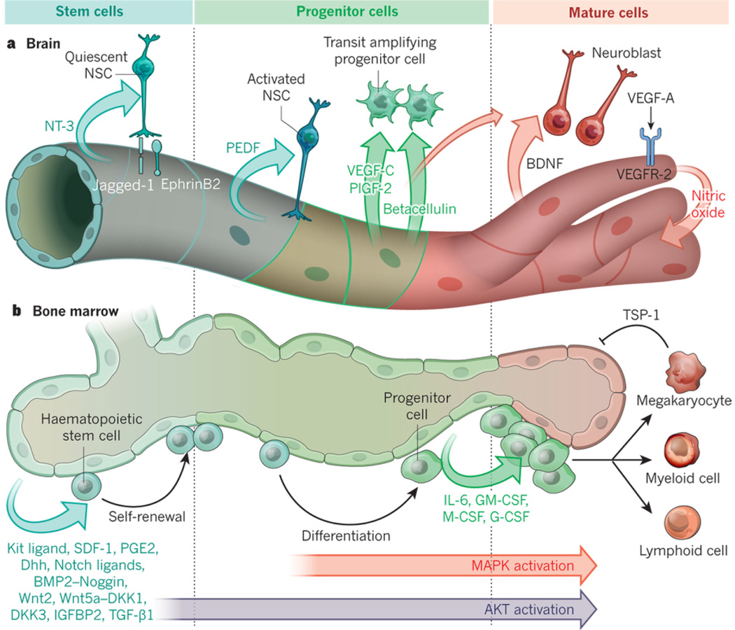
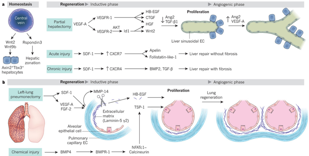
Endothelial cells that line capillaries are not just passive conduits for delivering blood. Tissue-specific endothelium establishes specialized vascular niches that deploy sets of growth factors, known as angiocrine factors. These cues participate actively in the induction, specification, patterning and guidance of organ regeneration, as well as in the maintainance of homeostasis and metabolism. When upregulated following injury, they orchestrate self-renewal and differentiation of tissue-specific resident stem and progenitor cells into functional organs. Uncovering the mechanisms by which organotypic endothelium distributes physiological levels of angiocrine factors both spatially and temporally will lay the foundation for clinical trials that promote organ repair without scarring.


Figures

Figure 1
Figure 1. Cross-talk of specialized capillary ECs with organ-specific parenchymal cells and their corresponding stem cells modulate homeostatic and regenerative processes
Each organ is arborized by an extensive network of specialized capillaries (A). Within each organ the capillaries assume unique structural, phenotypic, functional and angiocrine attributes. In the liver, hepatocytes are juxtaposed to fenestrated liver sinusoidal endothelial cells (LSECs) marked by the unique phenotype CD34-VEGFR1+VEGFR2+VEGFR3+VEcad+CXCR7+CD31low/-FactorVIII+ (B). In the hematopoietic organs such as bone marrow, the stem and progenitor cells are in direct cellular contact with arterial and fenestrated specialized sinusoidal vessels demarcated by VEGFR3+VEGFR2+VEcad+CD31+ ECs (C). In the lungs, the alveolar epithelial cells and their progenitors reside in the vicinity of continuous nonfenestrated pulmonary capillary endothelial cells (PCECs) defined by the signature VEGFR2+FGFR1+VEcad+CD31+ CD45 ECs (D). In the brain the majority of capillaries compose of tightly connected vessels with a common core phenotype of VEGFR2+VEcad+CD133+ thrombomodulin−/low ECs (E). At steady state conditions or during an angiogenic state upregulation of angiogenic factors, including VEGF-A through activation of its cognate tyrosine kinase receptors VEGFR2 and VEGFR1, FGF2 through FGFR1, angiopoietins through Tie2 not only modulate angiogenic processes, but also trigger or resolve the expression of tissue-specific angiocrine factors. Thrombospondins not only temper angiogenic response but also directly influence the proliferation and differentiation of the pancreatic islet and lung epithelial cells. Recruitment of pAkt-mTOR and MAPK/Raf signaling most likely play a role in choreographing the expression of the organotypic angiocrine factors (E).
Figure 2
Figure 2. Tissue-specific ECs by supplying membrane-bound and secreted angiocrine factors orchestrate self-renewal and regeneration of stem and progenitors cells
(A) Brain capillary ECs are strategically localized to the neural stem and progenitors cells. These ECs elaborate specific angiocrine factors, including membrane-bound Jagged-1 and EphrinB2 and NT-3 that establish the quiescence and survival of the Type-B1 quiescent NSCs (qNSCs) within the V-SVZ. ECs also deploy angiocrine factors, including PEDF, VEGF-C (mostly in SGZ) and PLGF2 that foster proliferation of Type-B1 activated NSCs (aNSCs). Angiocrine secretion of Betacellulin stimulates the amplification of Type-C progenitors (TAC). During angiogenesis, upregulation of VEGF-A through activation of VEGFR2 and induction of NO upregulates BDNF, which in conjunction with Betacellulin encourage the differentiation into neuroblast and mature neurons, leading to completion of neurogenesis. (B) Bone marrow arterial and sinusoidal ECs produce angiocrine factors that support the maintenance and regeneration of hematopoietic stem and progenitor cells (HSPCs) following myeloablative insult. At steady state conditions low levels of AKT activation in ECs stimulate production of Kit-ligand, Cxcl12, Notch-ligands, IGFBP2, Wnts, BMP2, Dhh and BMP4, which maintain and promote the self-renewal of hematopoietic stem cells. After myeloablative stress, inflicted by chemotherapy or irradiation, co-activation of AKT and MAPK initiates expression of progenitor active angiocrine factors, including IL6, GM-CSF, G-CSF, M-CSF, Metalloproteinases, chemokines and other factors forcing balanced differentiation of stem cells into lineage-committed progenitors. EC-derived Notch-ligands, (i.e. Jagged-1) prevent exhaustion of HSPCs. Induction of thrombospondin1 (TSP1) expression by maturing megakaryocytes puts a brake on ongoing hemangiogenesis finalizing the regeneration process.
Figure 3
Figure 3. Tissue-specific ECs by supplying membrane-bound and secreted angiocrine factors support regeneration of alveolar epithelial cells and hepatocytes
(A) At steady state Wnt2 and Wnt9b produced by liver central vein ECs sustains liver mass by replenishing the Axin2+Tbx3+ hepatic stem cell pools. After 70% partial hepatectomy (PH), VEGF-A via AKT activation induces Id 1 in LSECs upregulating HGF and Wnt2. VEGF-A through activation of VEGFR1 upregulates HGF, HB-EGF and CTGF. These angiocrine factors stimulate hepatocyte proliferation without provoking angiogenesis, (inductive phase). Four days after PH, increase in liver initiates proliferative angiogenesis. Upon PH downregulation of Ang2 in LSECs and TGF-β accelerates hepatic recovery. During resolution phase of liver regeneration (days 4–8 post-PH), VEGF-A and restoration of Ang2 stimulates angiogenesis and finalize hepatic reconstitution. Activation of CXCR7 on LSECs triggers pro-regenerative and anti-fibrotic angiocrine factors, including Apelin, follistatin-1-like that facilitates fibrosis-free healing. Chronic injury by persistent CXCR4 activation and CXCR7 suppression stimulates TGF-β and BMP4 leading to fibrosis. Balance of CXCR4 and CXCR7 in LSECs negotiates liver regeneration and fibrosis. (B) After removal of mice left lung (pneumonectomy, PNX), the PCECs in right lung express membrane-bound MMP14, which unmasks the cryptic EGF-receptor ligands from HB-EGF and Laminin5 γ-2. This inductive phase orchestrates the angiogenesis-independent compensatory alveolar epithelial regeneration. After chemical injury (i.e. bleomycin), BMP4 through engagement of its receptor Bmpr1 sets up NFATc1/Calcineurin-dependent transcription of TSP1. TSP1 facilitates differentiation of lung epithelial progenitors into functional epithelial cells. After PNX, upregulation of the VEGF-A, FGF2 and deposition of platelets on PCECs by production of SDF1 and CXCR4 activation induces MMP14 initiating alveolar regeneration. Increase in lung mass triggers angiogenic phase of lung regeneration.

References

    1. Carmeliet P, Jain RK. Molecular mechanisms and clinical applications of angiogenesis. Nature. 2011;473:298–307. - PMC - PubMed
    1. Ghesquiere B, Wong BW, Kuchnio A, Carmeliet P. Metabolism of stromal and immune cells in health and disease. Nature. 2014;511:167–176. - PubMed
    1. Butler JM, Kobayashi H, Rafii S. Instructive role of the vascular niche in promoting tumour growth and tissue repair by angiocrine factors. Nat Rev Cancer. 2010;10:138–146. - PMC - PubMed
    1. Nolan DJ, et al. Molecular signatures of tissue-specific microvascular endothelial cell heterogeneity in organ maintenance and regeneration. Dev Cell. 2013;26:204–219. In this article the authors developed an intra-vital labeling approach to purify non-lymphatic mouse organ-specific ECs and by employing molecular profiling demonstrated the remarkable angiocrine heterogeneity in ECs among various tissues

    1. Matsumoto K, Yoshitomi H, Rossant J, Zaret KS. Liver organogenesis promoted by endothelial cells prior to vascular function. Science. 2001;294:559–563. - PubMed

Publication types