Review
. 2016:129:109-74.
doi: 10.1016/bs.ai.2015.09.002. Epub 2015 Oct 26.

Forging T-Lymphocyte Identity: Intersecting Networks of Transcriptional Control

Affiliations
Review

Forging T-Lymphocyte Identity: Intersecting Networks of Transcriptional Control

Ellen V Rothenberg et al. Adv Immunol. 2016.

Abstract

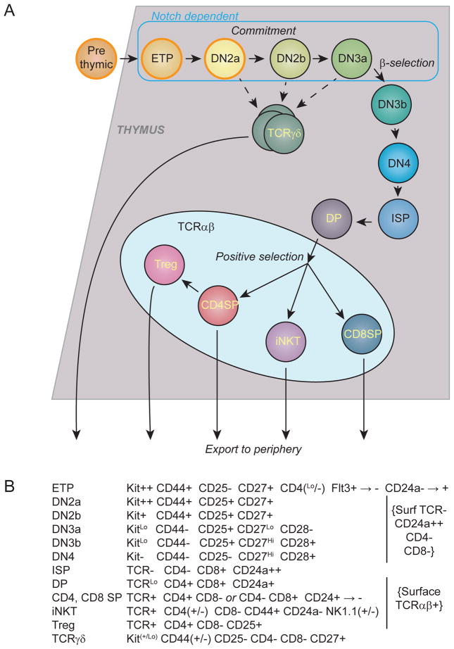
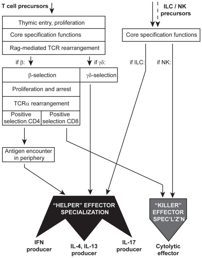
T-lymphocyte development branches off from other lymphoid developmental programs through its requirement for sustained environmental signals through the Notch pathway. In the thymus, Notch signaling induces a succession of T-lineage regulatory factors that collectively create the T-cell identity through distinct steps. This process involves both the staged activation of T-cell identity genes and the staged repression of progenitor-cell-inherited regulatory genes once their roles in self-renewal and population expansion are no longer needed. With the recent characterization of innate lymphoid cells (ILCs) that share transcriptional regulation programs extensively with T-cell subsets, T-cell identity can increasingly be seen as defined in modular terms, as the processes selecting and actuating effector function are potentially detachable from the processes generating and selecting clonally unique T-cell receptor structures. The developmental pathways of different classes of T cells and ILCs are distinguished by the numbers of prerequisites of gene rearrangement, selection, and antigen contact before the cells gain access to nearly common regulatory mechanisms for choosing effector function. Here, the major classes of transcription factors that interact with Notch signals during T-lineage specification are discussed in terms of their roles in these programs, the evidence for their spectra of target genes at different stages, and their cross-regulatory and cooperative actions with each other. Specific topics include Notch modulation of PU.1 and GATA-3, PU.1-Notch competition, the relationship between PU.1 and GATA-3, and the roles of E proteins, Bcl11b, and GATA-3 in guiding acquisition of T-cell identity while avoiding redirection to an ILC fate.

Keywords: E2A; GATA-3; Gene regulation; Notch; PU.1; T-cell development; Transcription factor.


Figures

Figure 1
Figure 1. Stages of intrathymic T cell development
A) Scheme of stages of mouse T-cell development discussed in the text, showing the stages affected by Notch-DLL4 signaling, intrathymic branchpoints in development, and the timing of three watershed events: commitment, β-selection, and positive selection. B) Markers used to define the stages shown in A.
Figure 2
Figure 2. Modularity of the T-cell developmental program: differential access to effector specialization functions depending on choice of T-cell or innate-cell lineage
The figure depicts a surprisingly common set of regulatory programming used to distinguish “killer” and different “helper” subtypes of effector T cells and innate lymphoid cells (ILC and natural killers, NK), and the layers of developmental programming that cells must undergo before gaining access to these common programs. Note that TCRαβ lineage T cells have a highly protracted, multistep pathway requirement before they can access the specialization functions, in contrast to ILC and NK cells. As described in the text, TCRγδ cells appear to be intermediate between the extreme of the longer path taken by CD4+ αβ T cells and the apparently short paths taken by ILCs.
Figure 3
Figure 3. Trajectories of gene regulation in T-cell precursors through commitment and β-selection
Data for (A) are RNA-seq analyses taken from (Zhang et al., 2012b), and data for (B, C) are highly curated microarray analyses taken from www.immgen.org (Mingueneau et al., 2013). A) Patterns of expression of T-cell identity genes and signaling components through the transition from ETP to DP. Data are presented on a log2 scale, with increases and decreases in expression plotted as changes relative to the geometric mean of values for each gene. Thus, genes with stable expression at high or low levels have little fold change. Precommitment stages are indicated by blue bars, postcommitment stages by yellow to red bars. Note that T-cell identity and signaling competence genes are drastically upregulated during commitment, while cytokine receptor genes are more diversely regulated. B) Notch target genes have developmentally distinct patterns of expression. Absolute microarray hybridization intensities at the indicated stages are presented on a log2 scale. All the genes shown are sharply affected by interruptions or increases in Notch signaling intensity in early T cells except the Notch signal-transducing regulatory gene Rbpj, which is shown to indicate the stability of the Notch response machinery. C) Little developmental change in expression of known Notch signal modulating genes despite dynamic target gene expression. The indicated modifiers are plotted as for the samples in (B), but the scale is expanded for greater sensitivity to change, with Rbpj shown again for reference. Note that only Lfng is highly regulated across these stages.
Figure 4
Figure 4. Dynamic regulatory stages in developing T-cell precursors
A) Diagram representing the major groups of regulatory factors at the stages when they are deployed in early T-cell development. The period dominated by Notch signaling is shown by shading. All the factors shown are transcription factors or cofactors except RAG1/2 (recombinases), Ptcra (TCRα surrogate chain used in β-selection), and Dtx1 (Notch pathway target gene). “Phase 1” regulators are the stem/progenitor-associated factors that are highly expressed in thymic immigrants but then silenced during commitment and immediately afterwards. “RUNX” refers to the constant presence of members of the Runx family, although Runx2 and Runx3 are more highly expressed in the ETP stages and Runx1 is more highly expressed at the DN3a stage. B) Phase 1 regulators have distinct patterns of usage in stem/progenitor cells and in other classes of lymphocytes. Patterns of expression are shown for two cytokine receptor genes (Kit and Flt3) and 22 phase 1 regulatory factor genes, using the ImmGen database (Heng et al., 2008; Mingueneau et al., 2013; Robinette et al., 2015) and interactive heatmap tool for “MyGeneSet” (http://rstats.immgen.org/MyGeneSet/)(July, 2015). Red and orange cells represent highly expressed genes in the indicated cells relative to their average, blue represents very low levels of expression relative to their average. Note that all these genes have their expression in T cells confined to the left end samples in the “T Cells” series, which correspond to the stages shown in Fig. 3B, C. However, they are all expressed in addition in at least some stem and progenitor cells (“Stem & Prog’n’r Cells”) and distinct groups are also shared with early B cells, B cells generally, and/or NK and ILC cells.
Figure 5
Figure 5. GATA-3 and TCF-1 DNA binding sites across the genome in different cellular contexts: higher developmental stability in sites of co-occupancy
The figure summarizes ChIP-seq data from EML-c1 progenitor cells (A), DP T-lineage cells (A–C), and ETP (DN1) pro-T cells (B–C). The Venn diagrams show the extent of overlap between the sequences recovered as binding sites for the two factors, or the same factors in different contexts. Data for GATA-3 in ETP and DP cells were taken from (Zhang et al., 2012b), data for TCF-1 in EML-c1 cells were from (Wu et al., 2012), and data for TCF-1 in DP cells were from (Dose et al., 2014). Peak calling of mm9 aligned sequences was performed with the HOMER package (Heinz et al., 2010) and filtered for a peak score ≥ 15. Peaks were identified using findPeaks.pl with the –style factor parameter and normalized to sequencing inputs; overlapping peaks were identified and Venn-diagram parameters were retrieved using mergePeaks.pl (default parameters with the Venn-diagram option). A) TCF-1 overlapping and non-overlapping ChIP-seq peaks in EML-C1 progenitor cells (Gene Expression Omnibus accession number GSE31221) and DP thymocytes (GSE46662). B) GATA-3 overlapping and non-overlapping ChIP-seq peaks in ETP (DN1) T-cell precursors and DP thymocytes (GSE31235). The poor overlap is also noted with a different graphical presentation in (Zhang et al., 2012b). C) Overlap between Gata3 sites in DP cells shared with TCF-1 bound sites in DP cells and the Gata3 sites in ETP cells shared with TCF-1 bound sites in DP cells. Note that these sites are far more likely to overlap than total GATA-3 sites. D) Evidence that occupancy involves recurrent partner factors: enrichment of motifs for GATA-3, TCF-1 [TCF (HMG)], and additional factors at GATA-3 and TCF-1 binding sites in DP cells. HOMER was used for de novo motif analysis within a 200 bp window of the sites of GATA-3 and/or TCF-1 occupancy in DP cells. Bold type identifies the most common co-enriched motifs, present at >15% of occupancy sites. Note the recurrent co-enrichment of ETS and Runx family motifs.
Figure 6
Figure 6. GATA-3 and PU.1 relationship in early T cells: not a simple bistable antagonism
A) Two diagrams contrasting the classic view of GATA/PU.1 interactions in hematopoiesis (left) with the relationship actually seen in early T-cell precursors (right). Note that the actual relationship is missing the positive autoregulation loops for GATA-3 and PU.1 and the categorical antagonism at the protein level. Instead, Notch signaling enables an asymmetric relationship between GATA-3 and PU.1 at the transcriptional level, and effects of PU.1 and GATA-3 on each other’s activities are specific for particular target genes, a small subset of their total functional targets. B) Genes subject to regulation by both PU.1 and GATA-3 in early T-cell precursors, shown with the direction of the effects of PU.1 and GATA-3 inferred from perturbation experiments in DN2 cells developed in vitro from fetal liver precursors (Champhekar et al., 2015; Scripture-Adams et al., 2014). Note that all combinations of effects are seen. The patterns of expression of these genes in stem/progenitor cells, T cells, and other lymphocytes, mostly from adult animals (Heng et al., 2008; Mingueneau et al., 2013; Robinette et al., 2015), are also shown as in Fig. 4B to indicate the diversity of developmental regulation patterns involved, not simply correlated with expression of PU.1 and GATA-3 themselves.

Similar articles

Cited by

References

    1. Albu DI, Feng D, Bhattacharya D, Jenkins NA, Copeland NG, Liu P, Avram D. BCL11B is required for positive selection and survival of double-positive thymocytes. J Exp Med. 2007;204:3003–3015. - PMC - PubMed
    1. Albu DI, VanValkenburgh J, Morin N, Califano D, Jenkins NA, Copeland NG, Liu P, Avram D. Transcription factor Bcl11b controls selection of invariant natural killer T-cells by regulating glycolipid presentation in double-positive thymocytes. Proc Natl Acad Sci U S A. 2011;108:6211–6216. - PMC - PubMed
    1. Alonzo ES, Sant’Angelo DB. Development of PLZF-expressing innate T cells. Curr Opin Immunol. 2011;23:220–227. - PMC - PubMed
    1. Anderson MK, Hernandez-Hoyos G, Diamond RA, Rothenberg EV. Precise developmental regulation of Ets family transcription factors during specification and commitment to the T cell lineage. Development. 1999;126:3131–3148. - PubMed
    1. Anderson MK, Weiss AH, Hernandez-Hoyos G, Dionne CJ, Rothenberg EV. Constitutive expression of PU.1 in fetal hematopoietic progenitors blocks T cell development at the pro-T cell stage. Immunity. 2002;16:285–296. - PubMed

Publication types

MeSH terms