. 2016 Jan 21:7:10473.
doi: 10.1038/ncomms10473.

The novel tumour suppressor Madm regulates stem cell competition in the Drosophila testis

Affiliations

The novel tumour suppressor Madm regulates stem cell competition in the Drosophila testis

Shree Ram Singh et al. Nat Commun. .

Abstract

Stem cell competition has emerged as a mechanism for selecting fit stem cells/progenitors and controlling tumourigenesis. However, little is known about the underlying molecular mechanism. Here we identify Mlf1-adaptor molecule (Madm), a novel tumour suppressor that regulates the competition between germline stem cells (GSCs) and somatic cyst stem cells (CySCs) for niche occupancy. Madm knockdown results in overexpression of the EGF receptor ligand vein (vn), which further activates EGF receptor signalling and integrin expression non-cell autonomously in CySCs to promote their overproliferation and ability to outcompete GSCs for niche occupancy. Conversely, expressing a constitutively activated form of the Drosophila JAK kinase (hop(Tum-l)) promotes Madm nuclear translocation, and suppresses vn and integrin expression in CySCs that allows GSCs to outcompete CySCs for niche occupancy and promotes GSC tumour formation. Tumour suppressor-mediated stem cell competition presented here could be a mechanism of tumour initiation in mammals.


Figures
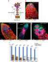
Figure 1
Figure 1. Madm functions in CySC to regulate GSC maintenance.
(a) A schematic diagram of Drosophila adult testis. GSCs and GBs (gonialblasts) contain the spherical spectrosomes (green dots), while spermatogonia and spermatocytes contain branched fusomes (green lines). GSCs are encysted by two CySCs (magenta). GBs and other germ cells (red) are encapsulated by cyst cells (lavender). (be) The confocal section of the testis apex from C587ts>control (b), and C587ts>MadmRNAi−1 (BL31644; ce) males after shifting the temperature from 18° to 29 °C for 1 to 15 days as adult flies. In control testis (b, 5 days at 29 °C, n=41) contain a rosette of GSCs around the hub (white dotted lines with one GSC is highlighted by an arrow), and CySCs are located away from the hub (yellow arrowhead). In C587ts>MadmRNAi testes (c, 1 day, n=40; (d,e), 3 days, n=39), the number of GSCs are gradually lost from the niche and differentiated (highlighted by white dotted lines in d,e), and CySCs moved into the niche (highlighted by yellow dotted lines in d). The testes were stained with antibodies against Vasa (red, marks all germ cells including GSCs), 1B1 (green, marks spherical spectrosomes and branched fusomes), Arm (green, hub cells at the tip). DAPI (blue) stains nuclei. Asterisks indicate hub cells. White arrows near hub indicate GSC. Green arrows (in d,e) and orange arrow (d,e) away from hub with white dotted lines indicate spermatogonial cells and spermatocytes, respectively. (f) A bar graph showing the number of GSCs per testis in C587ts>control, C587ts>MadmRNAi−1 (BL31644) and C587ts>MadmRNAi−2 (v27346) flies. Flies were cultured from 1 to 15 days at 29 °C before staining with antibodies against Vasa, 1B1 and Arm. Statistical significance determined by Student's t-test, ***P<0.0001. All values are mean±s.e.m. Scale bar, 10 μm (be).
Figure 2
Figure 2. Madm functions in CySCs to prevent CySCs from outcompeting GSCs for niche occupancy.
(ac) A confocal section of the testis apex containing C587ts>control (a) and C587ts>MadmRNAi (BL31644) (b, 2 days, n=25; c, 5 days, n=30) males after shifting the temperature from 18o to 29 °C for 2, 5 and 7 days as adult flies. The testes were stained with antibodies against Vasa (green, marks all germ cells including GSCs), Zfh-1 (red, marks CySCs and their immediate cyst cell daughters). DAPI (blue) stains nuclei. (d) A bar graph showing the number of Zfh-1+ cells per testis in C587ts>control, and C587ts>MadmRNAi−1 (BL31644) and C587ts>MadmRNAi−2 (v27346) flies at 2 and 7 days at 29 °C. Statistical significance determined by Student's t-test, ***P<0.0001; NS indicates not significant (P>0.05). All values are mean±s.e.m. (eg) The testes of C587ts>control (e) were stained with Zfh-1 (red) and Madm (green), highlighted in inset a co-stained of Zfh-1 and Madm. The testes of C587ts>control (f) and C587ts>MadmRNAi−1 (g) were stained with Madm (red), 1B1 and Arm (green), DAPI (blue). *indicate hub cells. GSCs are highlighted by white arrows and white dotted lines near hub cells in a and b, and by white arrows in e and f. Differentiated germ cells (away from the hub) in c are highlighted by white dotted line (green arrow). CySCs are highlighted by arrowhead (yellow) in a,b,e,f. Yellow dotted line in c highlighted Zfh-1-positive cells. Scale bar, 10 μm (ac, eg).
Figure 3
Figure 3. Madm functions in CySC non-cell autonomously and regulates the competition between GSCs and CySCs.
(a,b) Confocal sections through the apex of the testes containing FRT82B-Madm7L2 clones at 3 days ACI (a, n=45) and 12 days ACI (b, n=52). The testes were stained with the Zfh-1 (red), β-galactosidase (green), and DAPI (blue). The FRT82B-Madm7L2 CySC clones are β-galactosidase (green) negative and Zfh-1 (red) positive. β-galactosidase negative CySCs clones are highlighted by yellow arrowhead (a,b). (ch) GFP+ve clones were generated in the testes of wild-type control (FRT82B-PiM; c,d, n=56) or FRT82B-Madm3G5 (eh, n=62) flies using the MARCM technique, and were stained at 7 days ACI with GFP (green), Zfh-1 (red) and DAPI (blue). GFP-positive CySCs clones are highlighted by black arrowheads (ch). GFP-negative CySCs clones are highlighted by yellow arrowheads (eh). Green arrow in e indicates differentiated germ cells. Asterisks with orange dotted lines indicate hub cells. GSCs are highlighted by white arrows and white dotted lines near hub cells. Scale bar, 10 μm (ah).
Figure 4
Figure 4. Madm regulates integrin and p-dERK expression non-cell autonomously.
(a,b) GFP+ve clones were generated in the testes of wild-type control (FRT82B-PiM (a, n=27) and FRT82B-Madm3G5 (b, n=32) clones at 3 days ACI. βPS-integrin expression is markedly increased in Madm3G5 testes at the CySCs–hub interface compared with wild-type control testes. The testes were immunostained with βPS-integrin (red), GFP (green) and DAPI (blue). GSCs are highlighted by white arrows. GFP-positive clones are highlighted by arrowhead (yellow, a,b). (c) A bar graph showing the quantitation of fluorescence intensity of βPS-integrin in FRT82B-PiM and FRT82B-Madm3G5 testes at the CySC–hub interface. (d,e) GFP-positive clones were generated in the testes of wild-type control (FRT82B-PiM (d, n=35) and FRT82B-Madm3G5 (e, n=37) clones at 3 days ACI. p-dERK expression is markedly increased in FRT82B-Madm3G5 testes in both GFP-positive cells and GFP−ve cells compared with wild-type control testes. The testes were immunostained with p-dERK (red), GFP (green) and DAPI (blue). GFP-positive clones are highlighted by arrowhead (black, d,e) and GFP-positive cells are highlighted by arrowhead (yellow, d,e). GSCs are highlighted by white arrows. (f) A bar graph showing the quantitation of fluorescence intensity of p-dERK in FRT82B-PiM and FRT82B-Madm3G5 testes. Asterisks with orange dotted lines indicate hub cells. All values are mean±s.e.m. Statistical significance determined by Student's t-test, ***P<0.0001. Scale bar, 10 μm (a,b,d,e).
Figure 5
Figure 5. The Ras, rather than the JAK (Hop), regulates the competition between CySCs and GSCs for niche occupancy.
(ad) Confocal sections of the testes containing C587ts>Control (a, n=26), C587ts>hopTuml (b, n=25), C587ts>RasV12 (c, n=20), and C587ts>hopTuml/RasV12 (d, n=27). The testes were cultured for 6 days at 29 °C before they were immunostained with Zfh-1 (red), Vasa (green) and DAPI (blue). GSCs are highlighted by white arrows and white dotted lines near hub cells (ad). CySCs are highlighted by arrowhead (yellow, ad). (ek) GFP+ve clones were generated in the testes of FRT82B-PiM (e,f), FRT82B-UAS-RasV12 (g,h; n=45) and FRT82B-UAS-hopTuml (i,j; n=37) flies using the MARCM technique and were stained at 6 days ACI with antibodies against GFP (green) and βPS-integrin (red). DAPI (blue) stained nuclei. In FRT82B-UAS-RasV12 testes integrin is highly elevated in GFP+ve CySC clones (g,h); however, in FRT82B-UAS-hopTuml testes, integrin is weakly expressed (i,j). GSCs are highlighted by white arrows. (k) A bar graph showing the quantitation of fluorescence intensity of βPS-integrin in FRT82B-PiM, FRT82B-UAS-RasV12 and FRT82B-UAS-hopTuml testes. *indicate hub cells. All values are mean±s.e.m. Statistical significance determined by Student's t-test, ***P<0.0001. Scale bar, 10 μm (aj).
Figure 6
Figure 6. JAK signaling regulates the nuclear translocation of Madm, and Madm negatively regulates the EGFR/Ras/ERK signaling pathway by repressing vn expression.
(ad) The testes of C587ts> Control (6 days, a,b; n=15), C587ts>hopTuml (6 days; c,d; n=32) were immunostained with the Madm (red), Lamin (red) and DAPI (blue). *indicate hub cells (a,c). GSC is highlighted by white arrows and CySCs are highlighted by yellow arrowhead (a,c). (e) The testes of C587ts>hopTuml/MadmRNAi (6 days, n=25). Testes were cultured for 7 days at 29 °C before they were immunostained with Zfh-1 (red), Vasa (green) and DAPI (blue). (f,g) The testes of FRT82B-Madm3G5/vn-lacZ (n=33) were immunostained with β-galactosidase (vn-lacZ-red), GFP (green) and DAPI (blue). vn expression is markedly increased in GFP-positive Madm mutant CySCs (white dotted lines) compared with GFP-negative CySCs (yellow dotted lines). (h) A bar graph showing the quantitation of fluorescence intensity of vn-LacZ in FRT82B-Madm mutant clones. (ik). Confocal sections of the testes containing C587ts>vn (i (70%) and j (30%); n=32); and C587ts>MadmRNAi/vnRNAi (k, n=30). The testes were cultured for 7 days at 29 °C before they were immunostained with Zfh-1 (red), Vasa (green) and DAPI (blue). (l) The testes of FRT82B-Madm7L2/vnRNAi (n=22) were immunostained 4 days ACI with GFP (green, yellow arrowhead), Fas3 (green, hub cells), Vasa (red) and DAPI (blue). vn knockdown in GFP-marked MARCM CySC clones of Madm mutant significantly (Student's t-test, P<0.0001) suppress the phenotypes associated with Madm knockdown. Green arrows indicate differentiated germ cells (e,i,j). Yellow arrowhead indicate CySCs (e,il). GSC are highlighted by white arrows (white dotted lines, k,l). Yellow dotted lines indicate Zfh-1-positive cells (e,i,j). *indicate hub cells. All values are mean±s.e.m. Statistical significance determined by Student's t-test, ***P<0.0001. Scale bar, 10 μm (ah).
Figure 7
Figure 7. The overexpression of integrin rescued the GSC tumour phenotypes associated with the loss of EGFR-ERK signaling and overexpression of Zfh-1.
(ad) GSCs in the testes of C587ts>EgfrDN (a, n=22), C587ts>EgrfDN/PS1βPS (integrins) (b, n=32), C587ts>rlRNAi(c, n=30) and C587ts>rlRNAi/PS1βPS (integrins) (d, n=35). The testes were cultured for 7 days at 29 °C before they were immunostained with Vasa (red), 1B1 and Arm (green) and DAPI (blue). *indicate hub cells. GSCs are highlighted by white arrows (b,d). CySCs are highlighted by arrowhead (yellow, b,d). White dotted lines in a and c representing GSC tumour phenotype. (e) A model of how Madm regulates GSC and CySC competition in Drosophila testis. Details are described in the text. Scale bar, 10 μm (ad).

Similar articles

Cited by

References

    1. Bondar T. & Medzhitov R. p53-mediated hematopoietic stem and progenitor cell competition. Cell Stem Cell 6, 309–322 (2010) . - PMC - PubMed
    1. Issigonis M. et al. JAK-STAT signal inhibition regulates competition in the Drosophila testis stem cell niche. Science 326, 153–156 (2009) . - PMC - PubMed
    1. Jin Z. et al. Differentiation-defective stem cells outcompete normal stem cells for niche occupancy in the Drosophila ovary. Cell Stem Cell 2, 39–49 (2008) . - PMC - PubMed
    1. Martins V. C. et al. Cell competition is a tumour suppressor mechanism in the thymus. Nature 509, 465–470 (2014) . - PubMed
    1. Rhiner C. et al. Persistent competition among stem cells and their daughters in the Drosophila ovary germline niche. Development 136, 995–1006 (2009) . - PubMed

Publication types

Substances