. 2016 Apr 1;25(7):1328-44.
doi: 10.1093/hmg/ddw016. Epub 2016 Jan 19.

The ubiquitin ligase Ubr4 controls stability of podocin/MEC-2 supercomplexes

Affiliations

The ubiquitin ligase Ubr4 controls stability of podocin/MEC-2 supercomplexes

Markus M Rinschen et al. Hum Mol Genet. .

Abstract

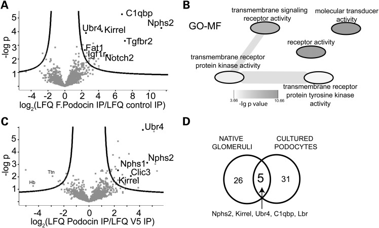
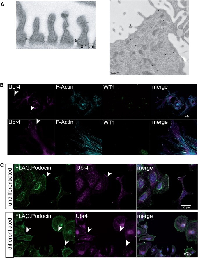
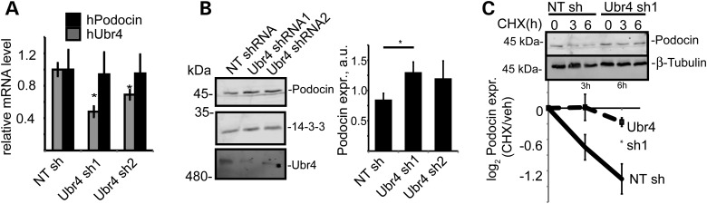
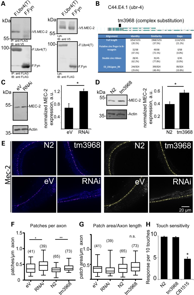
The PHB-domain protein podocin maintains the renal filtration barrier and its mutation is an important cause of hereditary nephrotic syndrome. Podocin and its Caenorhabditis elegans orthologue MEC-2 have emerged as key components of mechanosensitive membrane protein signalling complexes. Whereas podocin resides at a specialized cell junction at the podocyte slit diaphragm, MEC-2 is found in neurons required for touch sensitivity. Here, we show that the ubiquitin ligase Ubr4 is a key component of the podocin interactome purified both from cultured podocytes and native glomeruli. It colocalizes with podocin and regulates its stability. In C. elegans, this process is conserved. Here, Ubr4 is responsible for the degradation of mislocalized MEC-2 multimers. Ubiquitylomic analysis of mouse glomeruli revealed that podocin is ubiquitylated at two lysine residues. These sites were Ubr4-dependent and were conserved across species. Molecular dynamics simulations revealed that ubiquitylation of one site, K301, do not only target podocin/MEC-2 for proteasomal degradation, but may also affect stability and disassembly of the multimeric complex. We suggest that Ubr4 is a key regulator of podocyte foot process proteostasis.


Figures

Figure 1.
Figure 1.
Discovery of the podocin interactome and Ubr4 as a key interactor of podocin. (A) Analysis of the interactome of podocin expressed in podocytes. Podocytes were lysed, and FLAG-tagged podocin was pulled down using anti-FLAG antibody. The pull-down was analysed by MS/MS, and compared with a control pull-down. The negative decadic logarithm of the P-value (−logP) of the LFQ intensities is plotted against the log2-fold change between podocin pull-down and control pull-down. The proteins beyond the line are significant interactors (FDR = 0.2, s0 = 1). Ubr4 is a significant interactor. (B) Overrepresented GO-MF terms in the interacting proteins when compared with non-interacting proteins. Overrepresentation was analysed using a Fisher exact test (FDR < 0.05). GO-MF analysis of the podocin interactome revealed a strong association with signalling molecules. (C) Analysis of the podocin interactome purified from native glomeruli. Glomeruli from one mouse were lysed, and podocin was purified using an anti-podocin antibody. The pull-down was analysed by MS/MS and compared with a control (anti-V5) pull-down. The negative decadic logarithm of the P-value (−log P) of the LFQ intensities is plotted against the log2-fold change between podocin pull-down and control pull-down. Ubr4 was identified as a significant interactor. (D) Venn diagram comparing of significant interactors from glomeruli and cultured podocytes. Ubr4 and the known interactor Neph1 (Kirrel) are core components of the podocin interactome.
Figure 2.
Figure 2.
Ubr4 localizes to the slit diaphragm and leading edges of podocytes. (A) Immunogold labelling of Ubr4 in native mouse glomeruli. There was positive immunogold signal for Ubr4 at the slit diaphragm spanning between two cell processes. The right picture demonstrates a podocyte cell body with positive immunogold labelling in cytoplasma vesicles, multi-vesicular bodies and ER. (B) Immunostaining of Ubr4 in a glomerular outgrowth culture. The Ubr4 signal was detected at leading edges of podocytes. (C) Immunostaining of Ubr4 and F.Podocin in cultured mouse podocytes revealed an overlap in both undifferentiated (33°C) and differentiated (37°C) podocytes. Nuclei were counterstained with DAPI. An overlap is marked by white staining in the merged channel. Scale bars indicate 20 µm.
Figure 3.
Figure 3.
The effect of Ubr4 knock-down on cultured human podocytes expressing podocin. (A) mRNA expression of Ubr4 and podocin was measured using quantitative RT-PCR. A non-targeting (NT) shRNA served as control. n = 5 replicates, P < 0.05. (B) Protein expression of podocin was increased in Ubr4 knock-down cells. Whole-cell lysates were analysed using immunoblotting for podocin. N = 5 replicates, P < 0.01 in a two-tailed t-test. (C) Pulse chase assay with CHX incubation. Podocytes were incubated using CHX (c = 100 ng/ml) for the indicated time periods. Whole-cell protein lysates were analysed using immunoblotting and membranes were probed with an anti-podocin antibody. Stability of podocin was increased in Ubr4 knock-down cells. n = 5 replicates. Asterisk indicates significance (P < 0.05) in a two-tailed t-test. All error bars in this figure represent SEM.
Figure 4.
Figure 4.
The effect of mutation or knock-down of the Ubr4 C. elegans orthologue, C44E4.1 (ubr-4), on the C. elegans podocin orthologue, MEC-2. (A) An C-terminal truncation of Ubr4 interacts with MEC-2. FLAG-tagged Ubr4 truncation (containing the last 800 amino acids of human Ubr4), and V5-tagged MEC-2 were transfected in HEK293T cells. FLAG-tagged Fyn served as a control protein. Proteins were immunoprecipitated using an anti-FLAG pull-down lysates were separated using SDS–PAGE. Blots were probed with V5 and FLAG antibody. (B) Identification of the Ubr4 orthologue in C. elegans as C44E4.1, now termed ubr-4. A strain carrying a mutated allele (tm3968) was utilized for further studies. (C) The effect of knock-down of ubr-4 (RNAi) on MEC-2 abundance in nematode when compared with control RNAi [empty vector (eV)]. MEC-2 abundance was measured using immunoblotting and densitometry (n = 5 and P < 0.01 in a two-tailed t-test). (D) The effect of ubr-4 mutation (tm3968) on MEC-2 abundance when compared with wild-type (N2) worms. MEC-2 abundance was measured using immunoblotting (n = 5 and P < 0.01 in a two-tailed t-test). (E) Immunostaining of MEC-2 in ubr-4 mutant and knock-down worms. The left panel shows MEC-2-positive punctae in C. elegans heads. The right panel demonstrates thresholding analysis to quantify MEC-2-positive punctae. (F, G) Quantification of patch number and patch size. The number of quantified worm images (three independent grownups) is given in brackets. (H) Touch response of worms lacking ubr-4. Reaction of worms in response to 10 gentle touches was measured. No effect on touch sensitivity was found. The mutant CB1515 (Mec-10) served as a touch-hyposensitive control (14).
Figure 5.
Figure 5.
Ubiquitylomic analysis of native glomeruli. (A) Cartoon of Neph1 (Kirrel), Actn4 and Synpo proteins. Among > 600 ubiquitylation sites, ubiquitylation sites on Neph1, Actn4 and Synpo were discovered. The numbers indicate the lysine residues. (B) Proteomic discovery of the podocin ubiquitylation sites K301 and K370 (asterisk) in native glomeruli. The cartoon depicts membrane model of podocin. The podocin ubiquitylation site K301 localizes at the distal part of the PHB domain (spanning residues 125–301 according to PFAM). The distal part of the PHB domain is significantly enriched for ubiquitylation sites based on mass spectrometric evidence (***P = 0.0006).
Figure 6.
Figure 6.
Ubr4 ubiquitylates podocin at K301 to unfold the protein. (A) Analysis of the ubiquitylation of expressed podocin in the presence (black columns) and absence (white columns) of overexpressed Ubr4 by proteomics (n = 5 experiments). Cells were incubated with MG132 (10 µm, 1 h). Podocin was purified using an anti-FLAG antibody, and immunoprecipitates were analysed using LC-MS/MS. Ubiquitylation sites of podocin were identified as K-ε-Gly-Gly ubiquitin remnant peptides. MS1 intensities of site containing ubiquitylation sites were normalized against total protein intensity (based on quantification of 47 unique peptides). The majority of all ubiquitylation sites reside within the PHB domain. Error bars indicate SEM. (B) The model of the podocin PHB domain with an attached monoubiquitylation chain at the residue K301. K301 corresponds to K142. The PHB domain (grey) was modelled using the crystal structure template PDB 3bk6, the ubiquitin residue (cyan) was based on PDB 1ubq. (C) Melting curve of podocin ubiquitylation in the presence or absence of a K48-linked ubiquitin chain (4U) as determined by replica exchange molecular dynamics simulation. (D) Melting point determination of mono- and polyubiquitylated podocin by second derivative measurements. The melting points of each ubiquitylated proteoform are presented for the monoubiquitylated (1U spline, closed circles) and the polyubiquitylated (4U spline, open triangles) form of each proteoform. The residues K241, K261 and K301 (when attached to a mono- or polyubiquitylated residue) unfold the protein more easily when compared with ubiquitylation at other sites. See Supplementary Material, Figs. S8–S12 for further details of molecular dynamics simulation.
Figure 7.
Figure 7.
The Ubr4-dependent residues regulate podocin stability. (A) Measurement of podocin protein stability when expressed in HEK293T cells. Cells were incubated with CHX (c = 100 ng/ml) for the indicated periods. Densitometry quantification of WT and mutant (K301R and K370R) podocin are depicted as logarithmic ratios (n = 5; P < 0.01 in a two-tailed t-test). (B) Measurement of podocin protein stably expressed in podocyte cell lines. Podocytes were incubated with CHX for the indicated periods of time ratios (n = 5; P < 0.01 in a two-tailed t-test). Podocin expression was analysed using immunoblotting for FLAG. (C) Pulse labelling of podocytes with medium containing heavy isotope labelled amino acids. Podocin WT and mutant (K301R/K370R) was purified using an anti-FLAG antibody. Incorporation of heavy isotope labelled amino acids into podocin WT and mutant was measured. Each symbol represents a separate replicate of SILAC ratio quantification per protein. P = 0.05 in a two-tailed t-test. The following 10 peptides were analysed across the experiment: AASESLR, APAATATVVDVDEVR, ARPDAGAER, DMFIMEIDAVCYYR, LGHLLPGR, LPAGLQHSLAVEAEAQR, MAAEILSGTPAAVQLR, SLTEILLER, VALDAVTCIWGIK and VVQEYER. (D) Localization of FLAG-tagged podocin and podocin mutant forms in HeLa cells. Podocin and podocin mutants were transfected in HeLa cells. Podocin was stained using an anti-FLAG antibody. Nuclei were counterstained with DAPI. Podocin with K301R and K301R–K370R substitution colocalized with cotransfected Lamp1, a lysosome marker. Altered podocin localization was independent of co-transfection of Lamp1 (data not shown).

References

    1. Boute N., Gribouval O., Roselli S., Benessy F., Lee H., Fuchshuber A., Dahan K., Gubler M.C., Niaudet P., Antignac C. (2000) NPHS2, encoding the glomerular protein podocin, is mutated in autosomal recessive steroid-resistant nephrotic syndrome. Nat. Genet., 24, 349–354. - PubMed
    1. Tory K., Menyhárd D.K., Woerner S., Nevo F., Gribouval O., Kerti A., Stráner P., Arrondel C., Huynh Cong E., Tulassay T. et al. (2014) Mutation-dependent recessive inheritance of NPHS2-associated steroid-resistant nephrotic syndrome. Nat. Genet., 46, 299–304. - PubMed
    1. Pavenstädt H., Kriz W., Kretzler M. (2003) Cell biology of the glomerular podocyte. Physiol. Rev., 83, 253–307. - PubMed
    1. Huber T.B., Schermer B., Benzing T. (2007) Podocin organizes ion channel-lipid supercomplexes: implications for mechanosensation at the slit diaphragm. Nephron Exp. Nephrol., 106, e27–e31. - PubMed
    1. Huber T.B., Schermer B., Müller R.U., Höhne M., Bartram M., Calixto A., Hagmann H., Reinhardt C., Koos F., Kunzelmann K. et al. (2006) Podocin and MEC-2 bind cholesterol to regulate the activity of associated ion channels. Proc. Natl Acad. Sci. USA, 103, 17079–17086. - PMC - PubMed

Publication types

MeSH terms