

Association of Shiny White Blotches and Strands With Nonpigmented Basal Cell Carcinoma: Evaluation of an Additional Dermoscopic Diagnostic Criterion
- PMID: 26792406
- PMCID: PMC5037958
- DOI: 10.1001/jamadermatol.2015.5731
Association of Shiny White Blotches and Strands With Nonpigmented Basal Cell Carcinoma: Evaluation of an Additional Dermoscopic Diagnostic Criterion
Abstract
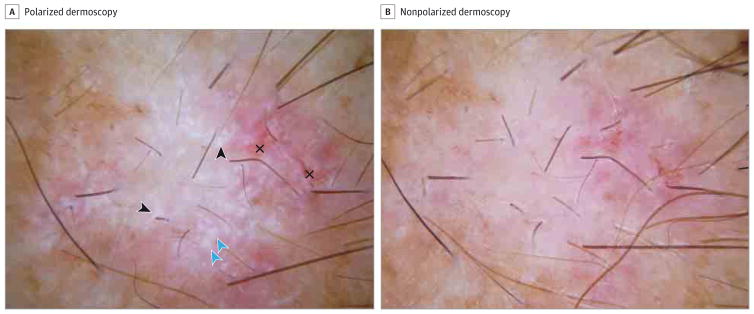
Importance: Basal cell carcinoma (BCC) is the most common type of skin cancer and is usually nonpigmented. Shiny white structures (SWSs) are frequently present in BCC.
Objective: To determine the diagnostic accuracy of various morphologies of SWSs for diagnosis of nonpigmented BCC.
Design, setting, and participants: Nonpigmented skin tumors, determined clinically and dermoscopically, were identified from a database of lesions consecutively biopsied over a 3-year period (January 2, 2009, to December 31, 2012) from a single dermatology practice. Data analysis was conducted from October 9, 2014, to November 15, 2015. Investigators blinded to histopathologic diagnosis evaluated the polarized dermoscopic images for the presence of SWSs, which were categorized as blotches, strands, short white lines, and rosettes. Measures of diagnostic accuracy for BCC were estimated. Participants included 2375 patients from a dermatologic clinic in Plantation, Florida. Review of the medical records identified 2891 biopsied skin lesions; 457 of these were nonpigmented neoplasms.
Main outcomes and measures: Diagnosis of BCC with dermoscopy compared with all other diagnoses combined was the primary outcome; the secondary outcome was diagnosis of BCC compared with amelanotic melanoma. We calculated diagnostic accuracy measured as odds ratios (ORs), sensitivity, and specificity of shiny white blotches and/or strands for the diagnosis of BCC.
Results: Of the 457 nonpigmented neoplasms evaluated, 287 (62.8%) were BCCs, 106 (23.2%) were squamous cell carcinoma, 39 (8.5%) were lichen planus-like keratosis, 21 (4.6%) were melanomas, and 4 (0.9%) were nevi. The prevalence of SWSs was 49.0% (n = 224). In multivariate analysis (reported as OR [95% CI]) controlling for age, sex, and anatomical location, the presence of any SWS was associated with a diagnosis of BCC (2.3 [1.5-3.6]; P < .001). Blotches (6.3 [3.6-10.9]; P < .001), strands (4.9 [2.9-8.4]; P < .001), and blotches and strands together (6.1 [3.3-11.3]; P < .001) were positively associated with BCC. Shiny white blotches and strands together had a diagnostic sensitivity of 30% and specificity of 91%.
Conclusions and relevance: The combined presence of shiny white blotches and strands is associated with high diagnostic specificity for nonpigmented BCC.
Conflict of interest statement
Disclosures: Dr Rabinovitz reported receiving financial compensation with equipment as a speaker and for testing dermatoscopes for 3-Gen, Canfield, and Heine. No other disclosures were reported.
Figures
References
-
- Lomas A, Leonardi-Bee J, Bath-Hextall F. A systematic review of worldwide incidence of nonmelanoma skin cancer. Br J Dermatol. 2012;166(5):1069–1080. - PubMed
-
- Chinem VP, Miot HA. Epidemiology of basal cell carcinoma. An Bras Dermatol. 2011;86(2):292–305. - PubMed
-
- Koh D, Wang H, Lee J, Chia KS, Lee HP, Goh CL. Basal cell carcinoma, squamous cell carcinoma and melanoma of the skin: analysis of the Singapore Cancer Registry data 1968–97. Br J Dermatol. 2003;148(6):1161–1166. - PubMed
Publication types
MeSH terms
Grants and funding
LinkOut - more resources
Full Text Sources
Other Literature Sources
Medical
