. 2016 Jul;41(8):2112-21.
doi: 10.1038/npp.2016.11. Epub 2016 Jan 21.

Neurofunctional Reward Processing Changes in Cocaine Dependence During Recovery

Affiliations

Neurofunctional Reward Processing Changes in Cocaine Dependence During Recovery

Iris M Balodis et al. Neuropsychopharmacology. 2016 Jul.

Abstract

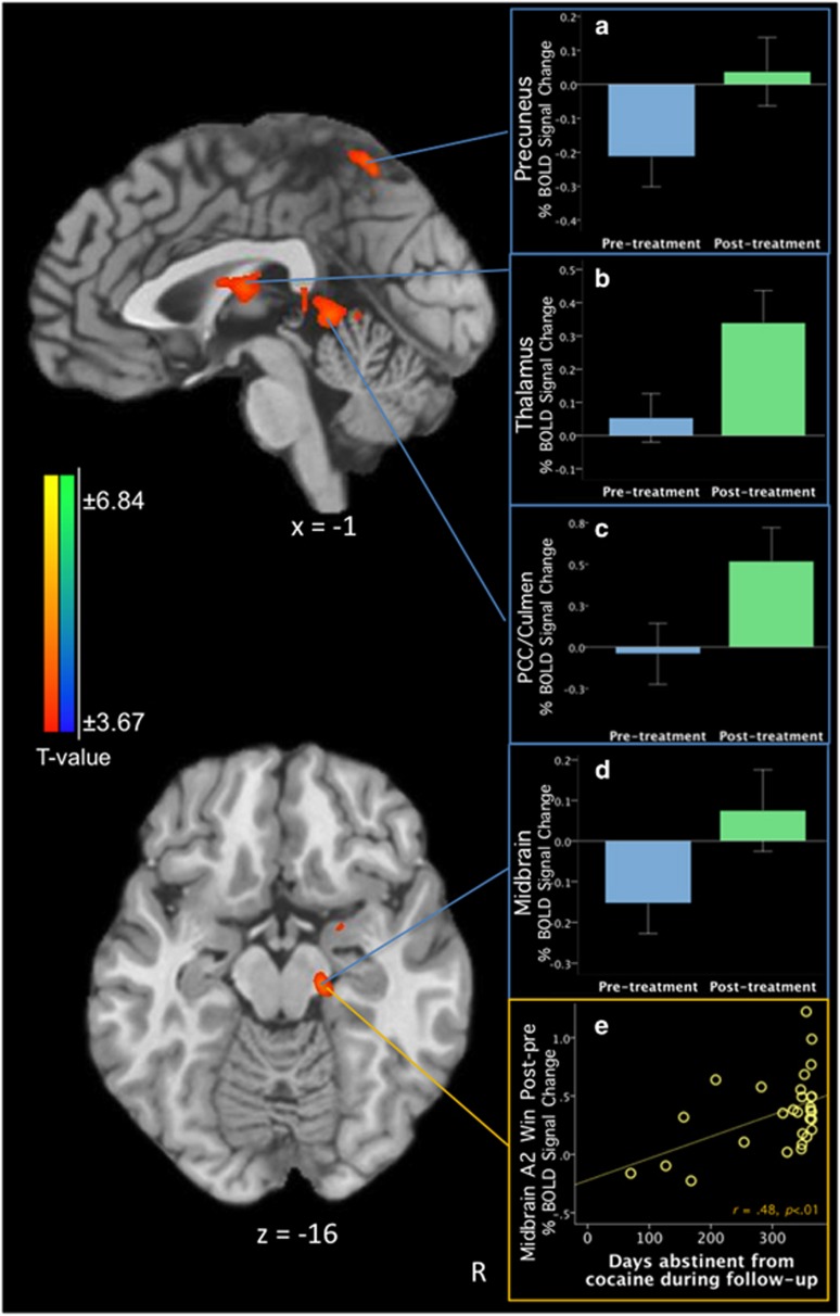
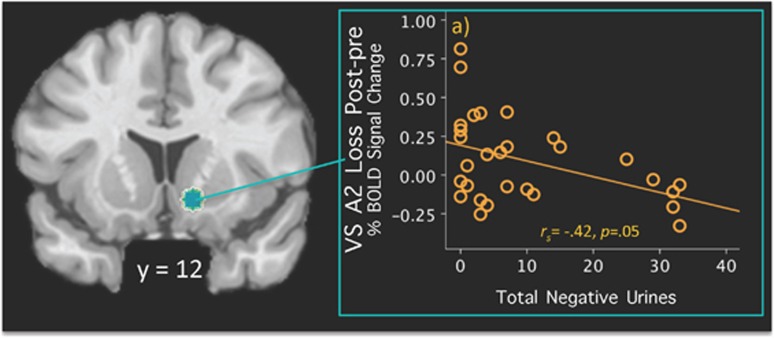
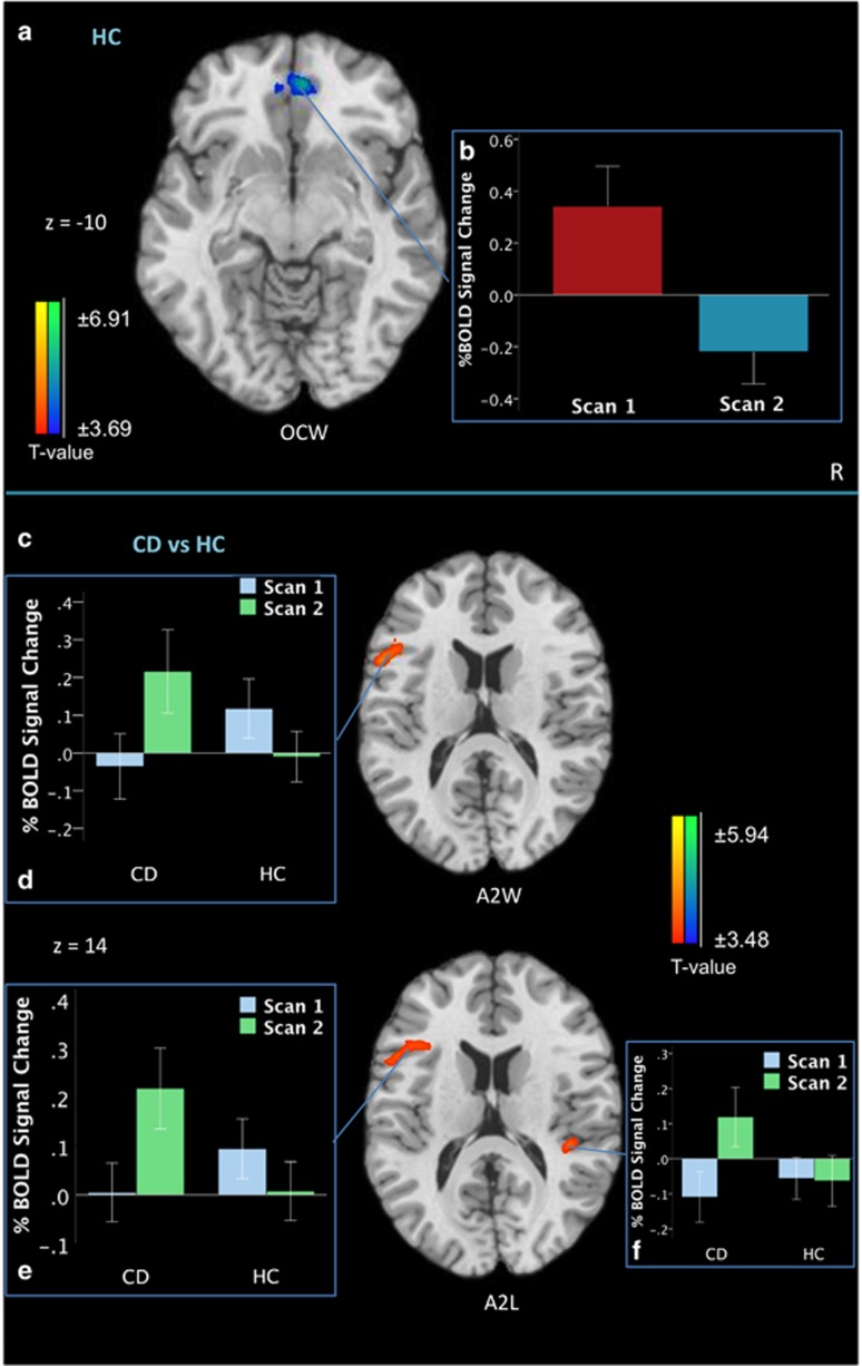
Although reward processing appears altered in addiction, few studies track neurofunctional changes following treatment or relate these to measures of reduced drug use. The current study examined neurofunctional alterations in reward processing in cocaine dependence (CD) pretreatment and posttreatment to determine whether these changes relate to clinically meaningful outcome indicators. Treatment-seeking CD outpatients (N=29) underwent functional magnetic resonance imaging while performing a monetary incentive delay task (MIDT) pretreatment and posttreatment. The MIDT parses anticipatory from outcome phases of reward/loss processing. Abstinence indicators (negative urines, days abstinent from cocaine during follow-up) were collected throughout treatment and up to 1 year later. Healthy control (HC) participants (N=28) were also scanned twice with the MIDT. Relative to pretreatment, at posttreatment CD participants demonstrated increased anticipatory reward activity in the midbrain, thalamus, and precuneus (pFWE<0.05). Increased midbrain activity correlated with cocaine abstinence during the 1-year follow-up. Ventral striatal (VS) activity during loss anticipation correlated negatively with negative urine screens. HC group test-retest results showed decreased ventromedial prefrontal cortex activity during winning outcomes. CD-HC group-by-time differences revealed increased left inferior frontal gyrus activity in the CD group during anticipatory phases at posttreatment. In CD participants, increased posttreatment activity in dopamine-innervated regions suggests lowered thresholds in anticipatory signaling for non-drug rewards. Midbrain and VS responses may represent biomarkers associated with CD abstinence. Abstinence-related neurobiological changes occur in similar regions implicated during active use and may possibly be used to track progress during short- and long-term recovery.


Figures

Figure 1
Figure 1
Posttreatment–pretreatment differences on the Monetary Incentive Delay Task in the cocaine-dependent (CD) group (n=29) during the A2 winning phase (A2W, associated with the anticipation of potentially winning money). (a) The percentage of blood-oxygen-level-dependent (BOLD) signal change in the precuneus cluster during pretreatment and posttreatment; (b) BOLD signal change in the thalamus cluster during pretreatment and posttreatment; (c) BOLD signal change in the posterior cingulate (PCC)/culmen cluster during pretreatment and posttreatment; (d) BOLD signal change in the midbrain cluster during pretreatment and posttreatment; (e) scatterplot demonstrating a positive correlation between the midbrain A2W cluster difference and days abstinent from cocaine during follow-up (r=0.48, p<0.01). All contrast maps are thresholded at an uncorrected level of p<0.001 two-tailed and family-wise-error-corrected at p<0.05. Blue color demonstrates areas where subjects show relatively less activation at posttreatment vs pretreatment, and red color indicates where participants show relatively greater activation at posttreatment vs pretreatment. The right side of the brain is on the right. A full color version of this figure is available at the Neuropsychopharmacology journal online.
Figure 2
Figure 2
Coronal view of the ventral striatal Region of Interest (ROI) using coordinates reported by Patel et al (2013). A blue spot indicates a 5-mm sphere around the ventral striatum (VS) on the right (12, 12, −9) side. Scatterplots depict the percentage of blood-oxygen-level-dependent (BOLD) signal change extracted from the 5-mm ROI during the A2L Loss phase (posttreatment>pretreatment) correlated with (a) the total negative urines during treatment period (rs=−0.42, p=0.05). A full color version of this figure is available at the Neuropsychopharmacology journal online.
Figure 3
Figure 3
Scan2–Scan1 differences on the Monetary Incentive Delay Task in (a) the HC group (n=28) during the outcome winning phase (OCW, associated with the receipt of reward); (b) depicts the percentage of blood-oxygen-level-dependent (BOLD) signal change in the ventromedial prefrontal cortex (vmPFC; z=−10) in the HC group for their first and second scans. (c) Scan2–Scan1 contrast between the CD group (n=29) and the HC group (n=28); (d) depicts the percentage of blood-oxygen-level-dependent (BOLD) signal change in the left inferior frontal gyrus (z=14) in the CD and HC groups for their first and second scans during the A2 winning phase (A2W, associated with the anticipation of potentially winning money); (e) depicts the percentage of blood-oxygen-level-dependent (BOLD) signal change in the left inferior frontal gyrus (z=14) in the CD and HC groups for their first and second scans during the A2 losing phase (A2L, associated with the anticipation of potentially losing money); (e) depicts the percentage of blood-oxygen-level-dependent (BOLD) signal change in the right superior temporal gyrus (z=14) in the CD and HC groups for their first and second scans during the A2 losing phase. All contrast maps are thresholded at an uncorrected level of p<0.001 two-tailed and family-wise-error-corrected at p<0.05. Blue color demonstrates areas where subjects show relatively less activation in the indicated contrast map, and red color indicates where participants show relatively greater activation. The right side of the brain is on the right. A full color version of this figure is available at the Neuropsychopharmacology journal online.

Similar articles

Cited by

References

    1. Asensio S, Romero MJ, Romero FJ, Wong C, Alia-Klein N, Tomasi D et al (2010). Striatal dopamine D2 receptor availability predicts the thalamic and medial prefrontal responses to reward in cocaine abusers three years later. Synapse 64: 397–402. - PMC - PubMed
    1. Balodis IM, Potenza MN (2015). Anticipatory reward processing in addicted populations: a focus on the monetary incentive delay task. Biol Psychiatry 77: 434–444. - PMC - PubMed
    1. Bari A, Robbins TW (2013). Inhibition and impulsivity: behavioral and neural basis of response control. Prog Neurobiol 108: 44–79. - PubMed
    1. Boileau I, Dagher A, Leyton M, Welfeld K, Booij L, Diksic M et al (2007). Conditioned dopamine release in humans: a positron emission tomography [11C]raclopride study with amphetamine. J Neurosci 27: 3998–4003. - PMC - PubMed
    1. Bustamante JC, Barros-Loscertales A, Costumero V, Fuentes-Claramonte P, Rosell-Negre P, Ventura-Campos N et al (2013). Abstinence duration modulates striatal functioning during monetary reward processing in cocaine patients. Addict Biol. 19: 885–894. - PubMed

Publication types