. 2017 Feb;16(1):45-56.
doi: 10.1177/1533034615626011. Epub 2016 Jul 8.

Advanced Small Animal Conformal Radiation Therapy Device

Affiliations

Advanced Small Animal Conformal Radiation Therapy Device

Sunil Sharma et al. Technol Cancer Res Treat. 2017 Feb.

Abstract

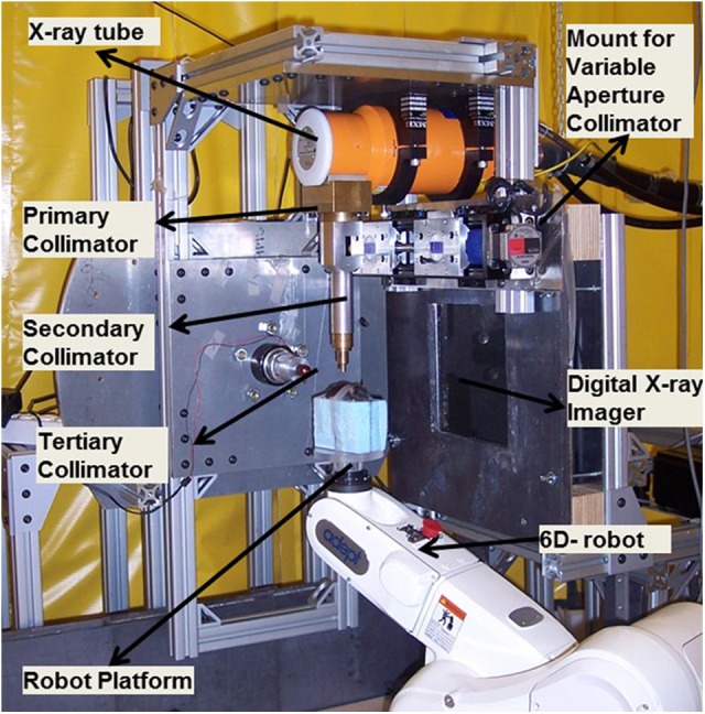
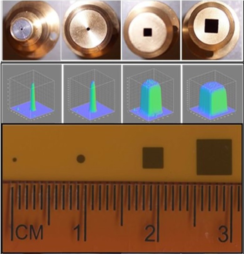
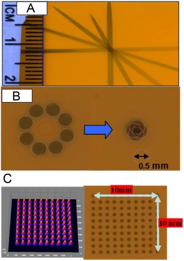
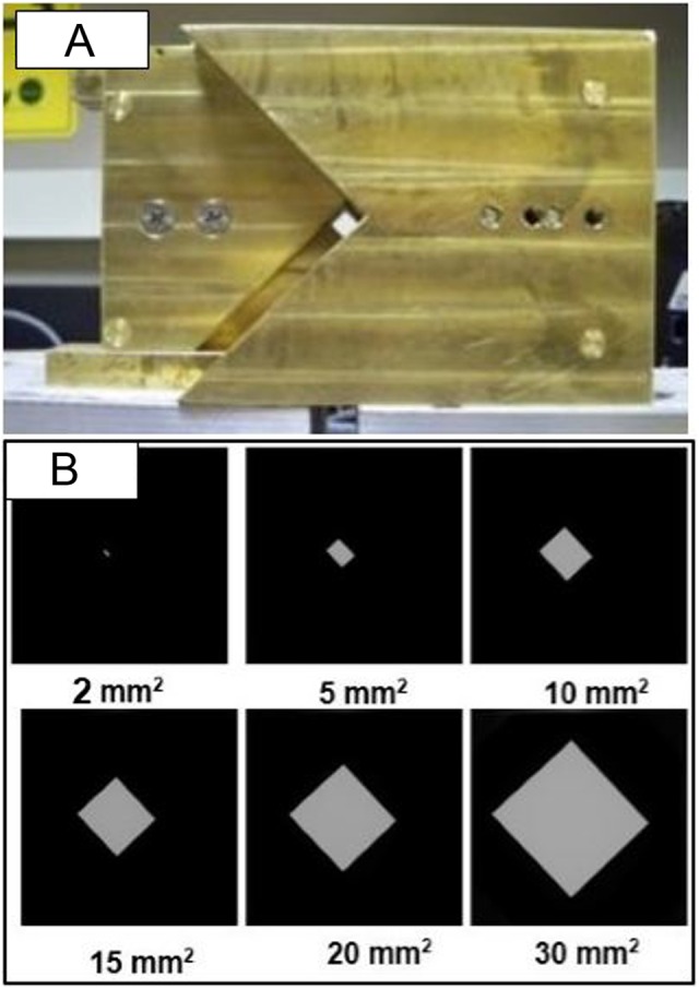
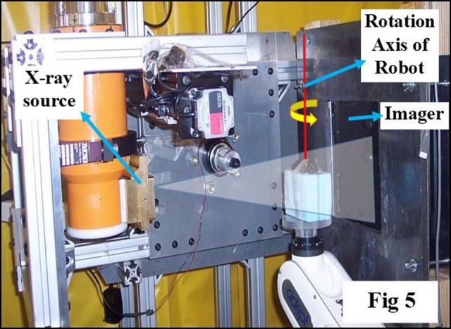
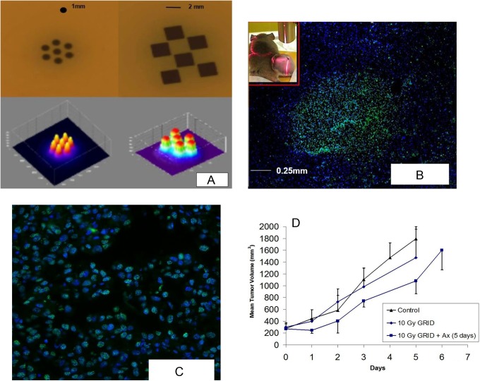
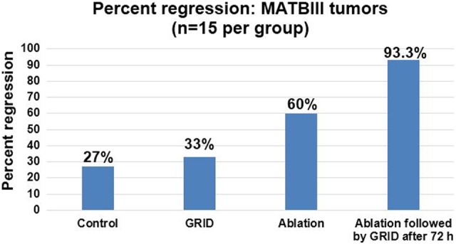
We have developed a small animal conformal radiation therapy device that provides a degree of geometrical/anatomical targeting comparable to what is achievable in a commercial animal irradiator. small animal conformal radiation therapy device is capable of producing precise and accurate conformal delivery of radiation to target as well as for imaging small animals. The small animal conformal radiation therapy device uses an X-ray tube, a robotic animal position system, and a digital imager. The system is in a steel enclosure with adequate lead shielding following National Council on Radiation Protection and Measurements 49 guidelines and verified with Geiger-Mueller survey meter. The X-ray source is calibrated following AAPM TG-61 specifications and mounted at 101.6 cm from the floor, which is a primary barrier. The X-ray tube is mounted on a custom-made "gantry" and has a special collimating assembly system that allows field size between 0.5 mm and 20 cm at isocenter. Three-dimensional imaging can be performed to aid target localization using the same X-ray source at custom settings and an in-house reconstruction software. The small animal conformal radiation therapy device thus provides an excellent integrated system to promote translational research in radiation oncology in an academic laboratory. The purpose of this article is to review shielding and dosimetric measurement and highlight a few successful studies that have been performed to date with our system. In addition, an example of new data from an in vivo rat model of breast cancer is presented in which spatially fractionated radiation alone and in combination with thermal ablation was applied and the therapeutic benefit examined.

Keywords: 3D conformal radiotherapy; GRID therapy; animal CBCT imaging; small animal irradiator; spatially fractionated radiation therapy.


Conflict of interest statement

Declaration of Conflicting Interests: The author(s) declared no potential conflicts of interest with respect to the research, authorship, and/or publication of this article.

Figures

Figure 1.
Figure 1.
Small animal conformal radiation therapy device (SACRTD) setup for conformal radiation therapy delivery.
Figure 2.
Figure 2.
The cone tips, beam profile, and film exposure for 0.5 mm and 1 mm diameter and 2 × 2 mm2 and 4 × 4 mm2 collimators at isocenter.
Figure 3.
Figure 3.
A, Gantry star shot image when the X-ray tube is rotated from the vertical to the horizontal position using 1-mm collimator. B, Image illustrates the 0.5-mm-diameter X-ray beam alignment with respect to the robot rotation axis. The beam intersects within ±0.20 mm at isocenter. C, Image shows microbeam square grid pattern from 0.5-mm-diameter collimator at isocenter (100 kV, 6 mA with 1 minute exposure) measured using Gafchromic film placed at 1 cm below isocenter. For 10 × 10 mm2 spatially fractionated radiation therapy (GRID), the rows are separated by 1 mm. The surface plot of relative optical density distribution of corresponding grid patterns is also shown.
Figure 4.
Figure 4.
A, Variable apertures collimation system mountable at the open end of the X-ray tube, which can produce field size up to 20 × 20 mm2 at isocenter. B, Images of various field sizes ranging from 2 × 2 mm2 to 30 × 30 mm2 recorded at digital imager.
Figure 5.
Figure 5.
Picture illustrating setup for cone-beam computed tomography (CBCT) imaging with horizontally oriented X-ray source, object immobilized on the robotic arm, and imaging detector at a fixed distance from the source. A software tool controls the robot motion, the X-ray activation, and data acquisition by the imager.
Figure 6.
Figure 6.
A, The calibration curve for dose delivered on the Gafchromic EBT-2 film exposed to clinical 6 MV X-ray beam using cubic polynomial fit, with error bar representing uncertainty in film measurement. Inset shows the similar plot for 4-mm collimator. B, Dose rate variation with depth in a solid water setup at 32.5 cm source to surface distance (SSD) for 0.5 and 1 mm diameter and 2 × 2 mm2 and 4 × 4 mm2 collimators. C, Relative beam profiles at 1 cm depth in a solid water setup at 32.5 cm SSD for 0.5 and 1 mm diameter and 2 × 2 mm2 and 4 × 4 mm2 collimators (225 kVp, 13 mA photon beam). D, Cross-beam profiles at depths of 0, 18.6, and 37.2 mm for a 0.5-mm-diameter collimator. E, Normalized beam profiles for 2 × 2 mm2 collimator at depths of 0, 18.6, and 37.2 mm.
Figure 7.
Figure 7.
A, Hexagonal spatially fractionated radiation therapy (GRID) pattern with 7 beams of 1 mm diameter separated by 2-mm center-to-center and a checkerboard GRID pattern with 6 squares of 2 × 2 mm2 collimators separated by 2.5 mm center-to-center. Exit beam pattern measured with Gafchromic XR-RV2 film by programming the robot to move the beam assembly in a step-and-shoot fashion. The corresponding surface plot is shown. B, Overlay image (×4) of BI6 murine melanoma stained with γ-H2AX (Bethyl Laboratories) to identify radiation-induced DNA strand breaks counterstained with 46′-diamidino-2-phenylindole-2 HCl (DAPI) staining for nuclear identification. The sharp bright region shows γ-H2AX staining, and the size of the bright region is approximately ∼1 mm diameter consistent with the diameter of the 1 mm beam used for irradiation. Insert, Mice with implanted tumor on the leg. C, Shows ×20 magnification of Figure 7B, showing γ-H2AX nuclear foci consistent with radiation-induced DNA breaks. D, Plot shows the growth of B16 tumors in rear limb of C57 mice after 10 Gy GRID alone or followed by 5 days of treatment with the antiangiogenic peptide anginex at 20 mg/kg intraperitoneally (IP). Each group contained 3 mice, and bars indicate 1 standard error of the mean volume per group.
Figure 8.
Figure 8.
Percentage regression in rat MAT B III orthotropic breast cancer in the 4 groups (n = 15 in each group).
Figure 9.
Figure 9.
The cone-beam computed tomography (CBCT) reconstructed slices of sagittal, coronal, and axial images of rat. Three hundred sixty projection images were cropped and downsampled to 512 × 512 pixels at 400 μm resolution. The resulting reconstructed matrix was 309 × 154 × 76 voxels with uniform spacing of 0.3 mm in the X, Y, and Z directions.
Figure 10.
Figure 10.
A, Cone-beam computed tomography (CBCT) reconstructed images of a tomotherapy resolution phantom from 360 projection images of 301 × 301 pixels at 200 μm resolution. The reconstructed matrix size was 141 × 141 × 199 voxels with uniform spacing of 0.3 mm in the X, Y, and Z directions. B, The microcomputed tomography (CT) of same phantom is also shown for comparison.
Figure 11.
Figure 11.
Immunohistochemical staining of nitrotyrosine (brown) of the (A) sham-irradiated heart and (B and C) irradiated hearts at 6 hours after irradiation (×40 magnification). Staining was observed at all time points after irradiation, mainly in cardiomyocytes. Minimal nitrotyrosine was detected after sham irradiation.

References

    1. Denekamp J. Tumour regression as a guide to prognosis: a study with experimental animals. Br J Radiol. 1977;50(592):271–279. - PubMed
    1. Augustine AD, Gondré-Lewis T, McBride W, Miller L, Pellmar TC, Rockwell S. Animal models for radiation injury, protection and therapy. Radiat Res. 2005;164(1):100–109. - PubMed
    1. DesRosiers C, Mendonca MS, Tyree C, et al. Use of the Leksell gamma knife for localized small field lens irradiation in rodents. Technol Cancer Res Treat. 2003;2(5):449–454. - PubMed
    1. Halperin EC, Sontag MR. Techniques of Experimental Animal Radiotherapy. Lab Anim Sci. 2004;44(5):417–423. - PubMed
    1. Holdsworth DW, Thornton MM. Micro-CT in small animal and specimen imaging. Trends Biotechnol. 2002;20:S34–S39.