. 2016 Feb 15;196(4):1449-54.
doi: 10.4049/jimmunol.1502396. Epub 2016 Jan 20.

Cutting Edge: Eomesodermin Is Sufficient To Direct Type 1 Innate Lymphocyte Development into the Conventional NK Lineage

Affiliations

Cutting Edge: Eomesodermin Is Sufficient To Direct Type 1 Innate Lymphocyte Development into the Conventional NK Lineage

Olga Pikovskaya et al. J Immunol. .

Abstract

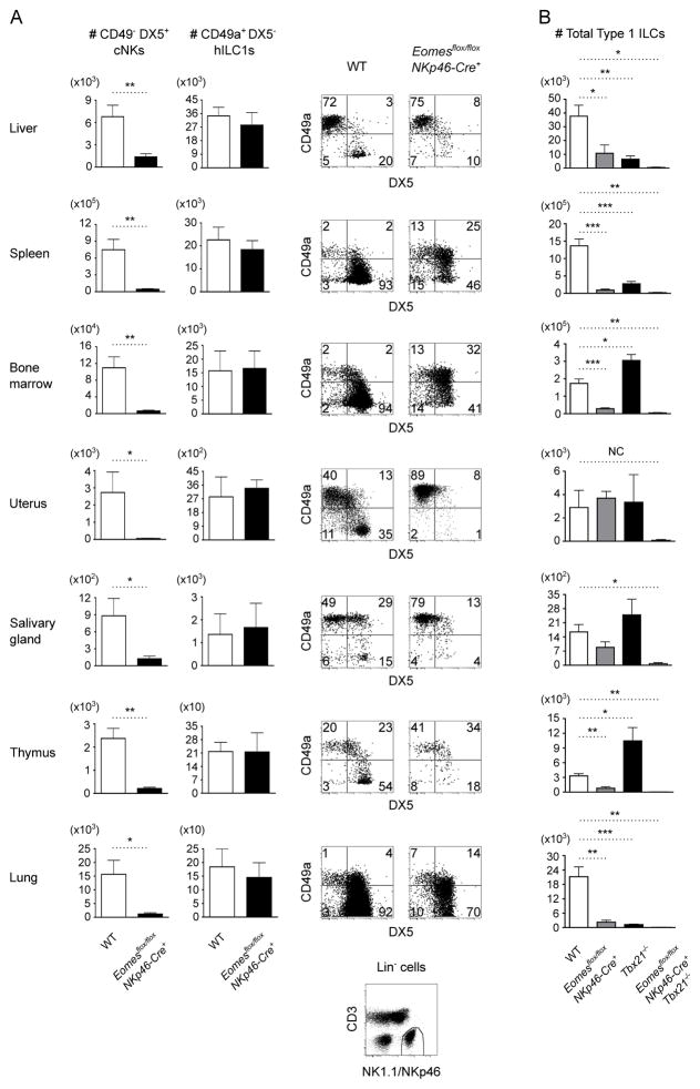
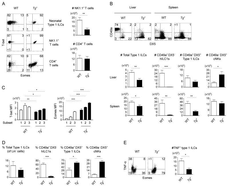
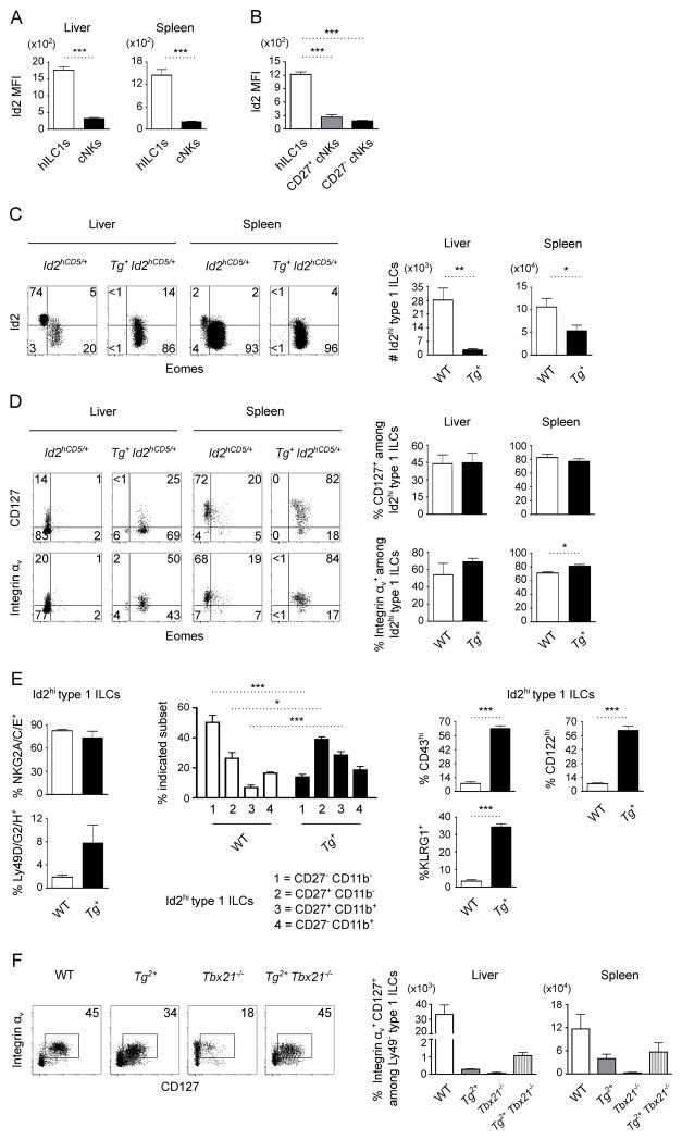
Type 1 innate lymphocytes comprise two developmentally divergent lineages, type 1 helper innate lymphoid cells (hILC1s) and conventional NK cells (cNKs). All type 1 innate lymphocytes (ILCs) express the transcription factor T-bet, but cNKs additionally express Eomesodermin (Eomes). We show that deletion of Eomes alleles at the onset of type 1 ILC maturation using NKp46-Cre imposes a substantial block in cNK development. Formation of the entire lymphoid and nonlymphoid type 1 ILC compartment appears to require the semiredundant action of both T-bet and Eomes. To determine if Eomes is sufficient to redirect hILC1 development to a cNK fate, we generated transgenic mice that express Eomes when and where T-bet is expressed using Tbx21 locus control to drive expression of Eomes codons. Ectopic Eomes induces cNK-like properties across the lymphoid and nonlymphoid type 1 ILC compartments. Subsequent to their divergent lineage specification, hILC1s and cNKs thus possess substantial developmental plasticity.


Conflict of interest statement

Disclosures

The authors have no financial conflicts of interest.

Figures

Figure 1
Figure 1. Loss of Eomes in NKp46+ ILCs impairs cNK development
(A) Representative flow cytometry of CD49a+ DX5 hILC1 and CD49a DX5+ cNK subsets among type 1 ILCs [Lin (CD3, Gr-1, TER-119, CD19) NK1.1+ NKp46+] from the indicated organs of wild-type (WT) and Eomesflox/flox NKp46-Cre+ mice (n=4–5 mice per genotype). Absolute numbers of hILC1s and cNKs are indicated. The NK1.1/NKp46 gate is shown at the bottom. (B) Absolute numbers of total type 1 ILCs from the indicated organs of WT, Eomesflox/flox NKp46-Cre+, Tbx21−/−, and Eomesflox/flox NKp46-Cre+ Tbx21−/− mice (n=3–5 mice per genotype). Statistics were not calculated (NC) for the difference between WT and Eomesflox/flox NKp46-Cre+ Tbx21−/− uterus because the Eomesflox/flox NKp46-Cre+ Tbx21−/− uterus group represents the average of 2 mice. All other data are mean ± SEM representative of 3–5 independent experiments; *p<0.05, **p<0.01, ***p<0.001.
Figure 2
Figure 2. Eomes induces cNK attributes among type 1 ILCs
(A) Transgenic (Tg+) Eomes expression in 3 cell populations (from at least 3 mice per genotype) that usually express T-bet but not Eomes: Lin NK1.1+ NKp46+ type 1 ILCs from 3-day old neonatal livers; NK1.1+ CD3+ T cells from adult livers; and CD4+ T cells from adult spleens after αCD3- and αCD28-stimulation for 48 hours. Absolute numbers of NK1.1+ T cells and CD4+ T cells are indicated. Transgenic Eomes does not affect the development of CD4+ T cells, but it results in moderate reduction of the number of hepatic NK1.1+ T cells. (B) Flow cytometry of CD49a+ DX5, CD49a+ DX5+, and CD49a DX5+ type 1 ILC subsets from the livers and spleens of WT and Tg+ mice (n=5 mice per genotype). (C) T-bet and Eomes expression by hepatic CD49a+ DX5 (1), CD49a+ DX5+ (2), and CD49a DX5+ (3) type 1 ILC subsets from WT and Tg+ mice (n=4 mice per genotype). (D) Frequency of total type 1 ILCs (among Lin cells) and frequency of indicated subsets (among total type 1 ILCs) from the livers of WT and Tg+ neonates (n=4 mice per genotype). Transgenic Eomes does not affect the overall frequency of neonatal type 1 ILCs. (E) Flow cytometry of TNF-α expression by hepatic type 1 ILCs from WT and Tg+ mice (n=4 mice per genotype) after stimulation with 200 ng/mL PMA and 5 μg/mL Ionomycin in the presence of GolgiPlug (Invitrogen) for 4 hours. All data were obtained from mice that have 1 copy of the transgene (Tg+). Data are mean ± SEM representative of 2–5 independent analyses; *p<0.05, **p<0.01, ***p<0.001.
Figure 3
Figure 3. Eomes is permissive to some hILC1 attributes
(A) Id2 expression by CD49a+ DX5 hILC1s and CD49a DX5+ cNKs among Lin NK1.1+ NKp46+ type 1 ILCs from the livers and spleens of Id2hCD5/+ mice (n=4 mice). (B) Id2 expression by CD49a+ Eomes hILC1s, CD27+ Eomes+ immature cNKs, and CD27 Eomes+ mature cNKs from the spleens of Id2hCD5/+ mice (n=4 mice). (C) Flow cytometry of Id2 and Eomes expression by NK1.1+ NKp46+ type 1 ILCs from the livers and spleens of Id2hCD5/+ and Tg+ Id2hCD5/+ mice (n=5 mice per genotype). Absolute numbers of Id2hi cells are indicated. (D) Flow cytometry of CD127 and integrin αv expression by Id2hi NK1.1+ NKp46+ type 1 ILCs from the livers and spleens of Id2hCD5/+ and Tg+ Id2hCD5/+ mice (n=3–4 mice per genotype). (E) Frequency of NKG2A/C/E, Ly49D/G2/H, CD27, CD11b, CD43, KLRG1, and CD122 expression by Id2hi NK1.1+ NKp46+ type 1 ILCs from the livers of Id2hCD5/+ and Tg+ Id2hCD5/+ mice (n=4 mice per genotype). (F) Flow cytometry of helper-like (CD127+ integrin αv+) cells among Ly49 NK1.1+ NKp46+ type 1 ILCs from the spleens of WT, Tg2+, Tbx21−/−, and Tg2+ Tbx21−/− mice. Absolute numbers of hILC1-like cells in the livers and spleens of n=2 mice per genotype are summarized. (A-F) Data were obtained from mice that have 1 (Tg+) or 2 (Tg2+) copies of the transgene as indicated. Data are mean ± SEM representative of 2–5 independent experiments; *p<0.05, **p<0.01, ***p<0.001.

References

    1. Gordon SM, Chaix J, Rupp LJ, Wu J, Madera S, Sun JC, Lindsten T, Reiner SL. The transcription factors T-bet and Eomes control key checkpoints of natural killer cell maturation. Immunity. 2012;36:55–67. - PMC - PubMed
    1. Daussy C, Faure F, Mayol K, Viel S, Gasteiger G, Charrier E, Bienvenu J, Henry T, Debien E, Hasan UA, Marvel J, Yoh K, Takahashi S, Prinz I, de Bernard S, Buffat L, Walzer T. T-bet and Eomes instruct the development of two distinct natural killer cell lineages in the liver and in the bone marrow. J Exp Med. 2014;211:563–577. - PMC - PubMed
    1. Fuchs A, Vermi W, Lee JS, Lonardi S, Gilfillan S, Newberry RD, Cella M, Colonna M. Intraepithelial type 1 innate lymphoid cells are a unique subset of IL-12- and IL-15-responsive IFN-gamma-producing cells. Immunity. 2013;38:769–781. - PMC - PubMed
    1. Peng H, Jiang X, Chen Y, Sojka DK, Wei H, Gao X, Sun R, Yokoyama WM, Tian Z. Liver-resident NK cells confer adaptive immunity in skin-contact inflammation. J Clin Invest. 2013;123:1444–1456. - PMC - PubMed
    1. Cortez VS, Fuchs A, Cella M, Gilfillan S, Colonna M. Cutting edge: Salivary gland NK cells develop independently of Nfil3 in steady-state. J Immunol. 2014;192:4487–4491. - PubMed

Publication types

MeSH terms

Substances