. 2016 Mar 1;196(5):2368-76.
doi: 10.4049/jimmunol.1501724. Epub 2016 Jan 20.

Epithelial Control of Gut-Associated Lymphoid Tissue Formation through p38α-Dependent Restraint of NF-κB Signaling

Affiliations

Epithelial Control of Gut-Associated Lymphoid Tissue Formation through p38α-Dependent Restraint of NF-κB Signaling

Celia Caballero-Franco et al. J Immunol. .

Abstract

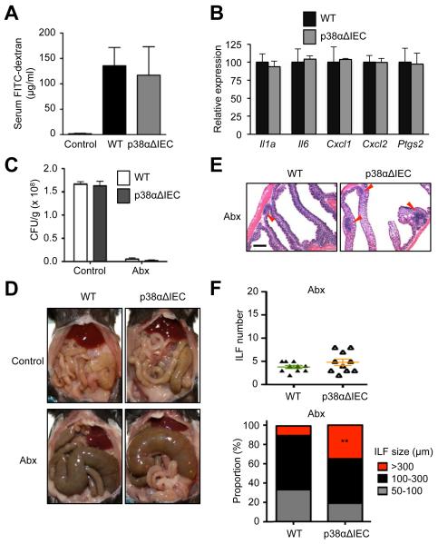
The protein kinase p38α mediates cellular responses to environmental and endogenous cues that direct tissue homeostasis and immune responses. Studies of mice lacking p38α in several different cell types have demonstrated that p38α signaling is essential to maintaining the proliferation-differentiation balance in developing and steady-state tissues. The mechanisms underlying these roles involve cell-autonomous control of signaling and gene expression by p38α. In this study, we show that p38α regulates gut-associated lymphoid tissue (GALT) formation in a noncell-autonomous manner. From an investigation of mice with intestinal epithelial cell-specific deletion of the p38α gene, we find that p38α serves to limit NF-κB signaling and thereby attenuate GALT-promoting chemokine expression in the intestinal epithelium. Loss of this regulation results in GALT hyperplasia and, in some animals, mucosa-associated B cell lymphoma. These anomalies occur independently of luminal microbial stimuli and are most likely driven by direct epithelial-lymphoid interactions. Our study illustrates a novel p38α-dependent mechanism preventing excessive generation of epithelial-derived signals that drive lymphoid tissue overgrowth and malignancy.


Figures

Figure 1
Figure 1. Mice lacking p38α in intestinal epithelial cells develop colonic lymphoid hyperplasia
Colon tissues were obtained from WT and p38αΔIEC mice at 12 to 16 weeks of age. (A) The number of colonic B220+ and CD3+ cells was determined by flow cytometry, and is shown as mean±SEM (n=3). (B, C) Colon tissue sections were analyzed by H&E staining (B), and immunostaining for B220 together with counter-staining of DNA (C). Red arrowheads indicate ILFs. Scale bar, 500 μm (B; and C). (D) The number of colonic ILFs (upper panel) and the proportions of their subsets grouped according to size (lower panel) were determined. **p<0.005. (E-G) Colon tissue sections were analyzed by PNA staining and immunostaining for B220 (E), and immunostaining for RORγt and B220 (F), and CD11c (G) together with counter-staining of DNA. White arrowheads indicate germinal centers (GC) and the crypt base (CB). Scale bar, 100 μm.
Figure 2
Figure 2. Epithelial p38α deficiency leads to colonic lymphoid hyperplasia independently of the influence of gut microbiota
(A) The concentration of FITC-dextran in serum of WT and p38αΔIEC mice was determined after its oral administration, and is shown as mean±SEM (n=3-4). (B) The expression of the indicated genes in the intestinal epithelium of WT and p38αΔIEC mice was analyzed by qPCR. Data are shown as mean±SEM. (C-F) WT and p38αΔIEC mice were subjected to long-term antibiotic treatment (Abx) and analyzed together with antibiotic-naïve counterparts (Control). Fecal bacterial counts were determined (C). The abdominal viscera were photographed (D). Colon tissue sections were analyzed by H&E staining (E). Red arrowheads indicate ILFs. Scale bar, 500 μm. The number of colonic ILFs (F, upper panel) and the proportions of their subsets grouped according to size (F, lower panel) were determined. **p<0.005.
Figure 3
Figure 3. Colitis-associated lymphoid hyperplasia occurs to a greater extent in mice lacking p38α in intestinal epithelial cells
The indicated mice were administered DSS in drinking water at the indicated concentrations for seven days. (A) Colon tissues were prepared on d 7 and analyzed by H&E staining. Red arrowheads indicate ILFs. Scale bar, 500 μm. (B) Survival was monitored daily (n=10).
Figure 4
Figure 4. Intestinal epithelial cells lacking p38α exhibit NF-κB hyperactivation
(A) Whole cell lysates were prepared from MODE-K cells expressing shRNA specific to p38α mRNA and control shRNA (V), and analyzed by immunoblotting. Numbers (#1-#6) denote shRNA clones with different target sequences. (B) Cytoplasmic (Cyto) and nuclear (Nuc) extracts were prepared from control (V and #5) and p38α-KD (#2 and #3) MODE-K cells at the indicated time points after treatment with TNF (50 ng/ml), and analyzed by immunoblotting. (C, D) Whole cell lysates were prepared from control (V) and p38α-KD (#2) MODE-K cells at the indicated time points after treatment with TNF (50 ng/ml), and analyzed by immunoblotting (C). The amount of phosphorylated (p-) TAK1 relative to that of total TAK1 was determined by densitometry (D). (E) Cytoplasmic and nuclear extracts were prepared and analyzed as in B. Where indicated, the cells were preincubated with the TAK1 inhibitor 5Z-7-Oz (2 μM) for 1 h before TNF exposure. (F) Colon tissues were prepared from WT and p38αΔIEC mice orally administered low-dose DSS (2.5%) as in Supplemental Fig. 2 and analyzed by immunostaining for RelA with counter-staining of DNA. Scale bar, 100 μm. Arrowheads indicate nuclei with strong RelA signals.
Figure 5
Figure 5. Mice with constitutive NF-κB activation in intestinal epithelial cells develop colonic lymphoid hyperplasia
Colon tissues were obtained from WT and IEC-IKKβEE mice at 35 to 45 weeks of age. (A, B) Colon tissue sections were analyzed by H&E staining (A), and immunostaining for B220 together with counter-staining of DNA (B). Red arrowheads indicate ILFs. Scale bar, 500 μm (A) and 100 μm (B). (C) The number of colonic ILFs (upper panel) and the proportions of their subsets grouped according to size (lower panel) were determined. **p<0.005. (D) The expression of the indicated genes in the intestinal epithelium of WT and IEC-IKKβEE mice was analyzed by qPCR. Data are shown as mean±SEM.
Figure 6
Figure 6. Loss of p38α augments NF-κB-driven chemokine gene expression in intestinal epithelial cells
(A) Intestinal epithelial cells from WT and IEC-IKKβEE mice (two animals for each genotype, #1 and #2) were subjected to DNA microarray analysis. Relative RNA amounts for differentially expressed genes are presented in color-coded arbitrary units. Select genes showing higher expression in cells from IEC-IKKβEE mice relative to WT counterparts are indicated on the right along with the ratios (fold change; FC) of their RNA amounts. (B) The expression of the indicated genes in the colonic epithelium of WT and p38αΔIEC mice was analyzed by qPCR. Data for the colon are shown as mean±SEM. (C-E) MODE-K cells were transfected with control and RelA- or p38α-specific siRNA (C, D), or with plasmids expressing control (V) and p38α-specific (#2) shRNA (E). Whole cell lysates were prepared and analyzed by immunoblotting (C). The expression of the indicated genes in TNF-treated cells was analyzed by qPCR (D, E).

Similar articles

Cited by

References

    1. Brewster JL, Gustin MC. Hog1: 20 years of discovery and impact. Sci. Signal. 2014;7:re7. - PubMed
    1. Trempolec N, Dave-Coll N, Nebreda AR. SnapShot: p38 MAPK signaling. Cell. 2013;152:656–656.e1. - PubMed
    1. Gupta J, Nebreda AR. Roles of p38α mitogen-activated protein kinase in mouse models of inflammatory diseases and cancer. FEBS J. 2015;282:1841–1857. - PMC - PubMed
    1. Hui L, Bakiri L, Mairhorfer A, Schweifer N, Haslinger C, Kenner L, Komnenovic V, Scheuch H, Beug H, Wagner EF. p38α suppresses normal and cancer cell proliferation by antagonizing the JNK-c-Jun pathway. Nat. Genet. 2007;39:741–749. - PubMed
    1. Ventura JJ, Tenbaum S, Perdiguero E, Huth M, Guerra C, Barbacid M, Pasparakis M, Nebreda AR. p38α MAP kinase is essential in lung stem and progenitor cell proliferation and differentiation. Nat. Genet. 2007;39:750–758. - PubMed

Publication types

MeSH terms

Substances

LinkOut - more resources