. 2016 Jun;53(6):389-96.
doi: 10.1136/jmedgenet-2015-103457. Epub 2016 Jan 20.

Deficiency of the alkaline ceramidase ACER3 manifests in early childhood by progressive leukodystrophy

Affiliations

Deficiency of the alkaline ceramidase ACER3 manifests in early childhood by progressive leukodystrophy

Simon Edvardson et al. J Med Genet. 2016 Jun.

Abstract

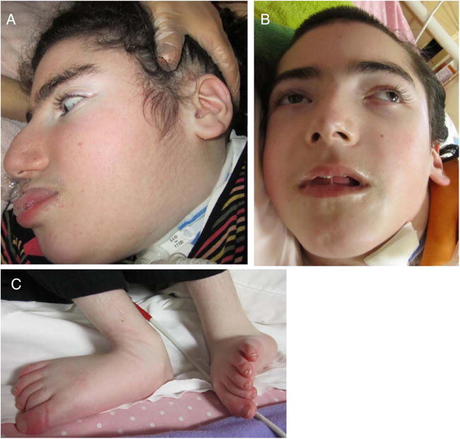
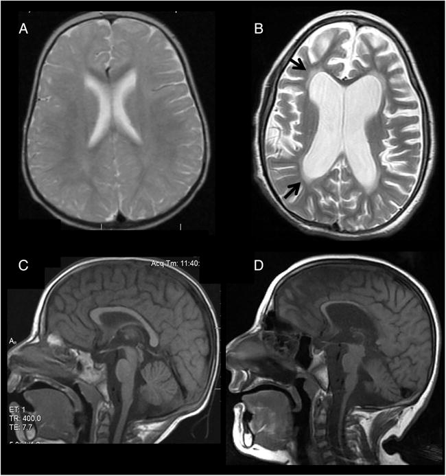
Background/aims: Leukodystrophies due to abnormal production of myelin cause extensive morbidity in early life; their genetic background is still largely unknown. We aimed at reaching a molecular diagnosis in Ashkenazi-Jewish patients who suffered from developmental regression at 6-13 months, leukodystrophy and peripheral neuropathy.

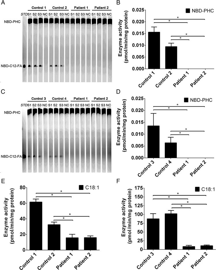
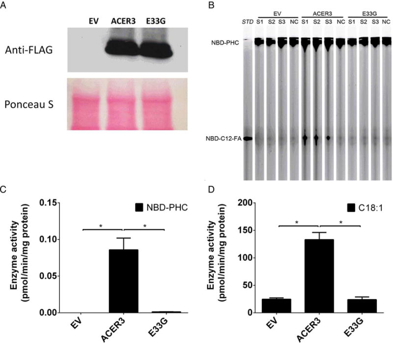
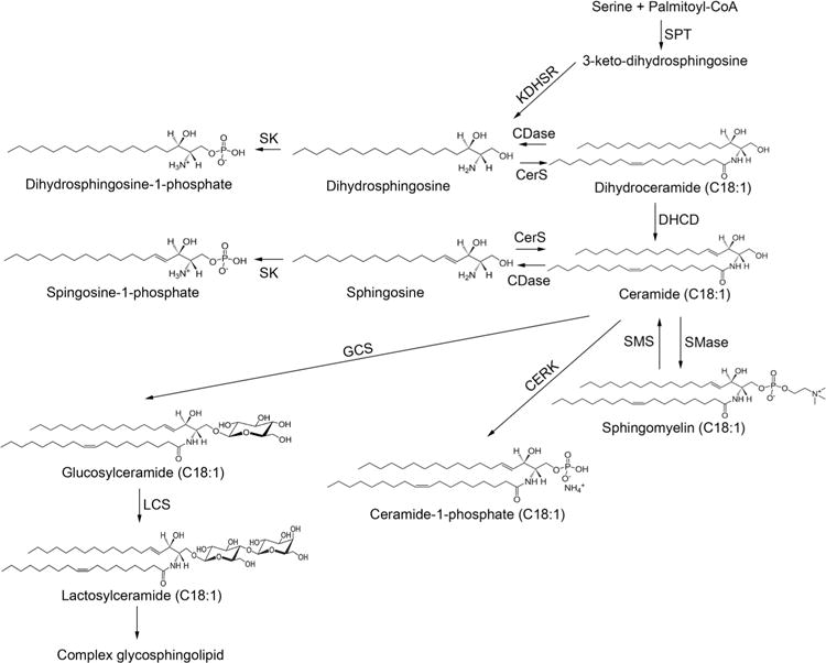
Methods: Exome analysis, determination of alkaline ceramidase activity catalysing the conversion of C18:1-ceramide to sphingosine and D-ribo-C12-N-(7-nitrobenz-2-oxa-1,3-diazol-4-yl) (NBD)-phytoceramide to NBD-C12-fatty acid using liquid chromatography-tandem mass spectrometry (LC-MS/MS) and thin layer chromatography, respectively, and sphingolipid analysis in patients' blood by LC-MS/MS.

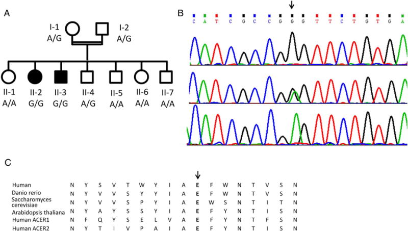
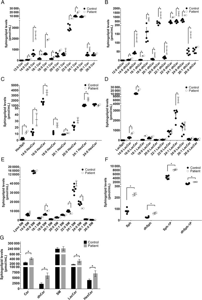
Results: The patients were homozygous for p.E33G in the ACER3, which encodes a C18:1-alkaline ceramidase and C20:1-alkaline ceramidase. The mutation abolished ACER3 catalytic activity in the patients' cells and failed to restore alkaline ceramidase activity in yeast mutant strain. The levels of ACER3 substrates, C18:1-ceramides and dihydroceramides and C20:1-ceramides and dihydroceramides and other long-chain ceramides and dihydroceramides were markedly increased in the patients' plasma, along with that of complex sphingolipids, including monohexosylceramides and lactosylceramides.

Conclusions: Homozygosity for the p.E33G mutation in the ACER3 gene results in inactivation of ACER3, leading to the accumulation of various sphingolipids in blood and probably in brain, likely accounting for this new form of childhood leukodystrophy.

Keywords: Neurology.


Conflict of interest statement

Conflicts of Interest: None declared.

Figures

Figure 1
Figure 1
Family pedigree and the c.A98G, p.Glu33Gly mutation in the ACER3 gene. (A) Family pedigree and mutation genotype; filled symbols represent affected individuals. (B) The mutation (arrow) in patient II-2 (upper), the mother I-1 (middle) and the healthy sister II-6 (lower chromatogram). (C) Conservation of the Glu33 residue throughout evolution and among alkaline ceramidase proteins.
Figure 2
Figure 2
Dysmorphic features of patients with ACER3 deficiency. Coarse facial features, sloping forehead, thick eye brows, prominent nose and prominent lower lip are shown in patient II-2 (A) and II-3 (B) as well as lower limb contractures (C).
Figure 3
Figure 3
Brain MRI of patient II-2 (A and B axial proton-density and T2-weighted images, respectively; C and D midsagittal T1-weighted images, respectively). Normal study is shown at age 1 year (A) and 2 years (C). At 7 years (B and D), there is diffuse supratentorial and infratentorial atrophy with thinning of the corpus callosum (D) and increased diffuse white matter signal (arrows) (B) consistent with progressive leukodystrophy.
Figure 4
Figure 4
ACER3activity is abolished in patients’ cells by the p.E33G mutation (A–F). Total membranes were isolated from skin fibroblasts (A, B and E) and lymphoblasts (C, D and F) from healthy individuals (Control 1, Control 2, Control 3 and Control 4) or from the patients (Patient 1 and Patient 2) and were subjected to alkaline ceramidase activity assays using either NBD-C12-phytoceramide (NBD-C12-PHC) (A, B, C and D) or C18:1-ceramide (E and F) as a substrate. The release of the fluorescent product NBD-C12-fatty acid (NBD-C12-FA) from the substrate NBD-C12-PHC was detected by thin layer chromatography (A and C) and quantified by densitometry (B and D). The release of sphingosine from C18:1 (E and F) was determined by HPLC/MS. Data represent mean values±SD, n=3. *p<0.05.
Figure 5
Figure 5
ACER3 activity is abolished in yeast cells by the p. E33G mutation (A–D). Yeast cells transformed with an empty vector (EV) and yeast cells expressing the FLAG-tagged ACER3 or its mutant, E33G, were homogenised in a lysis buffer as described in our previous study. The lysates were centrifuged at 1000g to pellet nuclei and unbroken cells and the resulting supernatants were centrifuged at 100 000g to pellet the membrane fractions, which were subjected to western blot analyses using anti-FLAG antibody following the protocol of the previous study or alkaline ceramidase activity assays using NBD-C12-phytoceramide (NBD-C12-PHC) (B and C) or C18:1-ceramide (D) as a substrate. The release of the fluorescent product NBD-C12-fatty acid (NBD-C12-FA) from the substrate NBD-C12-PHC was detected by thin layer chromatography (B) and quantified by densitometry (C). The release of sphingosine from C18:1-ceramide (D) was determined by HPLC/MS. Data represent mean values±SD, n=3. *p<0.05.
Figure 6
Figure 6
Sphingolipid metabolism ceramides, which are considered the central hub of sphingolipid metabolism, are synthesised via three different pathways (de novo biosynthesis, sphingomyelinase pathway and salvage pathway). They are then incorporated into various complex sphingolipids or broken down by ceramidases into sphingosine and dihydrosphingosine with subsequent production of sphingosine-1-phosphate and dihydrosphingosine-1-phosphate. CERK, ceramide kinase; CerS, ceramide synthase; DHCD, dihydroceramide desaturase; GCS, glucosylceramide synthase; KDHSR, 3-keto-dihydrosphingosine reductase; LCS, lactosylceramide synthase; SK, sphingosine kinase; SMase, sphingmyelinase; SMS, sphingomyelin synthase; SPT, serine palmitoyltransferase.
Figure 7
Figure 7
Sphingolipid profile in blood. Blood samples were collected from the five healthy individuals (Control) and the two patients (Patient). Each of the blood samples (100 μL) was subjected to liquid chromatography-tandem mass spectrometry (LC-MS/MS) for the levels of individual ceramides (A), individual dihydroceramides (B), individual monohexosylceramides (C), individual lactosylceramides (D), individual sphingomyelins (E), sphingoid bases and their phosphates (F) and total amount of each type of sphingolipids (G). Data represent mean values±SD, n=2 (patients) or 5 (controls). *p<0.05.

References

    1. Bonkowsky JL, Nelson C, Kingston JL, Filloux FM, Mundorff MB, Srivastava R. The burden of inherited leukodystrophies in children. Neurology. 2010;75:718–25. - PMC - PubMed
    1. Schiffmann R, van der Knaap MS. Invited article: an MRI-based approach to the diagnosis of white matter disorders. Neurology. 2009;72:750–9. - PMC - PubMed
    1. Mao C, Xu R, Bielawska A, Obeid LM. Cloning of an alkaline ceramidase from Saccharomyces cerevisiae. An enzyme with reverse (CoA-independent) ceramide synthase activity. J Biol Chem. 2000;275:6876–84. - PubMed
    1. Hu W, Xu R, Sun W, Szulc ZM, Bielawski J, Obeid LM, Mao C. Alkaline ceramidase 3 (ACER3) hydrolyzes unsaturated long-chain ceramides, and its down-regulation inhibits both cell proliferation and apoptosis. J Biol Chem. 2010;285:7964–76. - PMC - PubMed
    1. Mao C, Xu R, Szulc ZM, Bielawska A, Galadari SH, Obeid LM. Cloning and characterization of a novel human alkaline ceramidase. A mammalian enzyme that hydrolyzes phytoceramide. J Biol Chem. 2001;276:26577–88. - PubMed

Publication types