. 2016 Mar 18;291(12):6359-75.
doi: 10.1074/jbc.M115.686592. Epub 2016 Jan 20.

Norbin Stimulates the Catalytic Activity and Plasma Membrane Localization of the Guanine-Nucleotide Exchange Factor P-Rex1

Affiliations

Norbin Stimulates the Catalytic Activity and Plasma Membrane Localization of the Guanine-Nucleotide Exchange Factor P-Rex1

Dingxin Pan et al. J Biol Chem. .

Abstract

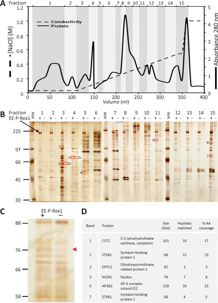
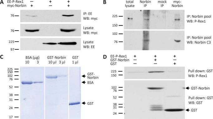
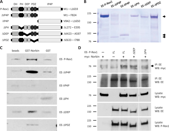
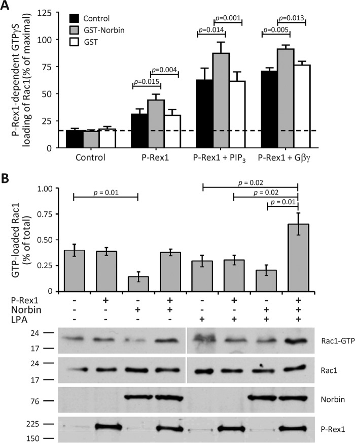
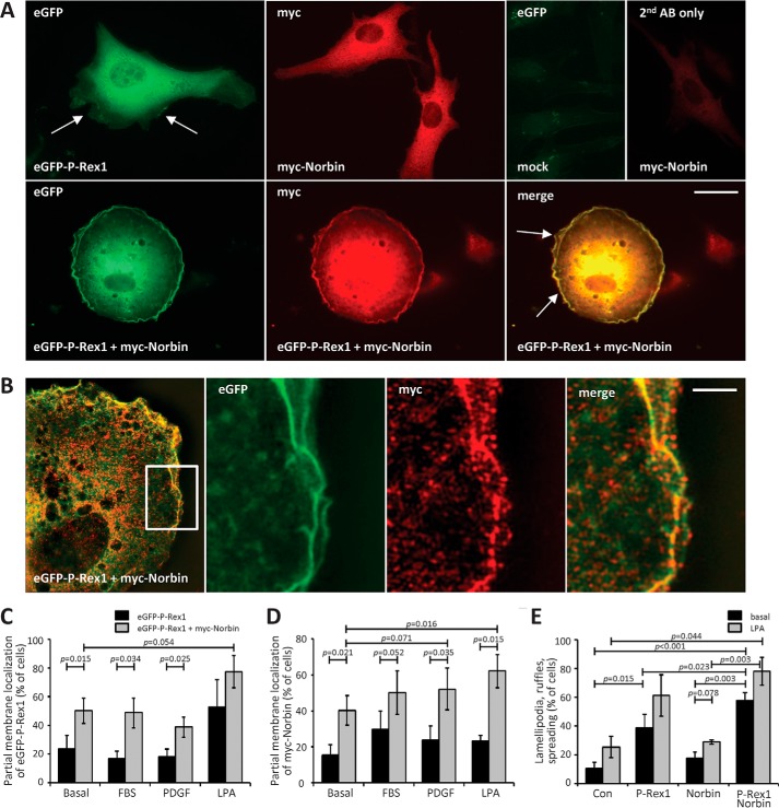
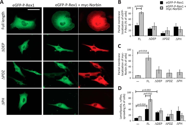
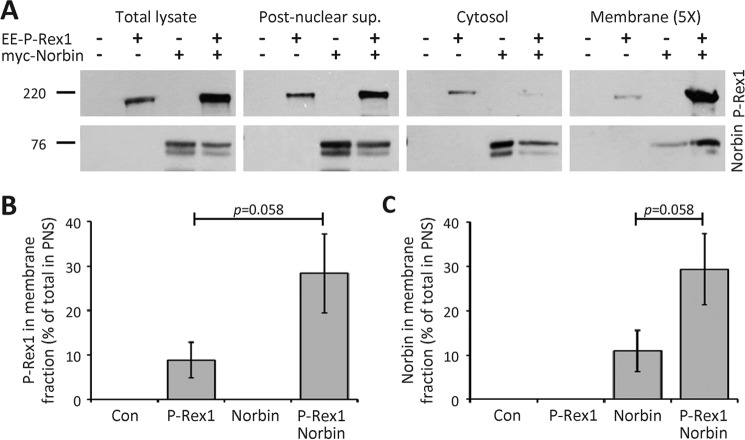
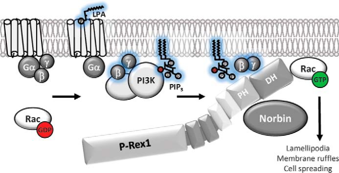
P-Rex1 is a guanine-nucleotide exchange factor (GEF) that activates the small G protein (GTPase) Rac1 to control Rac1-dependent cytoskeletal dynamics, and thus cell morphology. Three mechanisms of P-Rex1 regulation are currently known: (i) binding of the phosphoinositide second messenger PIP3, (ii) binding of the Gβγ subunits of heterotrimeric G proteins, and (iii) phosphorylation of various serine residues. Using recombinant P-Rex1 protein to search for new binding partners, we isolated the G-protein-coupled receptor (GPCR)-adaptor protein Norbin (Neurochondrin, NCDN) from mouse brain fractions. Coimmunoprecipitation confirmed the interaction between overexpressed P-Rex1 and Norbin in COS-7 cells, as well as between endogenous P-Rex1 and Norbin in HEK-293 cells. Binding assays with purified recombinant proteins showed that their interaction is direct, and mutational analysis revealed that the pleckstrin homology domain of P-Rex1 is required. Rac-GEF activity assays with purified recombinant proteins showed that direct interaction with Norbin increases the basal, PIP3- and Gβγ-stimulated Rac-GEF activity of P-Rex1. Pak-CRIB pulldown assays demonstrated that Norbin promotes the P-Rex1-mediated activation of endogenous Rac1 upon stimulation of HEK-293 cells with lysophosphatidic acid. Finally, immunofluorescence microscopy and subcellular fractionation showed that coexpression of P-Rex1 and Norbin induces a robust translocation of both proteins from the cytosol to the plasma membrane, as well as promoting cell spreading, lamellipodia formation, and membrane ruffling, cell morphologies generated by active Rac1. In summary, we have identified a novel mechanism of P-Rex1 regulation through the GPCR-adaptor protein Norbin, a direct P-Rex1 interacting protein that promotes the Rac-GEF activity and membrane localization of P-Rex1.

Keywords: G protein-coupled receptor (GPCR); Neurochondrin (NCDN); PREX1; PREX2; Rac (Rac GTPase); Rho (Rho GTPase); cell signaling; guanine nucleotide exchange factor (GEF); protein complex; small GTPase.


Figures

FIGURE 1.
FIGURE 1.
Identification of Norbin as a P-Rex1-binding protein. A, fractionation of P-Rex1−/− mouse brain cytosol. Desalted P-Rex1−/− mouse brain cytosol was fractionated by Source 15Q anion exchange chromatography with a linear 0.05–0.5 m NaCl gradient and a step to 1 m NaCl. 15 fractions ranging from the flow-through to the 1 m NaCl step and from 10 to 50 ml in volume were collected as indicated, and adjusted to 150 mm NaCl. B, isolation of putative P-Rex1-binding proteins from P-Rex1−/− mouse brain cytosol fractions. The salt-adjusted fractions from A were pre-cleared and half of each incubated with 10 μg of purified recombinant human EE-P-Rex1 immobilized on EE antibody-coupled protein G-Sepharose (48), the other mock-treated with EE antibody beads alone. Bound proteins were subjected to SDS-PAGE and silver staining. Protein bands seen in the presence but not absence of EE-P-Rex1, framed by red boxes, and those labeled “c” as controls, were subjected to LC/MS. The black arrow highlights full-length EE-P-Rex1. The vertical white lines show boundaries between individual gels. C, enlargement of fraction 4 from the silver gel in B. The red arrow highlights the band identified as Norbin by LC/MS. D, list of putative P-Rex1 binding proteins identified. The numbering of bands corresponds to the red-framed boxes shown in B. Bands 4 and 6 contained only keratin and trypsin, so were not considered further.
FIGURE 2.
FIGURE 2.
Norbin tissue distribution. A, Norbin distribution in the brain and up-regulation in some regions of P-Rex1−/− mouse brain. Total lysates of brain sections from adult P-Rex1+/+ (Wt) and P-Rex1−/− (Ko) mice (6, 10) were Western blotted and Norbin levels quantified by ImageJ and expressed as the ratio of P-Rex1−/− to corresponding P-Rex1+/+ samples. B, Norbin distribution in non-neuronal mouse tissues and cell types. Total lysates of organs, bone marrow cells, macrophages, and neutrophils isolated from adult wild-type mice (100 μg of tissue per lane) were Western blotted for Norbin expression. The vertical white lines show boundaries between individual gels. C, test of Norbin antibody. To confirm the specificity of the Norbin antibody, HEK-293 cells were treated with the indicated concentrations of siRNA1 and siRNA2 to down-regulate endogenous Norbin or a pool of 4 non-targeting siRNAs as a control. Norbin C1 antibody was used in A–C to detect endogenous Norbin, and β-actin antibody or Coomassie staining of the membranes to control for total protein loading.
FIGURE 3.
FIGURE 3.
Norbin binds P-Rex1 directly and interacts with P-Rex1 endogenously in cells. A, myc-Norbin interacts with EE-P-Rex1. COS-7 cells expressing EE-P-Rex1 and/or myc-Norbin were subjected to immunoprecipitation with immobilized EE antibody under stringent conditions (1% Triton X-100) and analyzed by Western blotting with myc and EE Abs as indicated. Blots shown are from 1 experiment representative of 3. B, endogenous P-Rex1 and Norbin interact. HEK-293 cell lysates were incubated with Norbin antibody pool (C1, N4, N7) or mock-treated with serum-IgG, washed stringently and Western blotted. Overexpression of myc-Norbin was used as a control. Blots shown are from 1 experiment representative of 3. C, purification of GST-Norbin. Coomassie gel showing purified recombinant GST-Norbin and GST (bead volumes are indicated) and BSA standards. The vertical white lines denote cropped lanes. D, direct interaction between EE-P-Rex1 and GST-Norbin. 10 pmol of immobilized purified recombinant GST-Norbin or GST, or beads alone, were incubated with 20 pmol of purified recombinant EE-P-Rex1, beads were washed stringently, and proteins were analyzed by Western blotting. Blots shown are from 1 experiment representative of 3.
FIGURE 4.
FIGURE 4.
The PH domain of P-Rex1 is required for Norbin binding. A, schematic of P-Rex1 mutants. B, purified mutant P-Rex1 proteins. Coomassie-stained SDS-PAGE showing 100 pmol of recombinant full-length or mutant EE-P-Rex1 proteins (50 pmol for EE-IP4P), purified from Sf9 cells. C, the PH domain of P-Rex1 is required for Norbin binding in vitro. 4.5 pmol of purified full-length or mutant EE-P-Rex1 proteins were incubated with 4.5 pmol of purified GST-Norbin or GST, or with beads alone, and Western blotted with EE antibody. Blots shown are from 1 experiment representative of 3 for the full-length protein and for ΔPH, ΔDEP, and ΔPDZ mutants and 5 for ΔIP4P and iIP4P. D, the PH domain of P-Rex1 is required for the interaction with Norbin in vivo. COS-7 cells expressing combinations of myc-Norbin full-length (FL) EE-P-Rex1, EE-ΔDEP, or EE-ΔPH were immunoprecipitated with immobilized EE antibody under stringent conditions (RIPA buffer) and analyzed by Western blotting with myc-HRP, EE, and P-Rex1 6F12 ABs. The white vertical lines show a cropped lane. Blots shown are from 1 experiment representative of 4.
FIGURE 5.
FIGURE 5.
Norbin stimulates the Rac-GEF activity P-Rex1. A, GST-Norbin directly stimulates the basal, PIP3- and Gβγ-dependent Rac-GEF activity of EE-P-Rex1. The ability of 50 nm purified full-length EE-P-Rex1 to activate (GTPγS-load) 100 nm EE-Rac1 within 10 min was tested ±200 nm purified GST-Norbin or GST protein, and with liposomes ±10 μm stearoyl-arachidonyl PIP3 or 0.3 μm1γ2, as indicated. Rac-GEF activity is expressed as % of EE-Rac1-GTPγS in EDTA controls. Data are mean ± S.E. of 10 independent experiments testing basal activity, 5 with PIP3 and 6 with Gβ1γ2; statistics are paired t test with Bonferroni/Holm multiple comparisons analysis. B, coexpression of P-Rex1 and Norbin activates endogenous Rac1 in LPA-stimulated HEK-293 cells. HEK-293 cells expressing EE-P-Rex1 and/or myc-Norbin, or mock-transfected cells, were serum-starved, stimulated with 50 nm LPA for 2 min, or mock-stimulated, and subjected to Pak-CRIB pulldown and Western blotting (1% of cell lysate loaded for total Rac1). Blots shown are from 1 experiment representative of 4. The vertical white lines show boundaries between gels run and blotted together, with the corresponding Rac1-GTP and total lysate Rac1 samples on one gel. Quantification of Rac1 activity in mock-stimulated (left-hand panel) and LPA-stimulated cells (right-hand panel) is expressed as mean ± S.E. of 4 experiments; statistics are unpaired t test.
FIGURE 6.
FIGURE 6.
P-Rex1 and Norbin promote the plasma membrane localization of each other and elicit cell spreading, lamellipodia formation and membrane ruffling. A, immunofluorescence micrographs. PAE cells expressing eGFP-P-Rex1 and/or myc-Norbin for 24 h were serum-starved for 6 h, fixed, permeabilized, and stained with myc antibody and Alexa Fluor 568 goat anti-mouse secondary antibody, or with secondary antibody alone, and subjected to widefield immunofluorescence microscopy. Arrows indicate lamellipodia and membrane ruffles. The scale bar represents 10 μm. Representative images from 1 of 4 independent experiments are shown. B, super-resolution structured illumination micrographs. Representative super-resolution SIM image of part of a PAE cell expressing eGFP-P-Rex1 and myc-Norbin, serum-starved and stained as in A (left-hand panel), with a zoom (white box) into membrane ruffles at the cell edge. One Z-plane of 0.12 μm depth is shown. The scale bar represents 2 μm. C–E, quantification of immunofluorescence microscopy. PAE cells expressing eGFP-P-Rex1 (black bars) or eGFP-P-Rex1 and myc-Norbin (gray bars) as in A, or mock-transfected cells, were serum-starved for 6 h and stimulated with 10% FBS, 10 ng/ml of PDGF, or 2 μg/ml of LPA for 5 min, or mock-treated, and stained as in A. Slides were blinded and analyzed by imaging for the percentage of cells showing partial membrane localization of eGFP-P-Rex1 (C) or myc-Norbin (D). E, co-expression of P-Rex1 and Norbin induces cell morphologies characteristic of active Rac1. Basal (serum-starved; black bars) or LPA-stimulated (gray bars) PAE cells expressing eGFP-P-Rex1 and/or myc-Norbin as in (C and D) were evaluated for the occurrence of lamellipodia, membrane ruffles, and cell spreading. In C–E, ≥100 transfected cells were scored per coverslip, with duplicate coverslips per condition and experiment. Data are mean ± S.E. from 7 independent experiments with basal cells, 3 with FBS and 4 with PDGF or LPA stimulation, respectively; statistics are paired t test with Bonferroni/Holm multiple comparisons analysis.
FIGURE 7.
FIGURE 7.
P-Rex1 domains required for Norbin-dependent plasma membrane localization and effects on cell morphology. A, immunofluorescence micrographs. PAE cells expressing full-length or mutant eGFP-P-Rex1, as indicated, with or without myc-Norbin for 40 h were serum-starved for 6 h, fixed, permeabilized, and stained with myc antibody and Alexa Fluor 568 goat anti-mouse secondary antibody, or with secondary antibody alone, and subjected to widefield immunofluorescence microscopy. The scale bar represents 20 μm. Representative images from 1 of 3 independent experiments are shown. B–D, quantification of immunofluorescence microscopy. PAE cells as in A expressing full-length (FL) or the indicated mutant forms of eGFP-P-Rex1 (black bars) or expressing these together with myc-Norbin (gray bars), or mock-transfected cells, were treated as described in A. Slides were blinded and analyzed for the percentage of cells showing partial membrane localization of eGFP-P-Rex1 (B), myc-Norbin (C), and cell morphologies (D) characteristic of active Rac1 (lamellipodia, membrane ruffles, and cell spreading). In B–D, ≥100 transfected cells were scored on duplicate coverslips per condition. Data are mean ± S.E. from 3 independent experiments; statistics are paired t test with Bonferroni/Holm multiple comparisons analysis.
FIGURE 8.
FIGURE 8.
P-Rex1 and Norbin promote the localization of each other in membrane fractions. A, HEK-293 cells expressing EE-P-Rex1 and/or myc-Norbin, and mock-transfected cells, were serum-starved, lysed, the total lysates were cleared of debris and nuclei, and the postnuclear supernatant fractionated into membrane and cytosol fractions. Aliquots from all stages of the fractionation were analyzed by Western blotting with P-Rex1 6F12 and Norbin C1 antibodies. Note that 5 times as much membrane fraction was loaded than of the other fractions, to allow direct comparison. Blots shown are from 1 experiment representative of 3. Total lysates and postnuclear supernatant were run on one gel, cytosol and membrane fractions on another; the vertical white lines denote cropped lanes. B and C, Western blots as in A were quantified by ImageJ and the amount of P-Rex1 (B) and Norbin (C) in the membrane fraction expressed as percent of the postnuclear supernatant. Data are mean ± S.E. from 3 independent experiments; statistics are unpaired t test.
FIGURE 9.
FIGURE 9.
Model of P-Rex1 regulation by Norbin. The GPCR adaptor protein Norbin interacts directly with P-Rex1 through the PH domain and stimulates the basal Rac1-GEF activity of P-Rex1. Norbin also proportionally increases the Rac1-GEF activity of P-Rex1 when stimulated through the lipid second messenger PIP3 that is generated by PI3K, or through the Gβγ subunits of heterotrimeric G proteins that are released upon GPCR stimulation. In response to cell stimulation with the GPCR ligand LPA, Norbin promotes the P-Rex1-dependent activation of endogenous Rac1. The interaction of Norbin with P-Rex1 also leads to a robust translocation of both proteins from the cytosol to the plasma membrane. Finally, coexpression of P-Rex1 and Norbin induces cell morphologies characteristic of active Rac1, namely lamellipodia formation, membrane ruffling, and cell spreading. We propose that the increased membrane localization of P-Rex1 caused by Norbin binding brings the Rac-GEF into closer contact with its other membrane-bound activators, PIP3 and Gβγ, and with its substrate Rac1, thereby promoting Rac1 activity and Rac1-dependent cell responses.

References

    1. Welch H. C., Coadwell W. J., Ellson C. D., Ferguson G. J., Andrews S. R., Erdjument-Bromage H., Tempst P., Hawkins P. T., and Stephens L. R. (2002) P-Rex1, a PtdIns(3,4,5)P3- and Gβγ-regulated guanine-nucleotide exchange factor for Rac. Cell 108, 809–821 - PubMed
    1. Welch H. C. (2015) Regulation and function of P-Rex family Rac-GEFs. Small GTPases 6, 49–70 - PMC - PubMed
    1. Damoulakis G., Gambardella L., Rossman K. L., Lawson C. D., Anderson K. E., Fukui Y., Welch H. C., Der C. J., Stephens L. R., and Hawkins P. T. (2014) P-Rex1 directly activates RhoG to regulate GPCR-driven Rac signalling and actin polarity in neutrophils. J. Cell Sci. 127, 2589–2600 - PubMed
    1. Lawson C. D., Donald S., Anderson K. E., Patton D. T., and Welch H. (2011) P-Rex1 and Vav1 cooperate in the regulation of fMLF-dependent neutrophil responses. J. Immunol. 186, 1467–1476 - PubMed
    1. Pan D., Amison R. T., Riffo-Vasquez Y., Spina D., Cleary S. J., Wakelam M. J., Page C. P., Pitchford S. C., and Welch H. C. (2015) P-Rex and Vav Rac-GEFs in platelets control leukocyte recruitment to sites of inflammation. Blood 125, 1146–1158 - PMC - PubMed

Publication types

Substances

LinkOut - more resources