. 2016 May 19;44(9):4147-62.
doi: 10.1093/nar/gkw008. Epub 2016 Jan 20.

Histone H3K36 trimethylation is essential for multiple silencing mechanisms in fission yeast

Affiliations

Histone H3K36 trimethylation is essential for multiple silencing mechanisms in fission yeast

Shota Suzuki et al. Nucleic Acids Res. .

Abstract

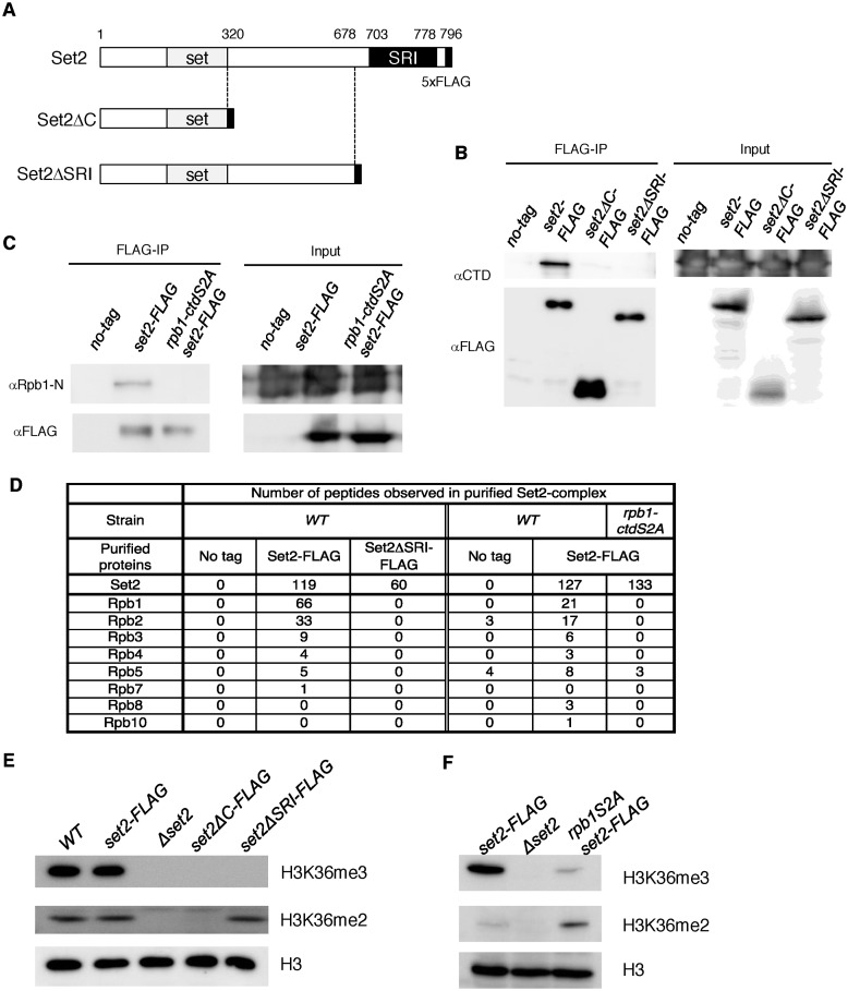
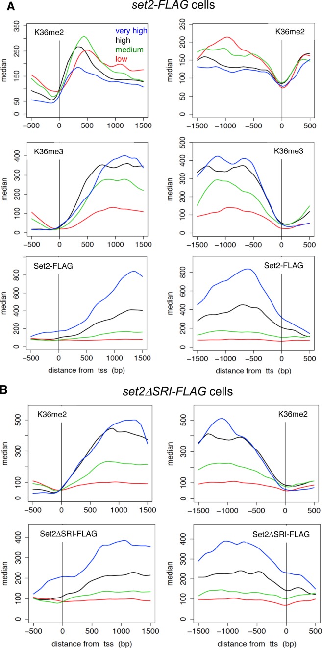
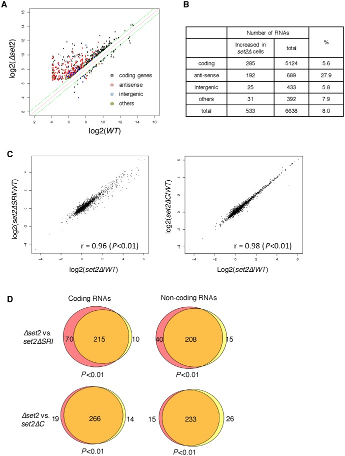
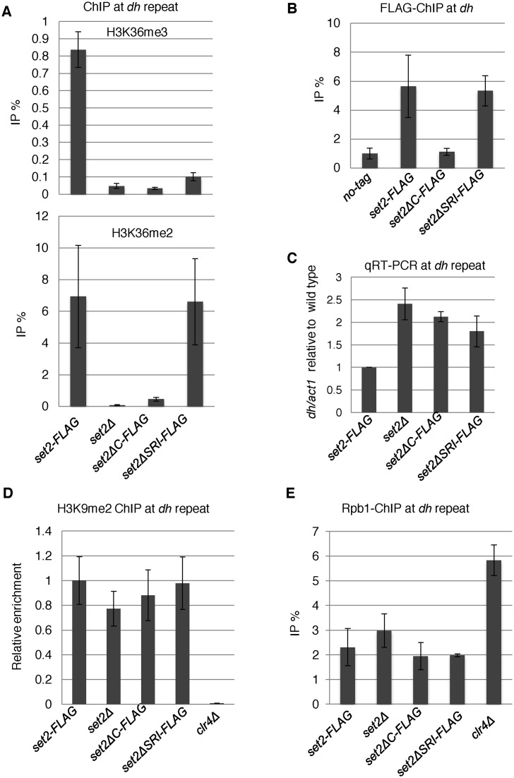
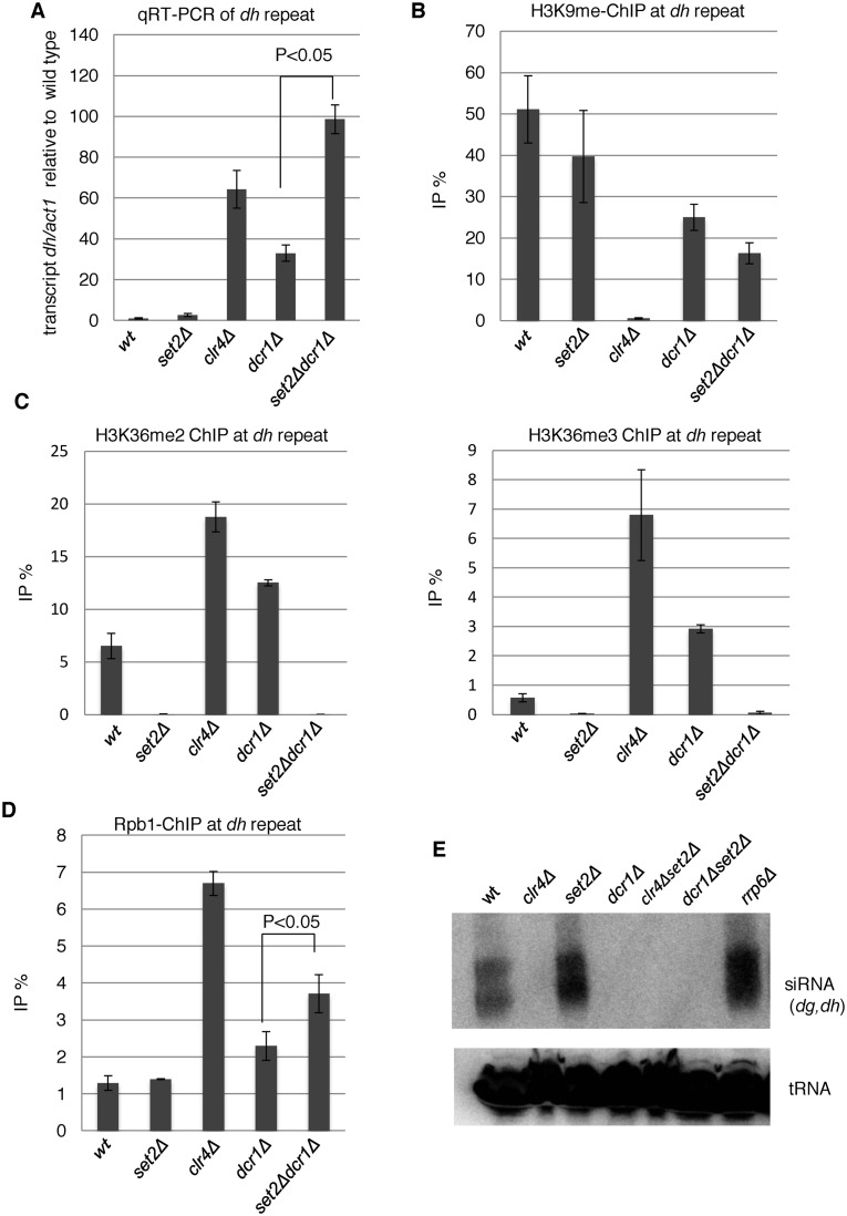
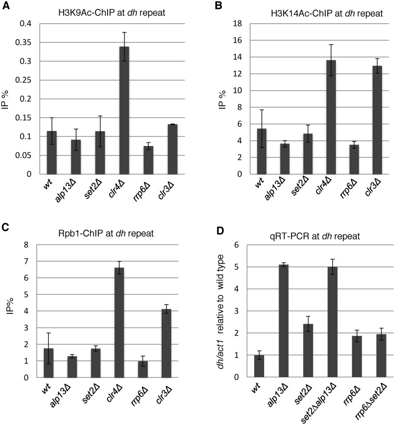
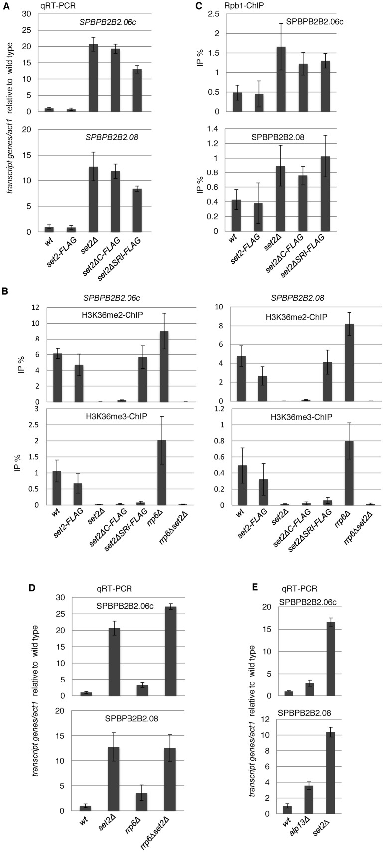
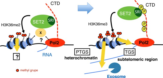
In budding yeast, Set2 catalyzes di- and trimethylation of H3K36 (H3K36me2 and H3K36me3) via an interaction between its Set2-Rpb1 interaction (SRI) domain and C-terminal repeats of RNA polymerase II (Pol2) phosphorylated at Ser2 and Ser5 (CTD-S2,5-P). H3K36me2 is sufficient for recruitment of the Rpd3S histone deacetylase complex to repress cryptic transcription from transcribed regions. In fission yeast, Set2 is also responsible for H3K36 methylation, which represses a subset of RNAs including heterochromatic and subtelomeric RNAs, at least in part via recruitment of Clr6 complex II, a homolog of Rpd3S. Here, we show that CTD-S2P-dependent interaction of fission yeast Set2 with Pol2 via the SRI domain is required for formation of H3K36me3, but not H3K36me2. H3K36me3 silenced heterochromatic and subtelomeric transcripts mainly through post-transcriptional and transcriptional mechanisms, respectively, whereas H3K36me2 was not enough for silencing. Clr6 complex II appeared not to be responsible for heterochromatic silencing by H3K36me3. Our results demonstrate that H3K36 methylation has multiple outputs in fission yeast; these findings provide insights into the distinct roles of H3K36 methylation in metazoans, which have different enzymes for synthesis of H3K36me1/2 and H3K36me3.


Figures

Figure 1.
Figure 1.
Set2 interacts with Pol2 through its C-terminal SRI domain in a CTD-S2P–dependent manner, and this interaction is required for the generation of H3K36me3 but not H3K36me2. (A) Schematic diagrams of the C-terminal truncation mutants of Set2 used in this study. The SET and SRI domains are indicated by gray and black boxes, respectively. The black box at the C-terminal end of each mutant indicates the 5×FLAG tag. (B) Set2 interacts with Pol2 through its C-terminal SRI domain. The interaction between Set2 and Pol2 was examined by co-immunoprecipitation. Extracts prepared from the indicated strains were incubated with an antibody against the FLAG tag. Immunoprecipitated fractions were analyzed by immunoblotting using an antibody against CTD of Pol2. Input represents 15% of extracts used for immunoprecipitation. (C) Set2–Pol2 interaction depends on Pol2 CTD-S2P. Set2–Pol2 was examined using extracts prepared from the indicated strains by the co-immunoprecipitation scheme described in (B), except that an antibody against the N-terminal region of Rpb1 (39) was used to detect Rpb1 to avoid possible influences of the rpb1-ctdS2A mutation on antibody recognition. (D) Subunits of RNA polymerase II observed in Set2 complexes analyzed by mass-spectrometry analysis. (E) The SRI domain is essential for H3K36me3 but not for H3K36me2. Whole-cell extracts from the indicated strains were analyzed by immunoblotting using antibodies against H3K36me2, H3K36me3 (37) and the C-terminal region of histone H3. (F) CTD-S2P is required for efficient H3K36me3 but not for H3K36me2. H3K36me2, H3K36me3 and histone H3 in whole cell extracts were analyzed as (E).
Figure 2.
Figure 2.
Distributions of methylation of H3K36 and Set2 proteins indicate co-transcriptional methylation by Set2. (A) ChIP-sequencing analysis of H3K36me2, H3K36me3 and Set2-FLAG in wild-type (set2-FLAG) cells. Positions (bp) relative to the transcription start site (TSS) (left panels) and termination site (TTS) (right panels) are shown on the x-axis. The gene sets (very high, high, medium, low) used were the same as those used in the previous study (42), which were classified according to the level of Pol2. The median tag counts of the indicated gene sets, which were transcribed at different levels, are shown on the y-axis in each upper panel. (B) ChIP-sequencing analysis of H3K36me2 and Set2ΔSRI-FLAG in set2ΔSRI-FLAG cells. Positions (bp) relative to the transcription start site (TSS) (left panels) and termination site (TTS) (right panels) are shown on the x-axis. The median tag counts of the indicated gene sets, which were the same as used in (A), are shown on the y-axis in each upper panel.
Figure 3.
Figure 3.
Comparison of change of RNA levels in set2 mutants. (A) RNAs whose levels changed in set2Δ cells more than 1.5-fold are plotted. The x-axis represents the log2 of the array signals in wild-type cells, and the y-axis represents the log2 of the array signals in set2Δ cells. RNAs are classified as coding, antisense, intergenic, and others, and shown by colored dots, as indicated in the inset. (B) A breakdown of RNAs that were elevated in set2Δ cells. (C) Scatter plots for comparisons of RNA levels in the indicated set2 mutants. The y-axis represents the log2 of the array signals in set2ΔSRI cells (left panel) or set2ΔC cells (right panel) over the array signals in wild-type, whereas the x-axis represents the log2 of the array signals in Δset2 over the array signals in wild-type. The correlation coefficients (r) are shown in the graph; P-values were calculated using Fisher's exact test. (D) Venn diagram showing the number of transcripts whose expression levels were more than 1.5-fold higher in the indicated mutants than in the wild-type. The transcripts were classified as coding and non-coding RNAs. Red color indicates transcripts up-regulated only in set2Δ, while yellow color shows transcripts up regulated only in set2ΔSRI cells (upper panels) or set2ΔC cells (lower panels). P-values were calculated using Fisher's exact test.
Figure 4.
Figure 4.
Loss of Set2 compromised heterochromatic silencing without affecting heterochromatin structure. (A, B and D) ChIP analyses of H3K36me2 and H3K36me3 (A), Set2-FLAG (B), H3K9me2 (D) and Rpb1 (E) at heterochromatic dh repeats were performed using the indicated strains. Error bars show the standard deviation of three independent experiments. (C) Quantitative RT-PCR (qRT-PCR) analysis of the forward transcript from dh repeats was performed using the indicated strains. Error bars show the standard deviation of three independent experiments.
Figure 5.
Figure 5.
Set2 functions at heterochromatin independently of RNAi. (A) qRT-PCR analyses of the forward transcripts from dh repeats were performed using the indicated strains. Error bars show the standard deviation of three independent experiments. P-values were determined using a two-sided Student's t-test to compare dcr1Δset2Δ cells with dcr1Δ cells. (B and D) ChIP analyses of H3K9me2 (B) and Rpb1 (D) at heterochromatic dh repeats were performed using the indicated strains. Error bars show the standard deviation of three independent experiments. P-values were determined using a two-sided Student's t-test to compare dcr1Δset2Δ cells with dcr1Δ cells. (C) H3K36 methylation was increased in mutants that compromised heterochromatin. ChIP analyses of H3K36me2 (left panel) and H3K36me3 (right panel) at heterochromatic dh repeats were performed in the indicated strains. Error bars show the standard deviation of three independent experiments. (E) siRNA analysis by northern blotting using oligonucleotide probes specific for dg and dh centromeric repeats (40). Oligonucleotide probe specific for tRNAAsn was used as a loading control.
Figure 6.
Figure 6.
Comparison of the states of heterochromatin among possible downstream factors of Set2. (A and C) ChIP analyses of H3K39Ac (A), H3K14Ac (B) and Rpb1 (C) at heterochromatic dh repeats in the indicated strains. Error bars show the standard deviation of three independent experiments. (D) Quantitative RT-PCR (qRT-PCR) analysis of the forward transcript from dh repeats was performed using the indicated strains. Error bars show the standard deviation of three independent experiments.
Figure 7.
Figure 7.
Set2 downregulates subtelomeric genes, SPBPB2B2.06c and SPBPB2B2.08 at the transcriptional level. (A, D and E) Quantitative RT-PCR (qRT-PCR) analyses of transcripts from both genes in the indicated strains were performed. Error bars show the standard deviation of three independent experiments. (B) Levels of H3K36 methylation at SPBBP2B2.06c and SPBPB2B2.08 in set2 mutants. ChIP analyses of H3K36me2 (upper panels) and H3K36me3 (lower panels) at SPBPB2B2.06c (left panels) and SPBPB2B2.08 (right panels) were performed in the indicated strains. Error bars show the standard deviation of three independent experiments. (C) ChIP analyses of Rpb1 at heterochromatic genes were performed using the indicated strains. Error bars show the standard deviation of three independent experiments.
Figure 8.
Figure 8.
Model of co-transcriptional methylation of H3K36 and its function in heterochromatic and subtelomeric regions in fission yeast. (Left panel) In the initial step of transcription, Set2 interacts with Pol2 or other factors with unknown mechanism and promotes H3K36me2 co-transcriptionally. The output of H3K36me2 is not yet known. (Right panel) When CTD-S2 (and probably CTD-S5) is phosphorylated, Set2 changes the binding mode to favor the interaction between the Set2 SRI domain and Pol2 CTD-S2,5P, thereby promoting formation of H3K36me3. H3K36me3 directs post-transcriptional gene silencing in heterochromatin, but assists in transcriptional gene silencing in the subtelomeric region.

Similar articles

Cited by

References

    1. Jenuwein T., Allis C.D. Translating the histone code. Science. 2001;293:1074–1080. - PubMed
    1. Tachibana M., Sugimoto K., Nozaki M., Ueda J., Ohta T., Ohki M., Fukuda M., Takeda N., Niida H., Kato H., et al. G9a histone methyltransferase plays a dominant role in euchromatic histone H3 lysine 9 methylation and is essential for early embryogenesis. Genes Dev. 2002;16:1779–1791. - PMC - PubMed
    1. Peters A.H.F.M., Mermoud J.E., O'Carroll D., Pagani M., Schweizer D., Brockdorff N., Jenuwein T. Histone H3 lysine 9 methylation is an epigenetic imprint of facultative heterochromatin. Nat. Genet. 2002;30:77–80. - PubMed
    1. Venkatesh S., Smolle M., Li H., Gogol M.M., Saint M., Kumar S., Natarajan K., Workman J.L. Set2 methylation of histone H3 lysine 36 suppresses histone exchange on transcribed genes. Nature. 2012;489:452–455. - PubMed
    1. Wagner E.J., Carpenter P.B. Understanding the language of Lys36 methylation at histone H3. Nat. Rev. Mol. Cell Biol. 2012;13:115–126. - PMC - PubMed

MeSH terms