

Size- and shape-dependent clinical and mycological efficacy of silver nanoparticles on dandruff
- PMID: 26792991
- PMCID: PMC4708193
- DOI: 10.2147/IJN.S86828
Size- and shape-dependent clinical and mycological efficacy of silver nanoparticles on dandruff
Erratum in
-
Erratum: Size- and Shape-Dependent Clinical and Mycological Efficacy of Silver Nanoparticles on Dandruff [Corrigendum].Int J Nanomedicine. 2021 Dec 6;16:7975-7976. doi: 10.2147/IJN.S350258. eCollection 2021. Int J Nanomedicine. 2021. PMID: 34908834 Free PMC article.
Abstract
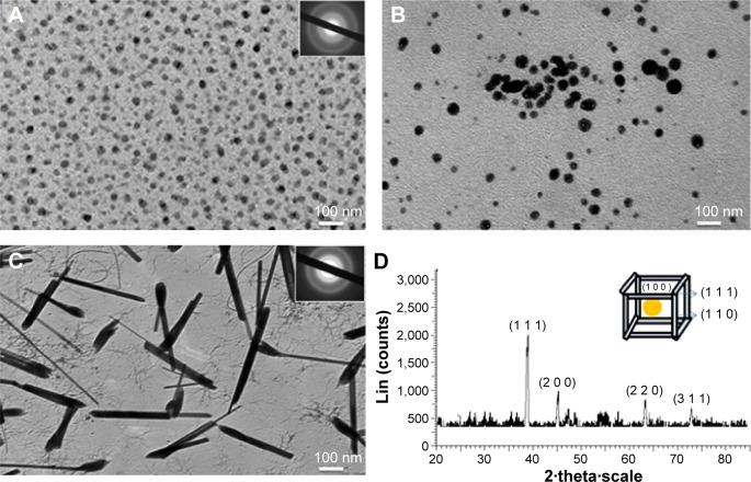
Dandruff is a prominent scalp problem caused by the growth of fungus Malassezia furfur, potentially cascading into dermal inflammation, itching, and tissue damage. The present work outlines a detailed analysis of the treatment of scalp infection using silver nanomaterials (Ag NMs), and focuses on biocidal activity owing to manipulation of size, shape, and structure. Monodisperse silver spherical nanoparticles (NPs) and nanorods (NRs) were synthesized by chemical routes that were characterized using analytical and spectroscopic techniques. Ag NMs demonstrated enhanced biocidal tendencies compared to market available drugs, itracanozole and ketoconazole, showing greater zones of inhibition. The obtained 20 nm and 50 nm spherical-shaped NPs and 50 nm NRs showed concentration-, size-, and shape-dependent antifungal activity, with 20 nm spherical-shaped NPs exhibiting excellent potency. Minimum inhibitory concentration for 20 nm was lowest at 0.2 mg/mL in comparison to 0.3 mg/mL for NRs. Primary irritation index was 0.33 and 0.16 for 20 nm and 50 nm spherical-shaped NPs, respectively, while 50 nm rod-shaped NMs exhibited negligible redness. An in vivo model for M. furfur infection was generated by passing fungi subcutaneously in rats' skin. Again, 20 nm particles showed best normalization of skin after 10 days on regular dosing, in comparison with bigger and rod-shaped particles. The statistical clinical score was highest for Ag nanorods, followed by 50 nm Ag NPs-treated animals. It was observed that 20 nm spherical particles exhibited the lowest score (0) compared with others as well as with antifungal drugs. Biochemical analysis performed by checking antioxidant enzymatic activities indicated tissue repair and normalization of enzymes and protein concentration by Ag NPs.
Keywords: Malassezia furfur; Wistar rat model; in vivo analysis; nanorods.
Figures


References
-
- University of Adelaide Mycology Online. 2015. [Accessed April 15, 2014]. Available from: http://www.mycology.adelaide.edu.au/Mycoses/Superficial/Malassezia_infec...
-
- Ravichandran G, Bharadwaj SV, Kolhapure SA. Evaluation of the clinical efficacy and safety of “anti-dandruff shampoo” in the treatment of dandruff. Antiseptic. 2004;201(5):5–8.
-
- Shuster S. The aetiology of dandruff and the mode of action of therapeutic agents. Br J Dermatol. 1984;111(2):235–242. - PubMed
-
- Sanfilippo A, English JC., 3rd An overview of medicated shampoos used in dandruff treatment. Pharm Ther. 2006;31:396–340.
-
- Cutsem VJ, Gerven VF, Fransen J. The in vitro antifungal activity of ketoconazole, zinc pyrithione, and selenium sulfide against pityrosporum guinea pigs. J Am Acad Dermatol. 1990;22(6):993–998. - PubMed
Publication types
MeSH terms
Substances
LinkOut - more resources
Full Text Sources
Other Literature Sources
Medical
