Gliotransmitter Release from Astrocytes: Functional, Developmental, and Pathological Implications in the Brain
- PMID: 26793048
- PMCID: PMC4709856
- DOI: 10.3389/fnins.2015.00499
Gliotransmitter Release from Astrocytes: Functional, Developmental, and Pathological Implications in the Brain
Abstract
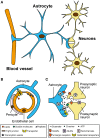
Astrocytes comprise a large population of cells in the brain and are important partners to neighboring neurons, vascular cells, and other glial cells. Astrocytes not only form a scaffold for other cells, but also extend foot processes around the capillaries to maintain the blood-brain barrier. Thus, environmental chemicals that exist in the blood stream could have potentially harmful effects on the physiological function of astrocytes. Although astrocytes are not electrically excitable, they have been shown to function as active participants in the development of neural circuits and synaptic activity. Astrocytes respond to neurotransmitters and contribute to synaptic information processing by releasing chemical transmitters called "gliotransmitters." State-of-the-art optical imaging techniques enable us to clarify how neurotransmitters elicit the release of various gliotransmitters, including glutamate, D-serine, and ATP. Moreover, recent studies have demonstrated that the disruption of gliotransmission results in neuronal dysfunction and abnormal behaviors in animal models. In this review, we focus on the latest technical approaches to clarify the molecular mechanisms of gliotransmitter exocytosis, and discuss the possibility that exposure to environmental chemicals could alter gliotransmission and cause neurodevelopmental disorders.
Keywords: astrocytes; exocytosis; glial cell; gliotransmitter; neurodevelopmental disorders; optical imaging; synaptic activity.
Figures


Similar articles
-
Regulated exocytosis from astrocytes physiological and pathological related aspects.Int Rev Neurobiol. 2009;85:261-93. doi: 10.1016/S0074-7742(09)85020-4. Int Rev Neurobiol. 2009. PMID: 19607976 Review.
-
Gliotransmission: focus on exocytotic release of L-glutamate and D-serine from astrocytes.Biochem Soc Trans. 2013 Dec;41(6):1557-61. doi: 10.1042/BST20130195. Biochem Soc Trans. 2013. PMID: 24256254 Review.
-
Expression and cellular function of vSNARE proteins in brain astrocytes.Neuroscience. 2016 May 26;323:76-83. doi: 10.1016/j.neuroscience.2015.10.036. Epub 2015 Oct 27. Neuroscience. 2016. PMID: 26518463 Review.
-
Gliotransmission and the tripartite synapse.Adv Exp Med Biol. 2012;970:307-31. doi: 10.1007/978-3-7091-0932-8_14. Adv Exp Med Biol. 2012. PMID: 22351062 Review.
-
Gliotransmission: Beyond Black-and-White.J Neurosci. 2018 Jan 3;38(1):14-25. doi: 10.1523/JNEUROSCI.0017-17.2017. J Neurosci. 2018. PMID: 29298905 Free PMC article. Review.
Cited by
-
Instructive roles of astrocytes in hippocampal synaptic plasticity: neuronal activity-dependent regulatory mechanisms.FEBS J. 2022 Apr;289(8):2202-2218. doi: 10.1111/febs.15878. Epub 2021 May 10. FEBS J. 2022. PMID: 33864430 Free PMC article. Review.
-
Sodium Butyrate Supplementation Modulates Neuroinflammatory Response Aggravated by Antibiotic Treatment in a Mouse Model of Binge-like Ethanol Drinking.Int J Mol Sci. 2022 Dec 10;23(24):15688. doi: 10.3390/ijms232415688. Int J Mol Sci. 2022. PMID: 36555338 Free PMC article.
-
Morphine Binds Creatine Kinase B and Inhibits Its Activity.Front Cell Neurosci. 2018 Dec 3;12:464. doi: 10.3389/fncel.2018.00464. eCollection 2018. Front Cell Neurosci. 2018. PMID: 30559651 Free PMC article.
-
GEARBOCS: An Adeno Associated Virus Tool for In Vivo Gene Editing in Astrocytes.bioRxiv [Preprint]. 2024 Oct 10:2023.01.17.524433. doi: 10.1101/2023.01.17.524433. bioRxiv. 2024. PMID: 36711516 Free PMC article. Preprint.
-
Synaptic plasticity and the role of astrocytes in central metabolic circuits.WIREs Mech Dis. 2024 Jan-Feb;16(1):e1632. doi: 10.1002/wsbm.1632. Epub 2023 Oct 13. WIREs Mech Dis. 2024. PMID: 37833830 Free PMC article. Review.
References
-
- Avendaño B. C., Montero T. D., Chávez C. E., Von Bernhardi R., Orellana J. A. (2015). Prenatal exposure to inflammatory conditions increases Cx43 and Panx1 unopposed channel opening and activation of astrocytes in the offspring effect on neuronal survival. Glia. 63, 2058–2072. 10.1002/glia.22877 - DOI - PubMed
Publication types
LinkOut - more resources
Full Text Sources
Other Literature Sources

