Review
. 2015 Dec;7(12):2139-50.
doi: 10.3978/j.issn.2072-1439.2015.10.69.

Echocardiographic assessment for ventricular assist device placement

Affiliations
Review

Echocardiographic assessment for ventricular assist device placement

Antolin S Flores et al. J Thorac Dis. 2015 Dec.

Abstract

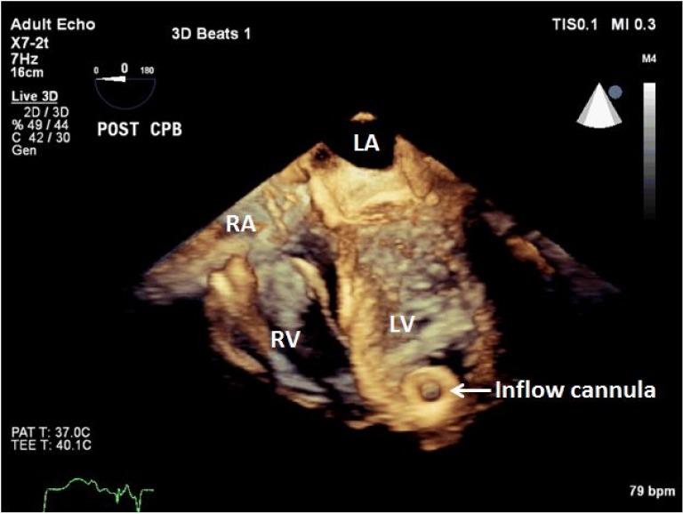
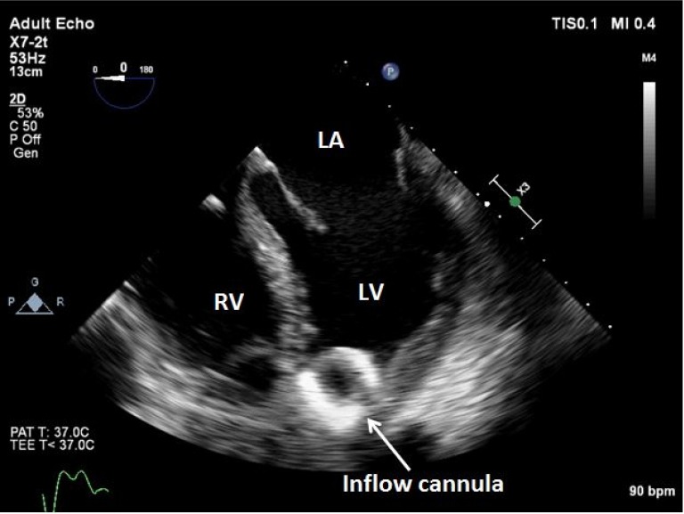
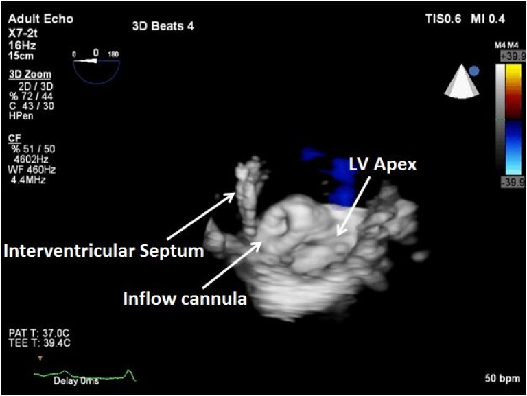
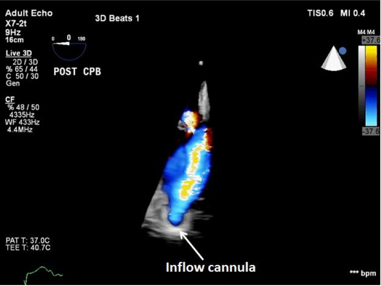
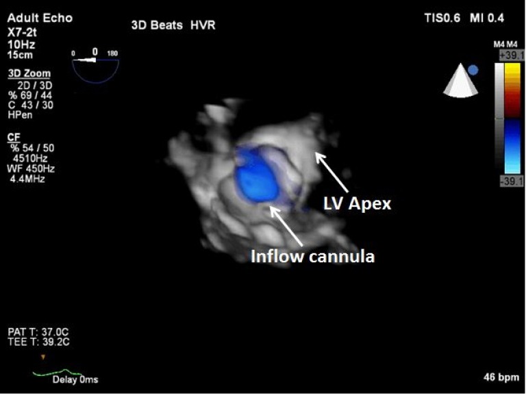
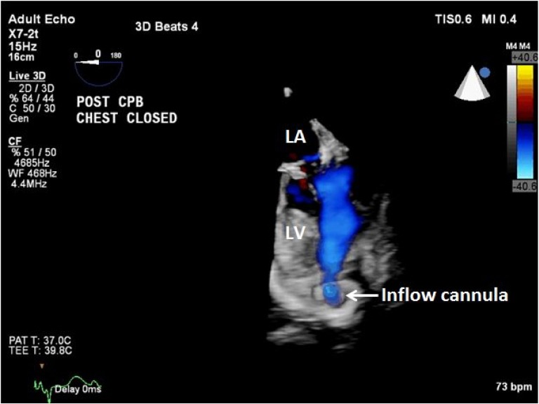
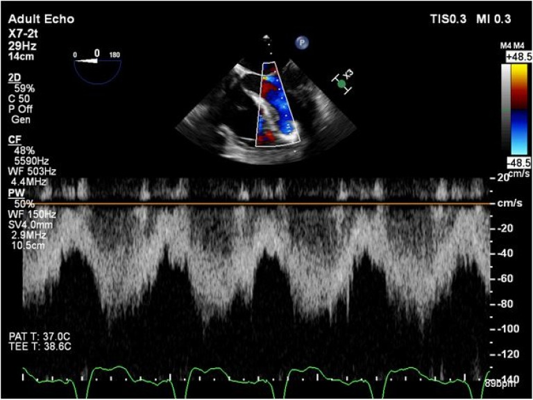
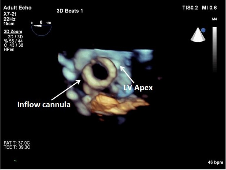
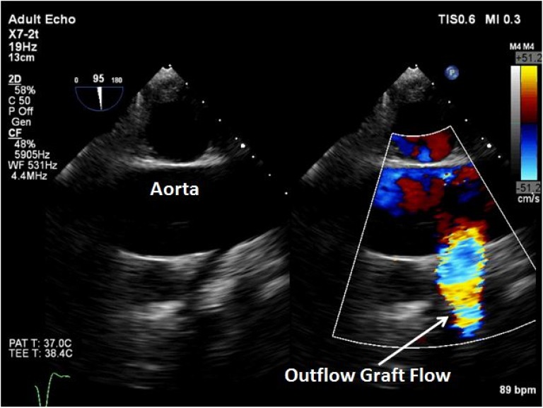
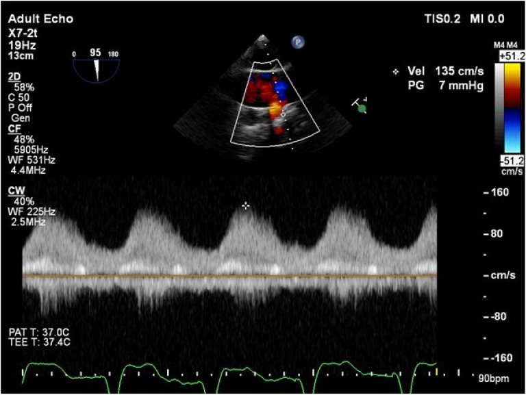
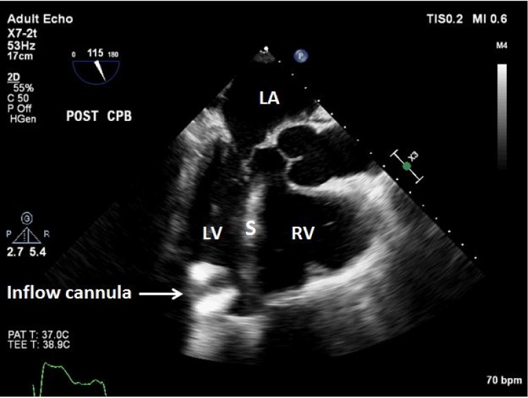
While many factors depend on successful implantation and outcome of left ventricular assist devices (LVAD), echocardiography remains an integral part and is vital to the success of this process. Transesophageal echocardiography (TEE) allows interrogation of all the cardiac structures and great vessels. The pre-implantation TEE exam establishes a baseline and may identify potential problems that need palliation. Among these, most significant are aortic insufficiency (AI), intracardiac thrombi, poor right ventricular (RV) function, and intracardiac shunts. The post-implantation exam allows for adequate de-airing of the heart and successful LVAD initiation. The position and flow profiles of the inflow and outflow cannulas of the LVAD may be assessed. Finally, it assists in the astute management and vigilant identification and correction of a number of complications in the immediate post-implantation period. TEE will continue to remain vital to the successful outcomes LVAD patients.

Keywords: Echocardiography; left ventricular assist devices (LVAD); transesophageal echocardiography (TEE); ventricular assist device.


Conflict of interest statement

Conflicts of Interest: The authors have no conflicts of interest to declare.

Figures

Figure 1
Figure 1
2D TEE mid-esophageal 4-chamber view showing significant biventricular and biatrial enlargement from end-stage heart disease. TEE, transesophageal echocardiography; LA, left atrium; RA, right atrium; LV, left ventricle; RV, right ventricle.
Figure 2
Figure 2
2D TEE mid-esophageal bicaval view with color flow Doppler demonstrating the absence of an interatrial shunt at a low Nyquist limit. TEE, transesophageal echocardiography; LA, left atrium; RA, right atrium; S, interatrial septum.
Figure 3
Figure 3
2D mid-esophageal long-axis view revealing moderate to severe aortic insufficiency (AI). LA, left atrium; LV, left ventricle; AV, aortic valve; RV, right ventricle.
Figure 4
Figure 4
2D mid-esophageal RV inflow-outflow view with color flow Doppler demonstrating moderate to severe tricuspid regurgitation (TR). LA, left atrium; RA, right atrium; RV, right ventricle.
Figure 5
Figure 5
3D TEE image after LVAD implantation demonstrating appropriate placement of the inflow cannula in the LV apex facing the mitral valve. TEE, transesophageal echocardiography; LVAD, left ventricular assist devices; CPB, cardiopulmonary bypass; LA, left atrium; RA, right atrium; RV, right ventricle; LV, left ventricle.
Figure 6
Figure 6
2D TEE mid-esophageal modified 4-chamber view showing the inflow cannula facing the mitral valve. TEE, transesophageal echocardiography; LA, left atrium; RV, right ventricle; LV, left ventricle.
Figure 7
Figure 7
3D TEE image showing the inflow cannula in the LV apex facing the interventricular septum. This TEE finding led to the repositioning of the inflow cannula. TEE, transesophageal echocardiography; LV, left ventricle.
Figure 8
Figure 8
3D TEE modified 4-chamber view demonstrating laminar blood flow with color flow Doppler in the inflow cannula. TEE, transesophageal echocardiography; CPB, cardiopulmonary bypass.
Figure 9
Figure 9
3D TEE en face view of the inflow cannula with color flow Doppler showing laminar blood flow. TEE, transesophageal echocardiography; LV, left ventricle.
Figure 10
Figure 10
3D TEE modified 4-chamber view demonstrating appropriate placement of the inflow cannula in the LV apex (facing the mitral valve), and laminar blood flow. TEE, transesophageal echocardiography; CPB, cardiopulmonary bypass; LA, left atrium; LV, left ventricle.
Figure 11
Figure 11
Pulsed-wave (PW) Doppler echocardiogram demonstrating the absence of cannula obstruction (peak velocities <1 m/s).
Figure 12
Figure 12
3D TEE en face view of the inflow cannula in the LV apex revealing the absence of tissue obstruction. TEE, transesophageal echocardiography; LV, left ventricle.
Figure 13
Figure 13
2D mid-esophageal long-axis view of the ascending aorta with color flow Doppler revealing laminar blood flow in the outflow graft.
Figure 14
Figure 14
Continuous wave Doppler echocardiogram demonstrating outflow graft patency.
Figure 15
Figure 15
2D TEE mid-esophageal long-axis view showing a suction event due to increased pump speed. The inflow cannula turned and faced the interventricular septum. TEE, transesophageal echocardiography; LA, left atrium; LV, left ventricle; S, interventricular septum; RV, right ventricle.

Similar articles

Cited by

References

    1. Stevenson LW, Miller LW, Desvigne-Nickens P, et al. Left ventricular assist device as destination for patients undergoing intravenous inotropic therapy: a subset analysis from REMATCH (Randomized Evaluation of Mechanical Assistance in Treatment of Chronic Heart Failure). Circulation 2004;110:975-81. - PubMed
    1. Rose EA, Gelijns AC, Moskowitz AJ, et al. Long-term use of a left ventricular assist device for end-stage heart failure. N Engl J Med 2001;345:1435-43. - PubMed
    1. Todaro MC, Khandheria BK, Paterick TE, et al. The practical role of echocardiography in selection, implantation, and management of patients requiring LVAD therapy. Curr Cardiol Rep 2014;16:468. - PubMed
    1. Lampert BC, Teuteberg JJ. Right ventricular failure after left ventricular assist devices. J Heart Lung Transplant 2015;34:1123-30. - PubMed
    1. Hahn RT, Abraham T, Adams MS, et al. Guidelines for performing a comprehensive transesophageal echocardiographic examination: recommendations from the American Society of Echocardiography and the Society of Cardiovascular Anesthesiologists. J Am Soc Echocardiogr 2013;26:921-64. - PubMed