Fully automated segmentation of the cervical cord from T1-weighted MRI using PropSeg: Application to multiple sclerosis
- PMID: 26793433
- PMCID: PMC4678307
- DOI: 10.1016/j.nicl.2015.11.001
Fully automated segmentation of the cervical cord from T1-weighted MRI using PropSeg: Application to multiple sclerosis
Abstract
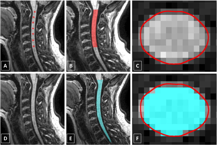
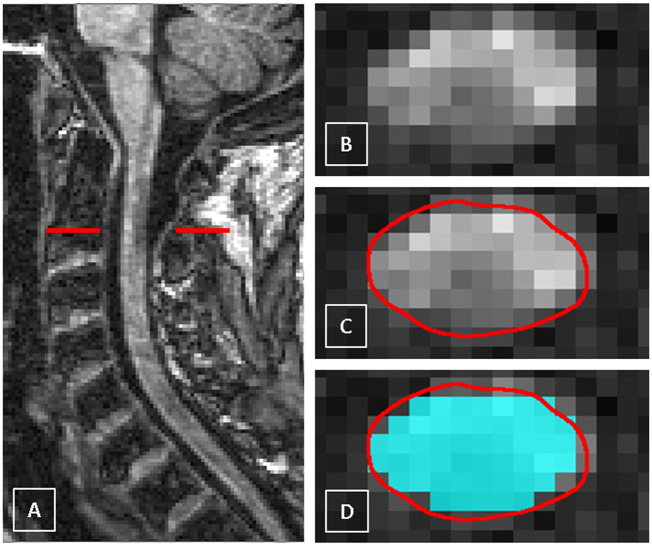
Spinal cord (SC) atrophy, i.e. a reduction in the SC cross-sectional area (CSA) over time, can be measured by means of image segmentation using magnetic resonance imaging (MRI). However, segmentation methods have been limited by factors relating to reproducibility or sensitivity to change. The purpose of this study was to evaluate a fully automated SC segmentation method (PropSeg), and compare this to a semi-automated active surface (AS) method, in healthy controls (HC) and people with multiple sclerosis (MS). MRI data from 120 people were retrospectively analysed; 26 HC, 21 with clinically isolated syndrome, 26 relapsing remitting MS, 26 primary and 21 secondary progressive MS. MRI data from 40 people returning after one year were also analysed. CSA measurements were obtained within the cervical SC. Reproducibility of the measurements was assessed using the intraclass correlation coefficient (ICC). A comparison between mean CSA changes obtained with the two methods over time was performed using multivariate structural equation regression models. Associations between CSA measures and clinical scores were investigated using linear regression models. Compared to the AS method, the reproducibility of CSA measurements obtained with PropSeg was high, both in patients and in HC, with ICC > 0.98 in all cases. There was no significant difference between PropSeg and AS in terms of detecting change over time. Furthermore, PropSeg provided measures that correlated with physical disability, similar to the AS method. PropSeg is a time-efficient and reliable segmentation method, which requires no manual intervention, and may facilitate large multi-centre neuroprotective trials in progressive MS.
Keywords: Cord cross-sectional area; Grey matter; Image segmentation; Magnetic resonance imaging; White matter.
Figures


Similar articles
-
Validation of mean upper cervical cord area (MUCCA) measurement techniques in multiple sclerosis (MS): High reproducibility and robustness to lesions, but large software and scanner effects.Neuroimage Clin. 2019;24:101962. doi: 10.1016/j.nicl.2019.101962. Epub 2019 Aug 6. Neuroimage Clin. 2019. PMID: 31416017 Free PMC article.
-
Cervical cord area is associated with infratentorial grey and white matter volume predominantly in relapsing-remitting multiple sclerosis: A study using semi-automated cord volumetry and voxel-based morphometry.Mult Scler Relat Disord. 2015 May;4(3):264-72. doi: 10.1016/j.msard.2015.04.003. Epub 2015 Apr 15. Mult Scler Relat Disord. 2015. PMID: 26008944
-
Reliable volumetry of the cervical spinal cord in MS patient follow-up data with cord image analyzer (Cordial).J Neurol. 2016 Jul;263(7):1364-74. doi: 10.1007/s00415-016-8133-0. Epub 2016 May 9. J Neurol. 2016. PMID: 27159989
-
Spinal Cord Atrophy in Multiple Sclerosis: A Systematic Review and Meta-Analysis.J Neuroimaging. 2018 Nov;28(6):556-586. doi: 10.1111/jon.12553. Epub 2018 Aug 13. J Neuroimaging. 2018. PMID: 30102003
-
Spinal cord imaging in multiple sclerosis.J Neuroimaging. 2005;15(4 Suppl):94S-102S. doi: 10.1177/1051228405283292. J Neuroimaging. 2005. PMID: 16385022 Review.
Cited by
-
Validation of mean upper cervical cord area (MUCCA) measurement techniques in multiple sclerosis (MS): High reproducibility and robustness to lesions, but large software and scanner effects.Neuroimage Clin. 2019;24:101962. doi: 10.1016/j.nicl.2019.101962. Epub 2019 Aug 6. Neuroimage Clin. 2019. PMID: 31416017 Free PMC article.
-
Cervical spinal cord atrophy: An early marker of progressive MS onset.Neurol Neuroimmunol Neuroinflamm. 2018 Jan 22;5(2):e435. doi: 10.1212/NXI.0000000000000435. eCollection 2018 Mar. Neurol Neuroimmunol Neuroinflamm. 2018. PMID: 29435472 Free PMC article.
-
MRI Atlas-Based Measurement of Spinal Cord Injury Predicts Outcome in Acute Flaccid Myelitis.AJNR Am J Neuroradiol. 2017 Feb;38(2):410-417. doi: 10.3174/ajnr.A5044. Epub 2016 Dec 15. AJNR Am J Neuroradiol. 2017. PMID: 27979798 Free PMC article.
-
Spinal Cord Gray Matter Atrophy in Amyotrophic Lateral Sclerosis.AJNR Am J Neuroradiol. 2018 Jan;39(1):184-192. doi: 10.3174/ajnr.A5427. Epub 2017 Nov 9. AJNR Am J Neuroradiol. 2018. PMID: 29122760 Free PMC article.
-
Evaluation of Intra- and Interscanner Reliability of MRI Protocols for Spinal Cord Gray Matter and Total Cross-Sectional Area Measurements.J Magn Reson Imaging. 2019 Apr;49(4):1078-1090. doi: 10.1002/jmri.26269. Epub 2018 Sep 9. J Magn Reson Imaging. 2019. PMID: 30198209 Free PMC article.
References
-
- Bartlett J., Frost C. Reliability, repeatability and reproducibility: analysis of measurement errors in continuous variables. Ultrasound Obstet. Gynecol. 2008;31:466–475. - PubMed
Publication types
MeSH terms
LinkOut - more resources
Full Text Sources
Other Literature Sources
Medical
Research Materials