

Tuberculous Constrictive Pericarditis
- PMID: 26793674
- PMCID: PMC4707979
- DOI: 10.5812/cardiovascmed.29614
Tuberculous Constrictive Pericarditis
Abstract
Introduction: Constrictive pericarditis is characterized by constriction of the heart secondary to pericardial inflammation. Cardiovascular magnetic resonance (CMR) imaging is useful imaging modality for addressing the challenges of confirming this diagnosis. It can be used to exclude other causes of right heart failure, such as pulmonary hypertension or myocardial infarction, determine whether the pericardium is causing constriction and differentiate it from restrictive cardiomyopathy, which also causes impaired cardiac filling.
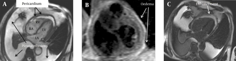
Case presentation: A 77-year-old man from a country with high incidence of tuberculosis presented with severe dyspnea. Echocardiography revealed a small left ventricle with normal systolic and mildly impaired diastolic function. Left heart catheterization revealed non-obstructive coronary disease, not felt contributory to the dyspnea. Anatomy imaging with cardiovascular magnetic resonance imaging (CMR) showed global, severely thickened pericardium. Short tau inversion recovery (STIR) sequences for detection of oedema/ inflammation showed increased signal intensity and free breathing sequences confirmed septal flattening on inspiration. Late gadolinium imaging confirmed enhancement in the pericardium, with all findings suggestive of pericardial inflammation and constriction.
Conclusions: CMR with STIR sequences, free breathing sequences and late gadolinium imaging can prove extremely useful for diagnosing constrictive pericarditis.
Keywords: Constrictive Pericarditides; Pericardial Effusion; Tuberculosis.
Figures
References
-
- Lower R. Treatment of Heart [in Latin]. London: Allestry; 1669.
-
- Myers RB, Spodick DH. Constrictive pericarditis: clinical and pathophysiologic characteristics. Am Heart J. 1999;138(2 Pt 1):219–32. - PubMed
-
- Mayosi BM, Wiysonge CS, Ntsekhe M, Volmink JA, Gumedze F, Maartens G, et al. Clinical characteristics and initial management of patients with tuberculous pericarditis in the HIV era: the Investigation of the Management of Pericarditis in Africa (IMPI Africa) registry. BMC Infect Dis. 2006;6:2. doi: 10.1186/1471-2334-6-2. - DOI - PMC - PubMed
Publication types
LinkOut - more resources
Full Text Sources
Other Literature Sources